Anus, Rectum, and Prostate Physical Exam
 Call Kai
 Learn
Learn Practice Test
 Spaced Repetition
 Match
Match Flashcards
 Knowt Play
1/119

There's no tags or description
Looks like no tags are added yet.
Name | Mastery | Learn | Test | Matching | Spaced | Call with Kai | Chat |
|---|
No analytics yet
Send a link to your students to track their progress
120 Terms
What is the typical length of the anal canal?
2.5 to 4 cm long.
Describe the appearance of visible tissue at the external margin of the anus.
Moist, hairless mucosa.
Which nerve type supplies the lower half of the anal canal?
Somatic sensory nerves.
Is the lower half of the anal canal sensitive to pain?
Yes, it is sensitive to pain.
Which nerve type controls the upper half of the anal canal?
Autonomic nervous system.
Is the upper half of the anal canal sensitive to pain?
No, it is relatively insensitive to pain.
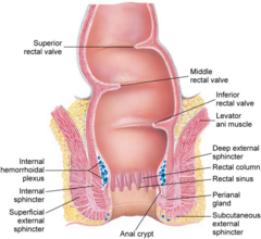
What is the length of the rectum?
Approximately 12 cm long.
Where is the rectum located relative to the anus?
Superior to the anus.
What structure is continuous with the proximal end of the rectum?
The sigmoid colon.
What is the primary function of the rectal ampulla?
To store flatus and feces.
What are the standard dimensions of the prostate gland?
4 × 3 × 2 cm.
What shape is the prostate gland commonly described as?
Heart-shaped.
Where is the prostate gland located relative to the bladder?
At the base of the bladder, surrounding the urethra.
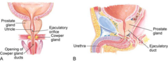
Which surface of the prostate gland is accessible via digital rectal examination?
The posterior surface.
Against which wall of the rectum does the prostate gland lie?
The anterior rectal wall.
How many lobes make up the prostate gland structure?
Three lobes.
Which structural marker divides the left and right lateral lobes of the prostate?
The median sulcus.
Can the median lobe of the prostate be felt during a DRE?
No, the median lobe is not palpable.
What key historical detail should be inquired regarding rectal symptoms?
Rectal pain, bleeding, and changes in bowel function.
Why is stool color an important history question during rectal evaluation?
It indicates potential sources of gastrointestinal bleeding.
What anal symptoms should be evaluated in HPI?
Anal itching, pain, and bleeding.
Why is sexual history relevant in a rectal exam HPI?
To assess risk related to receptive anal intercourse.
What obstructive voiding symptoms suggest prostatic pathology?
Difficulty starting urination and weak urine flow.
What frequency symptom is typical in prostate complaints?
Frequent urination, especially worse at night (nocturia).
What process is essential before screening for prostate cancer?
Informed decision making.
What two standard diagnostic methods screen for prostate issues?
Digital rectal examination (DRE) and PSA testing.
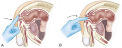
How should a skillfully performed rectal examination feel to the patient?
It should not be painful.
What clinician approach ensures a successful rectal examination?
Calm demeanor, clear explanation, gentleness, and slow movement.
In which age group is a rectal exam typically deferred if no complaints exist?
Adolescents.
Name one valid patient position for a rectal exam involving knees.
Knee-to-chest position.
Describe the left lateral position used for rectal examination.
Patient on left side with hips and knees flexed.
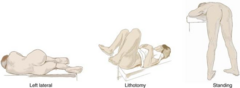
How is the standing position used for a rectal exam configured?
Hips flexed with upper body supported by the table.
What key cutaneous features should be inspected in the sacrococcygeal area?
Masses, rashes, inflammation, excoriation, and scars.
What congenital skin findings should be noted in the pilonidal area?
Pilonidal dimpling and tufts of hair.
What signs are sought during palpation of the sacrococcygeal area?
Tenderness and signs of inflammation.
Name two inflammatory conditions palpated in the perianal area.
Perianal abscess and anorectal fistula.
What localized tear can be identified during perianal assessment?
Anal fissure.
What condition causes severe perianal itching?
Pruritus ani.
What skin lesions or growth forms should be inspected around the anus?
Skin tags and warts.
What vascular abnormalities are visually inspected around the anus?
External hemorrhoids.

How are anal and rectal findings localized and described?
Using clock referents.
Where is 12 o'clock located on the anal clock reference system?
In the ventral midline.
Where is 6 o'clock located on the anal clock reference system?
In the dorsal midline.
How should finger entry into the anal canal begin during a DRE?
Press the finger pad gently against the anal opening.
When should the finger slip into the anal canal?
As the external sphincter relaxes.
What does reduced external sphincter tone potentially indicate?
A neurological deficit.
What can cause an extremely tight external anal sphincter?
Scarring, fissure spasticity, inflammation, or patient anxiety.
What does severe rectal pain almost always signify?
Local disease.
Name four causes of acute local rectal pain.
Irritation, rock-hard constipation, fissures, or thrombosed hemorrhoids.
Through which anatomical wall is the prostate palpated?
Anterior rectal wall.
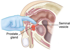
Which prostate features are assessed during digital palpation?
Size, contour, median sulcus, lateral lobes, consistency, tenderness.
What is the normal physical consistency of a healthy prostate gland?
Rubbery.
Is a normal prostate tender upon palpation?
No, it is normally non-tender.
In what standard position can a female rectal exam be done after a GU exam?
Lithotomy position.
What specific septum/wall is evaluated during a female rectovaginal exam?
The rectovaginal wall.
If a female rectal exam is standalone, what positioning is used?
The same positioning as described for male patients.
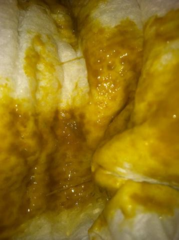
What general characteristics of stool should be routinely noted?
Color and consistency.
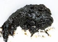
What pathological additives in stool should be identified visually or clinically?
Blood, pus, and mucus.
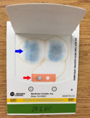
What testing tool checks for occult blood in stool samples?
Guaiac cards.
What causes hair penetration in pilonidal cyst formation?
Loose hairs penetrating skin in the sacrococcygeal area.
Where is a pilonidal cyst characteristically located on physical exam?
At the top of the gluteal cleft.
What physical signs indicate an active pilonidal cyst?
Painful, swollen, fluctuant area with potential sinus tracking.
What pathogen causes anal warts (Condyloma acuminata)?
Human Papillomavirus (HPV).
What physical appearance do condyloma acuminata present with?
Pedunculated warts along the anus and gluteal cleft.
What tissue spaces do perianal or perirectal abscesses infect?
Soft tissues or mucus-secreting anal glands surrounding the canal.

Where does abscess formation occur in perirectal infections?
In the deeper perianal/perirectal tissues.
What is the typical microbial nature of perianal abscesses?
Polymicrobial.
What is an anorectal fissure structurally?
A tear in the anal mucosa.
What history is classic for an anorectal fissure?
Pain with bowel movements, constipation, bright red blood on toilet paper.
What is seen on physical exam in a patient with an anorectal fissure?
Mucosal tear with or without active bleeding.
What is an anorectal fistula?
An inflammatory tract connecting anus/rectum to skin or other tissue.
What primary condition leads to the development of an anorectal fistula?
Drainage of a perianal or perirectal abscess.
What is the most common cause of pruritus ani in adults?
Fungal infection.
What is a frequent cause of pruritus ani in pediatric patients?
Parasitic infection (e.g., pinworms).
What anatomical landmark divides internal from external hemorrhoids?
The anorectal line (dentate/pectinate line).
Where do external hemorrhoids originate relative to the anorectal line?
Below the anorectal line.
What type of tissue covers external hemorrhoids?
Anal skin.
Where do internal hemorrhoids originate relative to the anorectal junction?
Above the anorectal junction.
What type of tissue covers internal hemorrhoids?
Rectal mucosa.
In which demographic group is rectal prolapse most typical?
Elderly women.
What is noted on physical examination of rectal prolapse?
Protruding mucosal prolapse.
How does rectal prolapse typically reduce?
Spontaneously or with manual assistance.
What histology constitutes the majority of anal cancers?
Squamous cell carcinoma.
Which viral infection is strongly linked to squamous cell anal cancer?
HPV infection.
What screening modality can be considered for high-risk anal cancer patients?
Anal Pap smear.
Where do adenocarcinomas of the anal region originate?
In the glands near the anus.
Name two rare malignant skin cancers that can occur at the anus.
Basal cell carcinoma and malignant melanoma.
What cell type forms the vast majority of colorectal cancers?
Adenocarcinoma.
Describe the progression pathway of colorectal adenocarcinoma.
Cell proliferation leading to adenoma and then invasive carcinoma.
Why are higher colonic carcinomas not detectable on digital rectal exam?
They are located beyond the reach of the examining finger.
What defines acute bacterial prostatitis?
Infectious inflammation of the prostate gland.
What combination of symptoms characterizes acute bacterial prostatitis?
Urinary symptoms and systemic symptoms (e.g., fever, chills).
How does a prostate feel on physical exam during acute prostatitis?
Tender, enlarged, or boggy.
Why must DRE be performed gently in suspected acute prostatitis?
To avoid extreme pain and bacteremia.
What is benign prostatic hypertrophy (BPH)?
Non-cancerous continuing enlargement of the prostate gland.
In what age group is BPH most commonly observed?
Men older than 50 years.
What histological origin accounts for 99% of prostate cancers?
Adenocarcinoma arising from gland cells.
Why is the somatic innervation of the lower anal canal clinically important?
It makes lesions in this area exquisitely painful.
Why are internal hemorrhoids often painless compared to external ones?
They lie above the dentate line under autonomic sensory control.
What orientation is used as the 3 o'clock position on the anal clock face?
Left lateral aspect of the anus.