Ch. 5 Skeletal system
 Call Kai
 Learn
Learn Practice Test
 Spaced Repetition
 Match
Match Flashcards
 Knowt Play
1/22

There's no tags or description
Looks like no tags are added yet.
Name | Mastery | Learn | Test | Matching | Spaced | Call with Kai | Chat |
|---|
No analytics yet
Send a link to your students to track their progress
23 Terms
Canal
Bone marking: large passageway (eg. optic canal)
Condyle
Rounded prominence providing support to hyaline cartilage and forms joints
Foramen
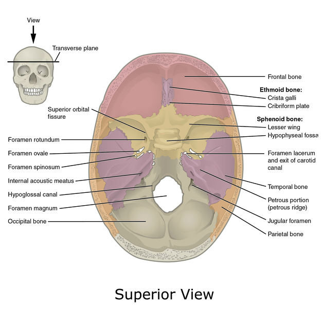
Hole in bone through which nerves and blood vessels pass
Fossa
Shallow depression in bone surface
Meatus
Tube-like channel that goes through bone, may provide passageway for nerves + vessel + sound

Process
Bony projection for muscle attachment, eg. spinal processes

Protuberance
A bump/outgrowth on bone

Compact bone structure + where is it found on the body
Osteon / Haversian system
Cylindrical bone matrix (concentric lamellae) w/ a central canal and bone cells + canaliculi in layers
Compact bone is in areas of high compressional stress and the outer portions of all bones
Spongy bone structure
“Cross beam” trabeculae with space (dipole) in btwn
Bone classifications
Long - long, medullary cavity w/ marrow (femur, tibia, fingers)
Short - approximately equal height/width, lack projections and holes (eg carpus and tarsus)
Flat - flat with thin spongy bone layer and no cavity (eg. ribs, sternum, scapula)
Irregular - many projections, do not fit other categories (vertebrae)
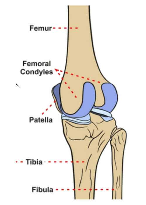
Sesamoid - small bones typically embedded in tendons (eg patella)
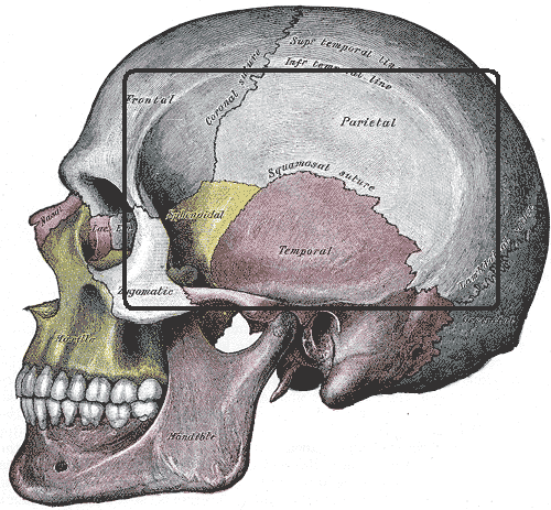
Sutural/wormian - extra bones variable across individuals that develop along cranial sutures
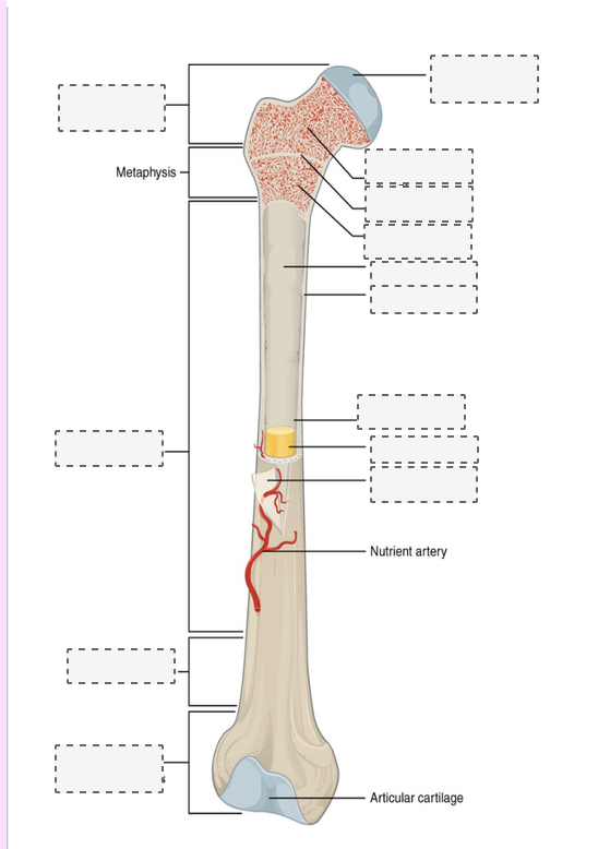
Long bone anatomy
red marrow - place of erythropoiesis
yellow - replaced with adipose with age

Periosteum and endosteum
Periosteum
primarily dense irregular connective; membrane surrounding bone composed of dense fibers extending from the inside to form a strong attachment point
continuous with synovial joint capsule
inner osteogenic layer: growth and repair
Endosteum
Membrane lining medullary cavities and trabeculae
Cells of the bone
Osteoprogenitor / osteogenic
Stem cells of perio + endosteum that differentiate into osteoblasts: growth and repair
Osteoblast
Cells actively secreting matrix in osteoid (soft uncalcified matrix)
Osteocyte
Mature cell in solid matrix, monitors stress + remodeling
Osteoclast
Macrophage WB stem cell-derived multinucleated cell that breaks down bone (remodeling)
Interstitial vs appositional growth
Interstitial: within/inside tissue, lengthens bone
Appositional: along circumference, increases diameter
Intramembranous vs endochondral growth
Intramembranous - No cartilage frame, bone grows around embryonic mesenchyme and forms trabeculae. Closed off then outer portions remodeled into compact bone while inside spongey
Endochondral - Cartilage frame, primary ossification center where calcium accumulates + chondrocytes die to leave behind cavities (lacunae) for osteoblasts and vessels to occupy. Secondary centers emerge and all cart except for joints and line replaced with bone
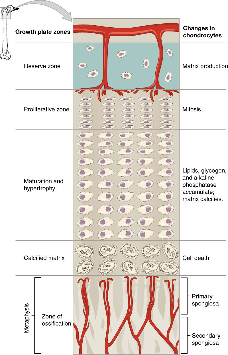
Growth plate zones
Resting - regular cartilage
Proliferating
Hypertrophic - chondrocytes stop dividing but incr in size
Calcification - calcium accumulates, chondrocytes die
Ossification - hardening of matrix
Types of fractures
+Pott’s (ankle)

Cranial bones
8 total:
Parietal, temporal (paired)
Frontal, occipital, sphenoid, ethmoid

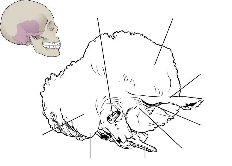

Temporal bone structures (inside of skull)
Petrous: Ridge on cranial cavity floor
Stylomastoid foramen: exit point for cranial nerves between styloid process and mastoid process
Carotid canal: passage for major brain artery, outside base of skull anteromedial to styloid process
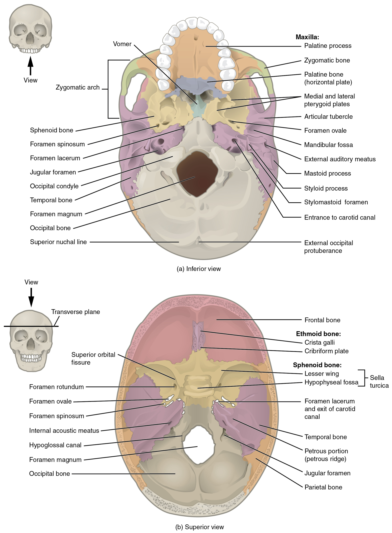
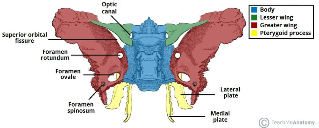
Sphenoid bone
“moth”like, keystone bone: joints with almost every other skull bone
Forms base of skull, has passageways for nerves, optic canals, arteries
Ethmoid bone
Located in midline of skull, embedded behind nasus
Forms roof and lateral wall of nasal cavity, part of orbit medial walls, upper nasal septum

Fontanelles
“Soft spots” on infant head: dense connective tissue separating fetal cranial bones to allow for growth and passage through the birth canal
Disappear around age 2 but bones remain separated at sutures before fusing