Biology - cell structure
1/103
There's no tags or description
Looks like no tags are added yet.
Name | Mastery | Learn | Test | Matching | Spaced |
|---|
No study sessions yet.
104 Terms
what are microscopes?
instruments that produce a magnified image of an object
how do lenses work more effectively?
if they are used in pairs in a compound light microscope
what the smallest distance between 2 objects that a light microscope can distinguish?
0.2 micrometres
what the smallest distance between 2 objects that an electron microscope can distinguish?
0.1 nanometres
what is the object of a microscope?
the material that is put under a microscope
what is the image of a microscope?
the appearance of the material when viewed under the microscope
what is magnification?
how many times bigger the image is when compared to the object
magnification =?
size of image / size of real object
what do you have to ensure when calculating magnification?
units are the same for the sizes measured
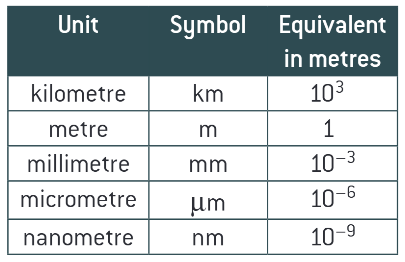
conversions table:
what is resolution/ resolving power of a microscope?
the minimum distance apart that 2 objects can be in order for them to appear separate
what does resolution depend on?
the wavelength/ form of radiation used
does increasing magnification increase resolution?
no - every microscope has a limit of resolution
what will happen at the limit of resolution?
increasing the magnification will just make the object appear more blurred
why do we want to obtain large numbers of isolated organelles?
to study structure and function
what is cell fractionation?
the process where cells are broken up and the different organelles they contain are separated out
what must happen before cell fractionation can begin?
the tissue must be placed in cold, buffered solution of the same water potential as the tissue
why must it be cold?
to reduce enzyme activity that might break down organelles
why must it be buffered?
so that the pH doesnt fluctuate. Any change in pH could alter the structure of the organelles or affect the functioning of enzymes
why must it be of the same water potential as the tissue?
to prevent organelles bursting or shrinking as a result of osmotic gain/ loss of water
what are the 2 stages to cell fractionation?
homogenation
ultracentrifugation
what is homogenation?
cells are broken up by a homogeniser. this releases the organelles from the cell. the resultant fluid (homogenate), is then filtered to remove any complete cells and large pieces of debris
what is ultracentrifugation?
the process by which the fragments in the filtered homogenate are separated in a machine called a centrifuge. this spices the tubes of homogenate at very high speed in order to create a centrifugal forces
what is the process for animal cells?
the tube of filtrate is placed in the centrifuge and spun at a slow speed
the heaviest organelles, the nuclei, are forced to the bottom of the tube, where they forms a thin sediment/ pellet
the fluid at the top of the tube (supernatant) is removed, leaving just the nuclei sediment
the supernatant is transferred to another tube and spun in the centrifuge at a faster speed than before
the next heaviest organelles, the mitochondria, are forced to the bottom of the tube
the process is continued in this way so that, at each increase in speed, the next heaviest organelle is sedimented and separated out
why is cell fractionation so good?
has allowed advances in biological knowledge by allowing a detailed study of the structure and function of organelles
what are the 2 main advantages of an electron microscope?
electron beam has a very short wavelength and the microscope can therefore resolve objects well - high resolution
as electrons are negatively charged the beam can be focused using electromagnets
what is the case because electrons are absorbed or deflected by molecules in air?
a near vacuum has to be created within the chamber of an electron microscope in order for it to work effectively
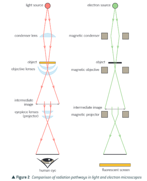
comparison of radiation pathways in light and electron microscopes?
what are the 2 types of electron microscope?
transmission electron microscope
scanning electron microscope
what is the TEM?
consists of an electron gun that produces a beam of electrons that is focused onto the specimen by a condensor electromagnet. the beam passes through a thin section of the specimen. parts of this specimen absorb electrons and therefore appear dark. other parts allow the electrons to pass through and so appear bright
what is the image produced on the screen that can be photographed called?
a photomicrograph
why is the resolving power of the TEM not always 0.1nm?
difficulties preparing the specimen limit the resolution that can be achieved
a higher energy electron beam is required and this may destroy the specimen
limitations of the TEM?
the system must be in a vacuum and therefore living specimens cant be observed
a complex staining process is required and even then the image isnt in colour
the specimen must be very thin
the image may contain artefacts
what are artefacts?
things that result from the way the specimen is prepared. Artefacts may appear on the finished micrograph but are not part of the natural specimen. it is therefore not always easy to be sure that what we see on a photomicrograph really exists in that form
why must the specimens be really thin?
to allow electrons to penetrate, resulting in a flat 2D image
how can you build the image up to being in 3D?
take a series of sections through the specimen and look at them through the photomicrographs produced
what is similar about the TEM and SEM?
same limitations, except the specimens dont need to be thin as electrons dont penetrate
what happens with the SEM?
the SEM directs a beam of electrons on to the surface of the specimen from above, rather than penetrating it from below. the beam is then passed back and forth across a portion of the specimen in a regular pattern. the electrons are scattered by the specimen and the pattern of this scattering depends on the contours of the specimen surface
how can you build up a 3D image?
by computer analysis of the pattern of scattered electrons and secondary electrons produced
whats the resolving power of a basic SEM?
20nm