Microbiology Exam 3, Dr. Collins
1/150
There's no tags or description
Looks like no tags are added yet.
Name | Mastery | Learn | Test | Matching | Spaced | Call with Kai |
|---|
No study sessions yet.
151 Terms
What are the two approaches to gene regulation?
1) Controlling Transcription: preventing transcription initiation or elongation.
2) Controlling Post-translation: Altering the activity of enzymes and proteins.
Constitutive genes:
Are housekeeping genes that are expressed continuously by the cell
Inducible genes:
Genes that code for inducible enzymes needed only in certain environments.
Enzymes that function in catabolic pathways are products of inducible genes.
These enzymes are only present when their substrate is available.
Example of an enzyme encoded by an inducible gene:
b-Galactosidase
Repressible genes:
Enzymes that function in biosynthetic pathways are products of repressible genes.
These enzymes are always present unless the end product in the biosynthetic pathway is available.
Example of an enzyme encoded by a repressible gene:
Enzymes from the trp operon
Catabolic enzymes:
Enzymes encoded by inducible genes.
Biosynthetic enzymes:
Enzymes encoded by repressible genes.
What are the two types of transcriptional control?
Negative control and positive control
Negative control:
Proteins that inhibit transcription initiation by binding to a DNA regulatory site.
mRNA expression is reduced.
Negative control has repressor proteins that exist in active and inactive forms.
Positive control:
Proteins that promote transcription initiation by binding to a DNA regulatory site.
mRNA synthesis increases.
Inactive protein is activated by inducer.
Active protein is inactivated by inhibitor.
What is used to control transcription initiation?
Regulatory proteins and DNA binding domains
What are the 3 inducible genes encoded for in Lac (lactose) Operon?
1) lacZ
2) lacY
3) lacA
lacZ
lacZ encodes for beta-galactosidase. This is an enzyme that will break down the lactose into galactose and glucose.
lacY
lacY encodes for lactose permease. This allows for the transport of lactose into the cell.
lacA
lacA encodes for galactoside transacetylase.
What is the regulatory gene in Lac operon?
lacI is the regulatory gene
lacI
lacI encodes for the repressor protein that will bind to the operator region and inhibit transcription. lacI only makes the repressor protein when lactose is NOT present.
lac operon repressor proteins:
Lac repressors are made of tetramers, which are made of 4 identical subunits.
The tetramers bind to 3 operator sites (O1, O2, O3).
This bends the DNA into a loop, preventing RNA polymerase from accessing promoter and transcribing it.
Lac operon is under both positive and negative control. Negative control of lac operon is mediated by inducible genes. What is positive control of lac operon mediated by?
Positive control of lac operon is regulated by catabolite activator protein (CAP).
How does CAP regulate positive control of lac operon?
CAP regulates in response to the presence or absence of glucose, which allows for the preferential use of glucose.
Catabolite Activator Protein (CAP):
Regulates operons that use the catabolic pathway. All catabolite operons contain a CAP-binding site.
CAP exists in 2 forms:
Active: when cAMP is bound to it.
Inactive: when cAMP is not bound to it.
CAP must be bound to this site before RNA polymerase can bind the promoter and begin transcription.
What regulates CAP activity?
cAMP
When there is NO glucose, there are HIGH levels of cAMP. What does this mean?
This means that cAMP can bind to CAP, thus activating it. When CAP is active it promotes the transcription of operons used for the catabolism of other sugars. This is why the organism preferentially uses glucose over other sugars.
What are the 5 transcriptional genes of the trp operon?
trpA,B,C,D, and E. These genes encode for enzymes needed to synthesize tryptophan.
trp repressor:
Regulates negative transcriptional control of repressible genes
The trp operon only functions in the _______ of tryptophan.
absence
What is Arabinose and what does the Arabinose operon do:
Arabinose is a sugar that the cell can use to produce ATP. The arabinose operon can either promote or inhibit transcription.
What is the regulatory gene of the Arabinose operon?
The regulatory gene is araC.
araC:
araC is a regulatory gene that can act as an activator or a repressor.
What are the 3 transcriptional genes of the Arabinose operon?
araA, araB, and araD
When is the arabinose operon active or inactive?
araC is active when arabinose is absent.
araC is inactive when arabinose is present.
This activity depends on environmental conditions.
What are the components of the pGLO plasmid?
1) Origin of replication.
2) GFP: green fluorescent protein.
3) bla: encodes for beta-lactamase, which causes ampicillin resistance.
4) araC: arabinose regulator protein, which regulates GFP expression.
Characteristics of Helicobacter pylori:
Gram-negative bacteria.
Curved rod shaped.
Microaerophilic (only requires a
small amount of oxygen to grow).
Forms biofilms.
Where is H. pylori found, and what survival mechanism does it use to allow for its survival there?
H. pylori is found in the stomach, and it uses Urease activity to increase the pH of the stomach, making it less acidic and allowing the bacteria to survive.
Who is most susceptible to H. pylori infections and why?
H. pylori typically infects children because their stomachs are less acidic, which the bacteria prefer.
Symptoms of H. pylori infection:
Burning pain in your abdomen
Nausea
Loss appetite
Frequent burping
Bloating
Unintentional weight loss
What percent of individuals with H. pylori infection are asymptomatic?
80%
How is H. pylori infection transmitted?
It is transmitted person-person via contact with saliva, feces, or vomit, or from contaminated food and water.
Prevalence of H. pylori is _______ in developing countries that don’t have good water treatment, and _________ in the US and industrialized countries.
70%, 30-40%
4 ways to diagnosis H. pylori infection:
1) Blood test: If antibodies are found that is an indication of an infection.
2) Breath test: Urease test to see if patient has urease activity occuring, which indicates the disease.
3) Stool test: Fecal antigen test.
4) Scope test: Biopsy.
Treatment of H. pylori infection:
Proton pump inhibitors (stop acid production).
Omeprazole and the antibiotics clarithromycin and amoxicillin.
Histamine blockers.
Bismuth subsalicylate (Peptobismul).
What change within organisms did the scientist Griffith observe in 1928?
He observed the change of non-virulent organisms (don’t cause disease) into virulent ones as a result of “transformation”
What did the scientists MacLeod and McCarty discover in 1944 in regards to transformation?
These two scientists showed that DNA was the transforming principle.
What did Griffith demonstrate with his mice experiment?
Griffith showed that DNA could transform the virulence of a cell by injecting virulent and non-virulent bacteria into mice. A mixture of virulent and non-virulent strains were obtained from the deceased mice.
What did scientists Hershey and Chase demonstrate in their 1952 experiment?
They determined that only DNA entered the cell, but both new DNA and protein coats were synthesized and incorporated into new viruses. This indicates that DNA had the genetic information for synthesis of both of these viral components.
Vertical transfer
Parent to progeny. This includes mutations.
Horizontal transfer:
Independent cell to another independent cell. This is recombination.
Mutations:
Stable, heritable changes in sequence of bases in DNA
Induced mutations:
Caused by external agents that directly damage DNA:
Exposure to base analogs.
Exposure to DNA modifying agents.
Exposure to intercalating agents.
How do Base analogs cause induced mutations?
They are structurally similar to normal bases but cause mistakes when they are added to a growing polypeptide chain.
How do DNA modifying agents cause induced mutations?
They alter a base, causing it to miss-pair.
How do intercalating agents cause induced mutations?
They get between the base pairs of DNA. This distorts the DNA to induce single nucleotide pair insertions and deletions.
How does UV radiation cause induced mutations?
UV radiation puts small cuts into the DNA. This causes mutations called thymine dimers.
Spontaneous mutations:
Arise without exposure to external agents.
May result from errors in DNA replication.
May also result from the action of mobile genetic elements such as transposons.
What is a forward mutation?
Wild type —> mutant form
What is a reversion mutation?
Mutant phenotype —> wild type phenotype.
When does a suppressor mutation occur?
Occurs when the second mutation is at a different site than the original mutation
Point mutations are the most common type of mutation. What are the 4 types of point mutations?
1) Silent
2) Missense
3) Nonsense
4) Frameshift
Which of the 4 point mutations is the worst?
Frameshift
Silent mutations:
Change the nucleotide sequence of the codon, but not the encoded amino acid
Missense mutations:
A single base substitution that changes the codon for one amino acid into the codon for another amino acid.
Nonsense mutations:
Converts a sense codon to a stop codon
Frameshift mutations:
Results from the insertion or deletion of one or two base pairs in the coding region of the gene
Recombination:
The process in which one or more nucleic acids are rearranged or combined to produce a new nucleotide sequence (recombinants)
Is recombination vertical or horizontal transfer?
Horizontal
What are the 3 mechanisms of horizontal transfer?
1) Conjugation
2) Transformation
3) Transduction
Conjugation
Requires DIRECT cell to cell contact, mediated by the F pilus.
Unidirectional transfer of DNA that goes from donor to recipient.
F+ strain have pili and are the donors.
F- strain are the receptors.
F factor:
Circular plasmid of extracellular DNA found in bacteria
What is the role of the F factor in bacteria?
Codes for the formation of the sex pilus, which is what attaches the F+ cell to the F- cell for DNA transfer.
A copy of the F factor is transferred to the recipient and does not integrate into the host chromosome.
Have insertion sequences (IS) that assist in plasmid integration.
How is the F factor replicated?
By the rolling circle method
What are the characteristics of HFr conjugation?
The donor HFr cell has F factor integrated into its chromosome.
Donor genes are transferred to recipient cell.
A complete copy of the F factor is usually not transferred.
Gene transfer can be clockwise or counterclockwise.
Transformation:
The uptake of naked DNA by a competent cell followed by incorporation of the DNA into the recipient cell’s genome
Transduction:
The transfer of bacterial genes via viruses (bacteriophages).
What are the 3 types of phages involved in transduction?
1) Virulent bacteriophages
2) Latent prophages
3) Temperate bacteriophages
Virulent bacteriophages:
Carry out the lytic cycle in which the host cell is destroyed.
Latent prophages:
Integrate viral DNA into the host genome.
Temperate bacteriophages:
Remain inactive for several generations. They are lysogenic but can be induced to become lytic
Generalized transduction:
Any part of the bacterial genome can be transferred.
Occurs during the lytic cycle of virulent phages.
During viral assembly, fragments of host DNA are mistakenly packaged into phage head.
Specialized transduction:
Only a specific portion of the bacterial genome is transferred.
Occurs when the prophage is incorrectly excised.
Is carried out only by temperate phages that have established lysogeny.
Viruses are a major cause of disease, but what things are they important for?
Important as a new source of therapy. Specific phages can infect certain bacteria, which can be used beneficially.
Important members of the aquatic world.
Important in genetic variation.
Important model systems in molecular biology.
A complete virus particle is called a _________
virion
Virion structure:
Virions contain a nucleocapsid which is composed of nucleic acid (DNA or RNA) and a protein coat (capsid).
Some virion also have an envelope.
Size range is ~10–400 nm in diameter and most viruses must be viewed with an electron microscope.
Viruses are ________, which means they do not contain the typical cellular components.
acellular
Enveloped viruses:
Virions having envelopes
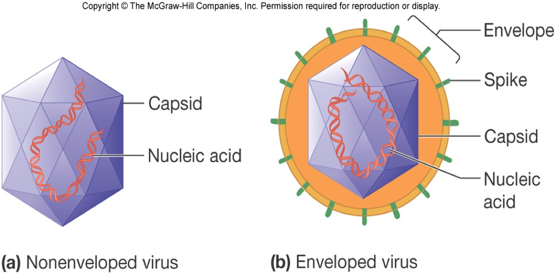
Naked viruses:
Virions lacking envelopes

Are most viruses eukaryotic or prokaryotic?
Eukaryotic
Capsids:
Large macromolecular structures that serve as the protein coat of viruses
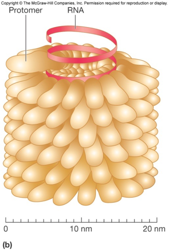
What is the structure of capsids?
They are made of protein subunits called protomers, and can be made into a helical, icosahedral, or complex shape.
Capsid function:
Protects viral genetic material and aids in its transfer between host cells.
Helical capsids:
Are shaped like hollow tubes with protein walls.
Protomer subunits self-assemble.
The size of capsid is a function of
nucleic acid.
Icosahedral capsids:
An icosahedron is a geometric shape with 20 equilateral faces and 12 vertices.
Most viruses have this type of capsid shape.
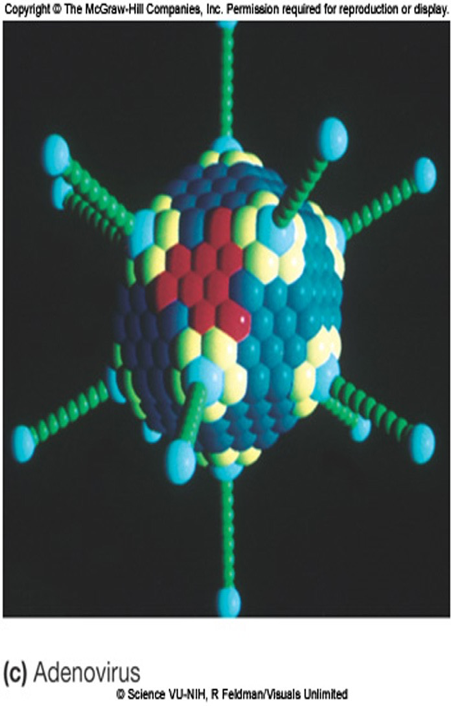
Icosahedral capsid structure:
Icosahedral capsids are made of two types of capsomeres: pentamers and hexamers
Complex shape capsids:
Viruses that do not fit into the category of having helical or icosahedral capsids.
What are 2 examples of viruses that have envelopes?
Rabies and herpes
Envelope proteins:
Are viral encoded and may project from the envelope surface as spikes or peplomers
Spikes:
Spikes are envelope proteins involved in viral attachment to a host cell.
Spikes are used for virus identification.
May play a role in nucleic acid replication.
May have enzymatic or other activity.
Example of a virus with spikes:
An example is hemagglutin of influenza virus.
Do virions have enzymes, Y/N?
Yes, virions have enzymes associated with the envelope or capsid but most are within the capsid.
What shape are viral genomes?
Viral genomes can be segmented or circular