Skeletal System
 Call Kai
 Learn
Learn Practice Test
 Spaced Repetition
 Match
Match Flashcards
 Knowt Play
1/34

There's no tags or description
Looks like no tags are added yet.
Name | Mastery | Learn | Test | Matching | Spaced | Call with Kai | Chat |
|---|
No analytics yet
Send a link to your students to track their progress
35 Terms
The skeletal system forms the…
internal protective framework of the body
The skeleton itself is composed exclusively of _______, primarily in the form of ___________.
connective tissue; bone and cartilage
“Organs” of the skeletal system include _________, which together constitute _____________.
bones and joints; a system of levers upon which skeletal muscles work to effect movement
The skeletal system is divided into an axial skeleton, which consists of ______________, and an appendicular skeleton that includes ______________.
the skull, hyoid bone, vertebrae and ribs; the bones of the pectoral and pelvic girdles and the extremities
Bones are classified by their…
shape (e.g., long, short, flat, irregular)
What is a special type of bone?
Sesamoid bones are a special type; these typically small nodules of bone (resembling a sesame seed) are embedded within the tendons of muscles where they cross synovial joints.
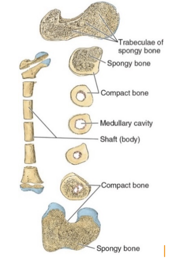
Bones are composed of two types of bone tissue
compact or cortical bone, and spongy or trabecular bone
compact vs. spongy bone
Vary with respect to their amount of solid matter and the number and size of marrow spaces they contain
Compact bone appears as a solid mass
Trabecular bone consists of a network of bony spicules or trabeculae delimiting spaces occupied by bone marrow
All bones have a superficial thin layer of compact bone around a central mass of trabecular bone, except where the latter is replaced by a large marrow cavity
Due to its density, compact bone is readily observable in conventional radiographic images
Head
large, round articular end
examples: head of the femur, head of the mandible
Neck
relatively narrow portion connecting the head to the shaft
example: femoral neck
Facet
smooth flat area, usually covered with cartilage, where a bone articulates with another bone
examples: costal facets on thoracic vertebrae
Condyle
knuckle-shaped region of a bone; usually associated with an articulation
examples: medial & lateral condyles of the femur, occipital condyles of the skull
Protuberance
a bulge or projection of bone
example: external occipital protuberance
Process
an extension or projection serving a particular purpose
examples: ulnar styloid process, uncinate process of cervical vertebrae, mastoid process of skull
Spine
thorn-like process
examples: spine of the scapula; ischial spine of the pelvis
Crest
ridge/edge of bone
examples: iliac crest, petrous crest
Line
linear elevation, sometimes called a ridge
examples: soleal line of the tibia; superior nuchal line of skull
Tubercle
small, raised eminence
examples: greater tubercle of the humerus, costal tubercle
Tuberosity
large, rounded elevation
examples: calcaneal tuberosity, ischial tuberosity of pelvis
Foramen
passage through a bone
examples: obturator foramen, foramen magnum
Fossa
hollow or depressed area
examples: olecranon fossa of the humerus, iliac fossa of the pelvis, hypophyseal (pituitary) fossa of the skull
Groove
elongated depression or furrow
examples: radial groove of the humerus, groove for the sigmoid sinus in the skull
Cartilage
Located at strategic points in the skeleton
Cartilage provides flexible support of tissues in locations such as the external nose and the thoracic cage
In the joints, cartilage withstands great pressures and distributes the forces generated by movement fairly evenly across its surface
During growth, cartilage provides a means of tissue expansion in the presence of direct pressure
Joints
A joint, or articulation, is defined as the union between two or more bones. There are three main categories of joints
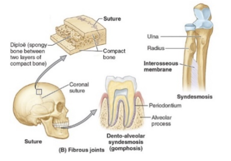
Fibrous Joints
Bones are physically connected to one another by fibrous connective tissue
Little, if any, movement occurs at these joints
examples include the sutures of the skull, the ligamentum flavum of the vertebral column, and the interosseous membrane between the tibia and fibula
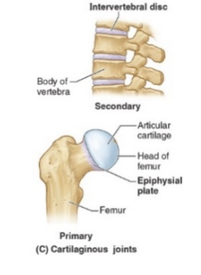
Cartilaginous Joints
In these joints, bones are physically connected to one another by cartilage
examples include — intervertebral (IV) discs and the pubic symphysis, both of which are formed of fibrocartilage, and the epiphyseal plates (growth plates) of bones, which are formed of hyaline cartilage
Little or no movement is typically allowed at these joints
Synovial Joints
Most of the joints of the body are synovial joints
Bones are not physically connected to one another at all —> their cartilage-covered articulating surfaces are held apposed to one another by associated connective tissues and muscles
Synovial joints are highly movable joints
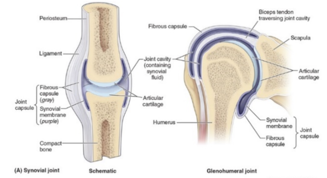
The articulating surfaces (facets) of the bones in a synovial joint (and there may be more than just two) are covered in…
articular cartilage – a specialized form of hyaline cartilage
The ends of the bones entering into a synovial joint are enclosed within a…
connective tissue capsule
This fibrous joint capsule encloses a…
sealed joint cavity that contains synovial fluid – a colorless, transparent, viscous fluid that both nourishes and lubricates the articular cartilage
some synovial joints contain…
accessory ligaments — intrinsic and extrinsic (collateral) ligaments
articular discs
Intrinsic Ligaments
Part of, or within (between the fibrous capsule and the synovial membrane) the fibrous joint capsule
Examples of intrinsic ligaments include iliofemoral, pubofemoral, and ischiofemoral ligaments of the hip joint, and the lateral ligament of the temporomandibular joint
Extrinsic (collateral) Ligaments
outside and independent of the fibrous capsule
Examples of extrinsic ligaments include the lateral and medial collateral ligaments of the elbow and knee
Articular discs, labra (sing. = labrum), and menisci (sing. = meniscus)
these structures, usually composed of fibrocartilage, divide or partially divide joint cavities, and modify the shape of articulating surfaces so as to enhance joint stability