Radiology Lab 8 - Musculoskeletal Imaging
1/20
There's no tags or description
Looks like no tags are added yet.
Name | Mastery | Learn | Test | Matching | Spaced |
|---|
No study sessions yet.
21 Terms
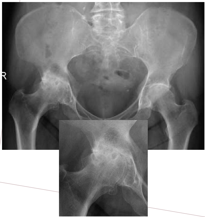
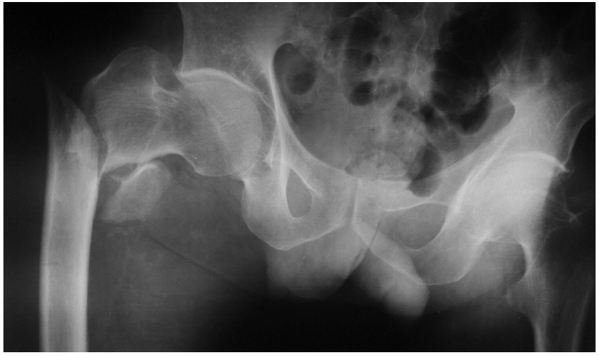
What is this X-ray Image?
Hip Osteoarthritis
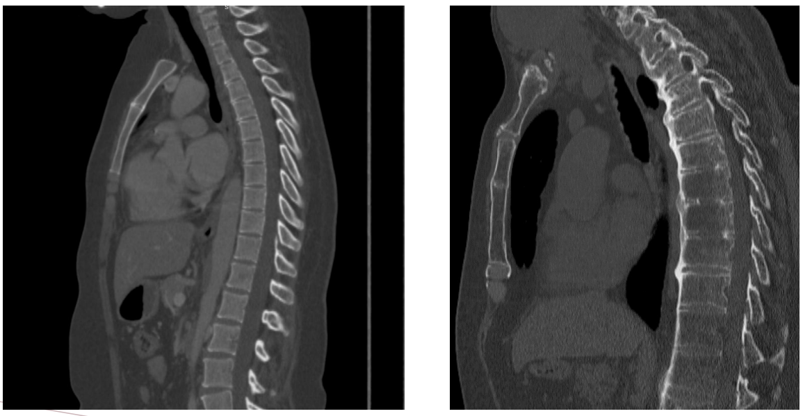
What is this CT bone window imaging presenting?
Diffuse osteoporosis - level of Sternum and Spine
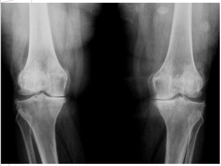
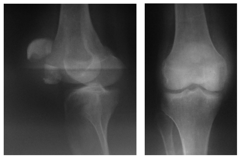
What is this X-ray image showing?
Knee osteoarthritis
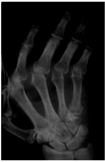
What is this X-ray image showing?
Rheumatoid arthritis
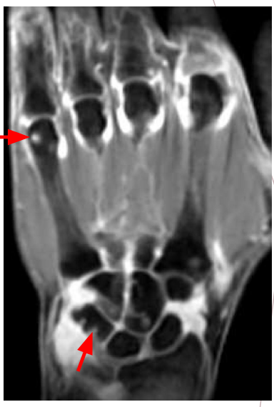
What is this MRI image showing?
Tenosynovitis of the wrist and MCP joints + bone erosions
This X-ray image shows a lateral and anterior view of what?
Patellar fracture
What does this X-ray image show?
Fracture with upward displacement of the right femoral neck
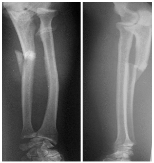
This X-ray image in the anterior and lateral view shows?
Comminuted Ulnar fracture with displacement
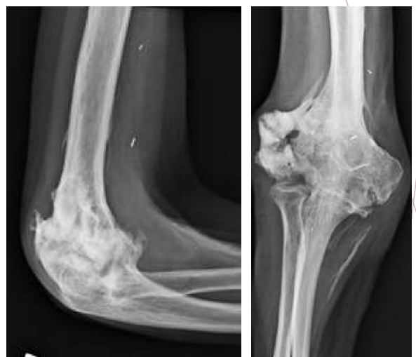
What does this image show?
Improper healing of an intra-articular fracture at the level of the lateral epicondyle
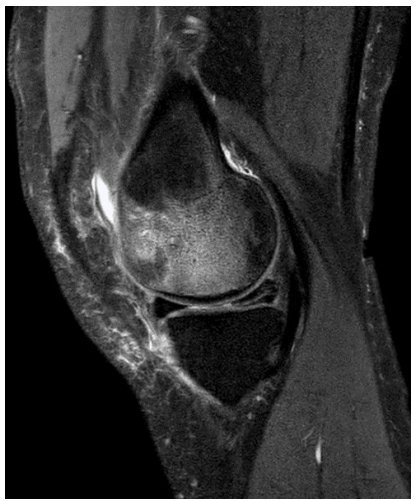
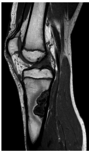
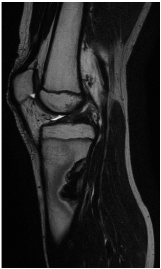
What does this MRI image show?
Horizontal meniscal tear of the posterior horn
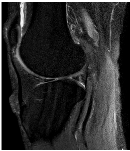
What does this MRI image show?
Longitudinal Meniscal tear of the posterior horn
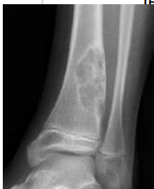
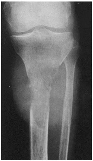
What is shown here?
Lytic type of bone changes
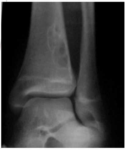
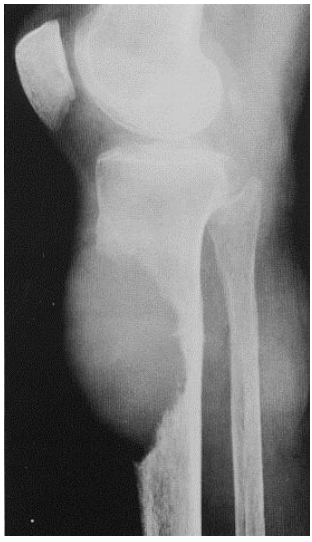
What is shown here?
Osteogenic type of bone change
What do we see in the contour of this lesion?
Sclerotic
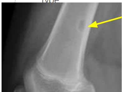
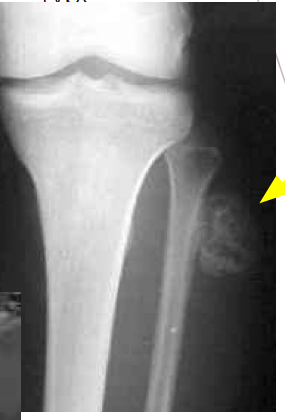
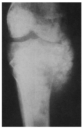
What does this X-ray image show?
Non-ossifying fibroma (in children)
What phase is this?
T1
What phase is this?
T2
What is shown in the image here?
Osteosarcoma: osteogenic
What is shown in the image here?
Osteosarcoma: osteolytic
What is shown in the image here?
Osteosarcoma: osteolytic
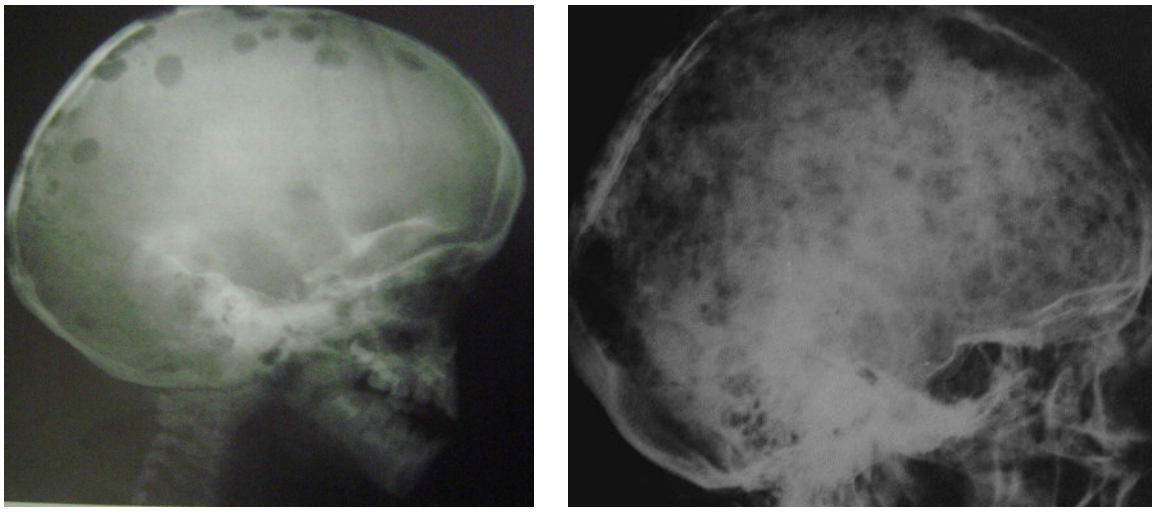
What is this?
Multiple Myeloma