Test 2 Bio Lab
1/39
There's no tags or description
Looks like no tags are added yet.
Name | Mastery | Learn | Test | Matching | Spaced | Call with Kai |
|---|
No analytics yet
Send a link to your students to track their progress
40 Terms
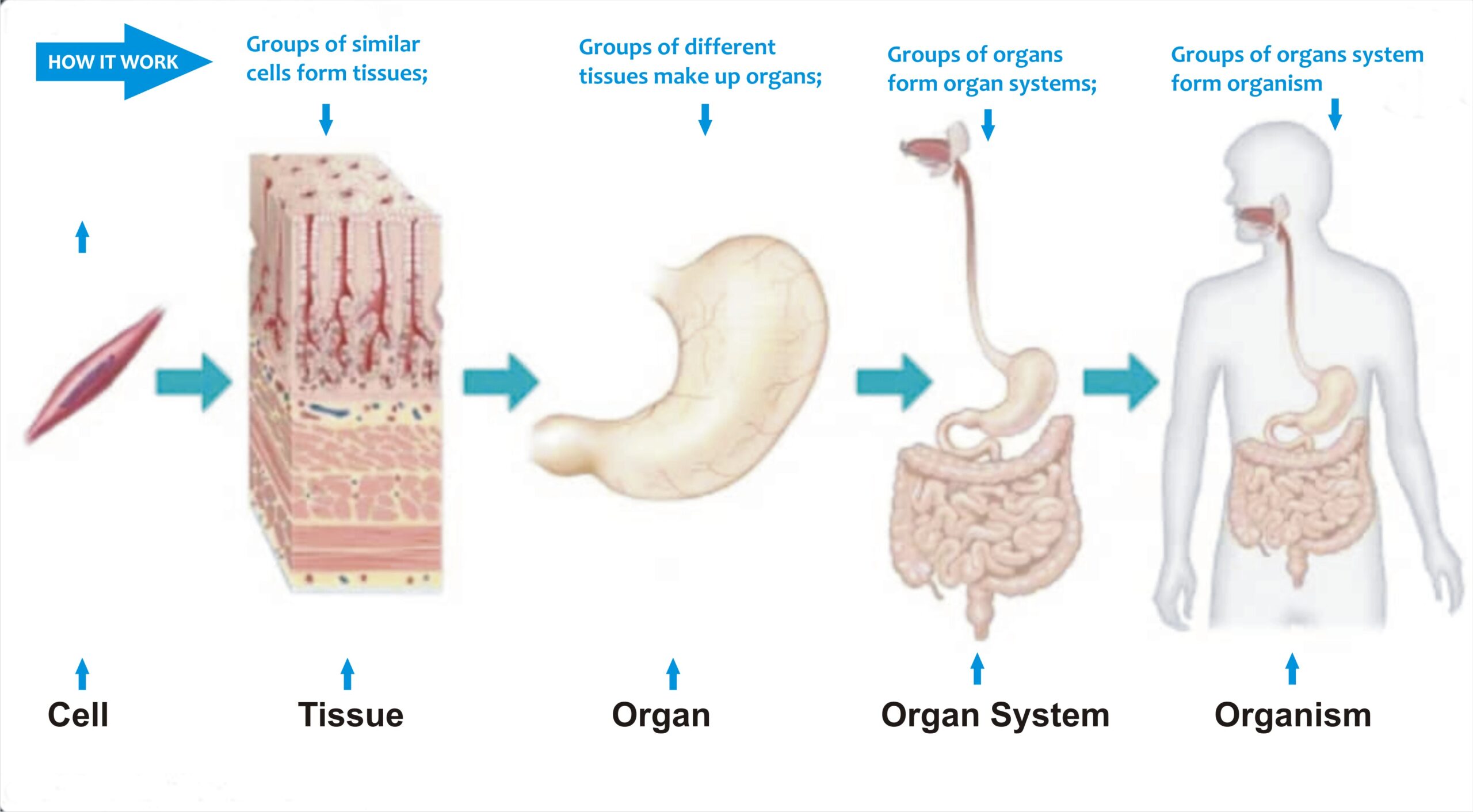
LEVELS OF ORGANIZATION OF THE HUMAN BODY
Cell(cardiac muscle cell) to Tissues(cardiac muscle) to Organs(heart) to System(circulatory system) to Organism(living individual)


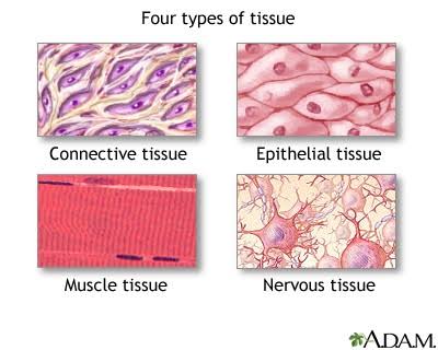
Primary tissue types in vertebrate animals
Epithelial Tissue, Connective Tissue, Muscle Tissue(Muscular), Nervous Tissue
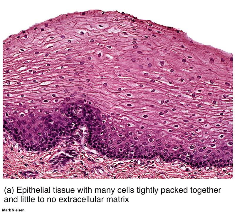
Epithelial Tissue
Epithelial cells provides critical functions like protection, secretion, and absorption. They cover the exterior of an organism, line the gut and other cavities, and line the coelomic cavity. 1)Protect underlying tissues from dehydration and mechanical damage. 2)Provide a selectively permeable barrier that facilitates or impedes passage of materials. 3)Provide sensory surfaces. 4)Secretes fluids.
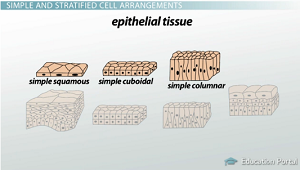
Shapes of Epithelial Tissues
Squamous-flat, irregular(like fried eggs)
Cuboidal-cubes
Columnar-tall and rectanglar
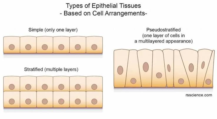
Layers of Epithelial Tissues
Simple is 1 layer
Stratified is multiple layers
Pseudo-stratified is a single layer of cells that appear stratified because the nuclei appear in different positions within the columnar cells

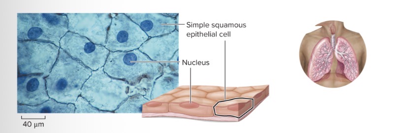
Simple Squamous
Characteristic Feature: epithelial cells; single cell layer
Cell Shape/Characteristics: irregular and flattened; minimum barrier to diffusion
Function: cells form layer across which diffusion can readily occur
Location: line the alveoli of the lungs, capillary walls, the filtration system of the kidneys, and major cavities of the body
*Relatively inactive and are associated with the sites of passive movement of water, electrolytes, and other substances

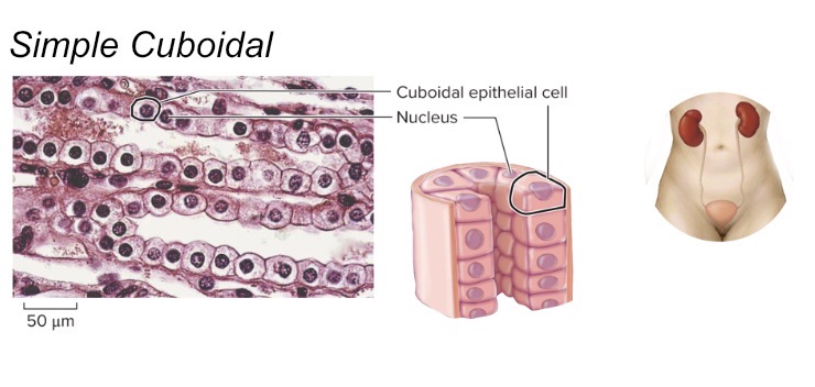
Simple Cuboidal
Characteristic Feature: gland cells; appear more full
Cell shape/Characteristics: as tall as they are wide; cube-like shape; has cilia
Function: provide functions in secretion and absorption
Location: line the kidney tubules, some glands, and cover the ovaries
These cells are rich in specific transport channels

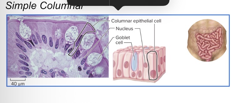
Simple Columnar
Characteristic Feature: epithelial cells; appear more full
Cell shape/Characteristics: taller than they are wide; column-like shape; has cilia
Function: provides protection and functions in secretion and absorption
Location: in the intestines, lining of the stomach, and parts of the respiratory tract
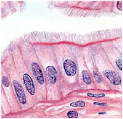
Simple Ciliated Columnar
epithelium contains cells with cilia, motile, hair-like processes that help to move fluids or particles along a surface. Most often seen in fallopian tubes and bronchioles.
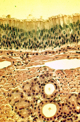
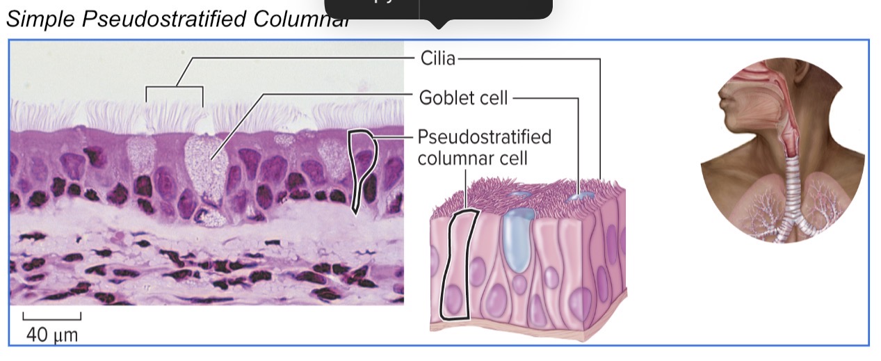
Pseudo-stratified Ciliated Columnar
Lines the respiratory tract and provides protection
Secretes mucus, dense with cilia that aid in movement of mucus
Found from the trachea and bronchii
Stratified Epithelium
animal tissue made up of 2 or more layers of stacked cells

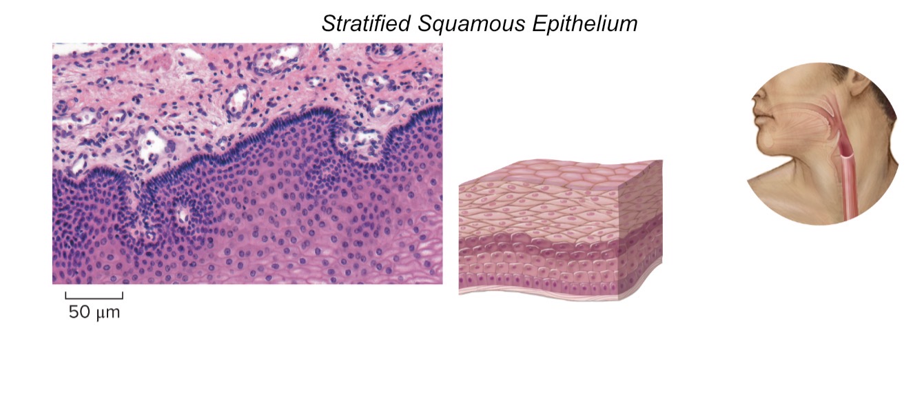
Stratified Squarmous
Characteristic Feature: epithelial cells; several layers thick
Cell shape/Characteristics: upper layer, squamous; middle layer, cuboidal; basal (bottom) layer, columnar
Function: tough layers of cells providing protection to underlying tissues
Location: outer layer of skin and lining of mouth
NonKeratinized Stratified Squamous
Characteristic Feature: epithelial cells; several layers thick
Cell shape/Characteristics: upper layer, squamous; middle layer, cuboidal; basal (bottom) layer, columnar
Function: tough layers of cells providing protection to underlying tissues
Location: outer layer of skin and lining of mouth
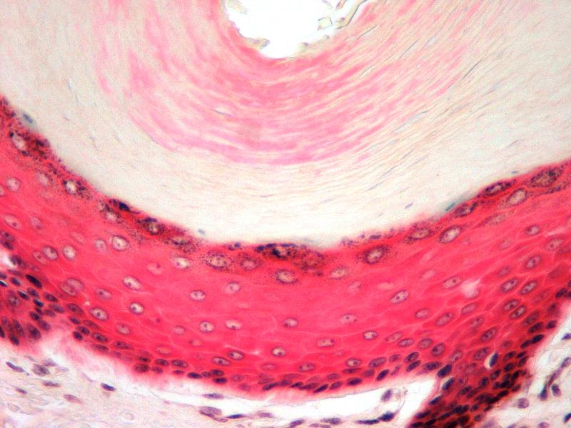
Keratinized Stratified Squamous
epithelium, a tough layer of keratin (a protein resistant to friction and repels bacteria) is deposited in the surface cells. This is located on skin, hair and nails. This tissue will form a cutaneous membrane.
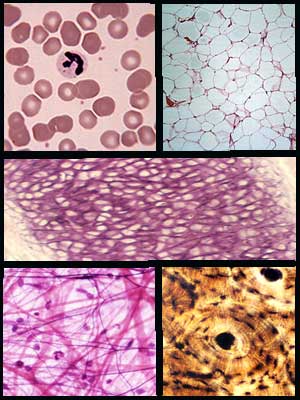
Connective Tissue
supports the body, fight pathogens, and store energy; these cells are loosely packed, and are typically suspended in an extracellular matrix of fibers
The most diverse and abundant tissues in animal bodies
They connect and support all other tissues
2 major classes: connective tissue proper & specialized connective tissue
Proper - divided into loose and dense connective tissues
Special - includes blood, cartilage, and bone
Bone Model(Connective Tissue)

Connective tissue proper
Loose(three types): Areolar, adipose, and reticular tissue. These tissues contain:
Fibroblasts-cells that secretes fibrous proteins that form the extracellular matrix.
Collagen-strong fibrous proteins
Elastin=allows to stretch(extensibility) and to recoil(elasticity)
Ground substance-protein sugar combination that can be a liquid, solid, or semisolid and fills spaces between cells and fibers
Dense(three types): Dense regular
Special connective tissue
Liquid(blood and lymph)
Cartilage(three types)
Bone(two types)
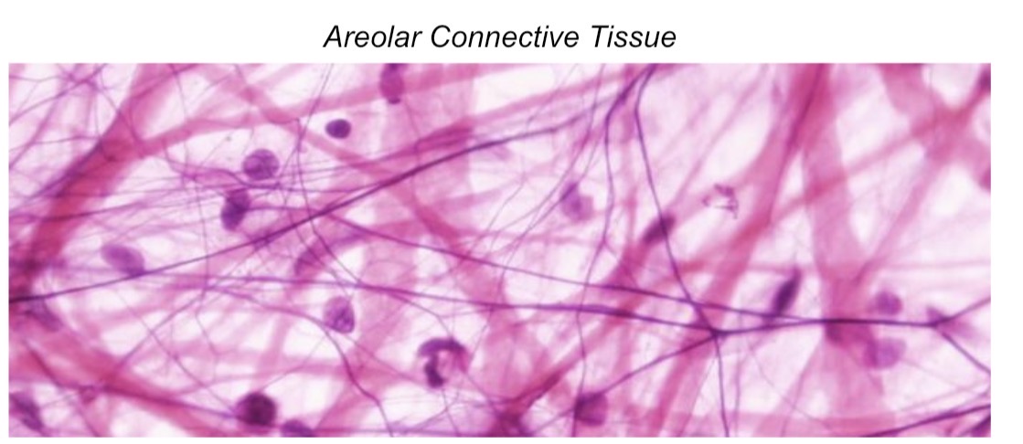
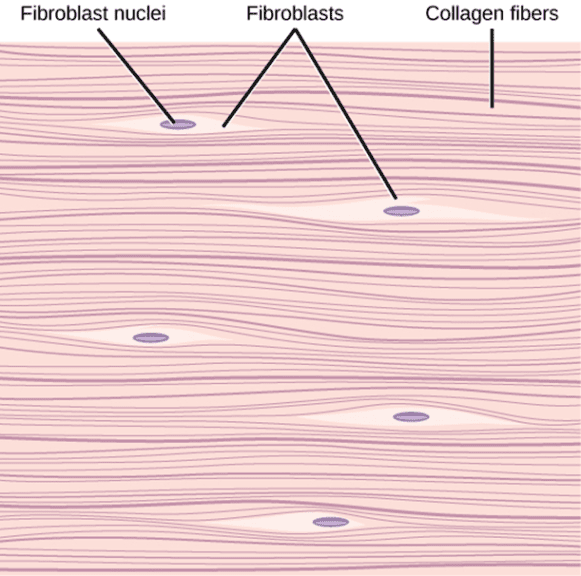
Areolar(Loose)Connective Tissue
Characteristic Feature: loose connective tissue; extracellular matrix
Cell shape/Characteristics: cells imbedded within a mesh-like fiber network
Function: protective; wraps around blood vessels, nerves, and organs; connects skin to the underlying muscle
Location: beneath epithelial tissue
Fibroblasts- cells that secretes fibrous proteins that form the extracellular matrix.
Collagen- strong fibrous proteins
Elastin= allows to stretch(extensibility) and to recoil(elasticity)
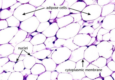
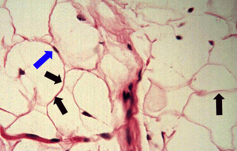
Adipose(Loose) Connective Tissue
Characteristic Feature: loose connective adipose tissue, fat cells, fibroblasts (active cells that create the extracellular matrix)
Cell shape/Characteristics: can develop in large groups; fixed amount that enlarge and shrink; looks like bubbles
Function: used for nutrient storage; hydrolyzes stored triglycerides and secretes fatty acids into blood for oxidation
Location: under the skin, in bone marrow, and around kidneys
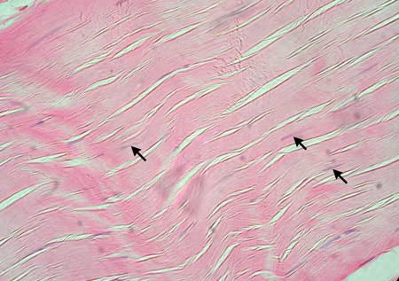
Dense Regular Connective Tissue
Characteristic Feature: loose connective adipose tissue, fat cells, fibroblasts (active cells that create the extracellular matrix)
Cell shape/Characteristics: can develop in large groups; fixed amount that enlarge and shrink; looks like bubbles
Function: used for nutrient storage; hydrolyzes stored triglycerides and secretes fatty acids into blood for oxidation
Location: under the skin, in bone marrow, and around kidneys
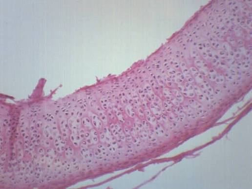
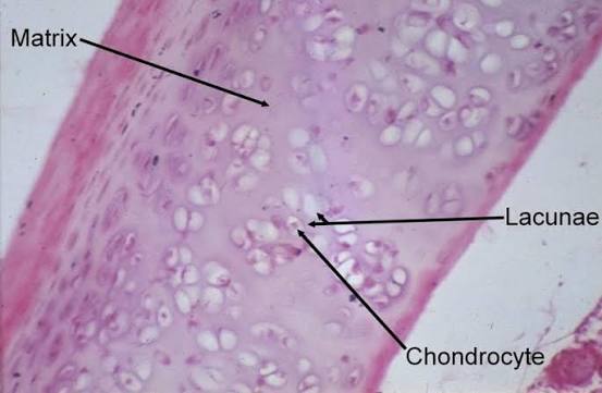
Hyaline Cartilage Connective Tissue
Characteristic Feature: chondrocytes in lacunae (cavities) surrounded by chondrin extracellular matrix.
Cell shape/Characteristics: pockets of cells.
Function: provides flexible support, shock absorption, and reduction of friction on load-bearing surfaces.
Location: in lacunae cavities within cartilage ground substances, spinal disks, knees, joints, ear, nose.
Chondrocytes-cells that maintain the cartilage daily activity. The cell secretes protein-carbon hydrate complex that forms the extracellular matrix.
Lacuna-cavities in cartilage where chondrocytes reside.
Chondrin Sulfate-is the ground substance- gelatin-like (rubber-like) extracellular matrix of cartilage.
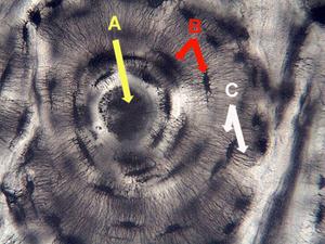
Compact Bone
Characteristic Feature: surrounded by osteocytes - bone cells made of fibrous, crystalline extracellular matrix
Cell shape/Characteristics: looks like the rings or bark of a tree
Function: protects internal organs, provides rigid support for muscle attachment
Location: Most of the skeleton
A is Haversian Canal-contain blood and nerves vessels
B is Lacuna- cavities in the cartilage where chondrocytes reside
C is Canaliculi- tiny passages that allow chondrocytes in lacunae to communicate with other cells
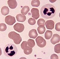
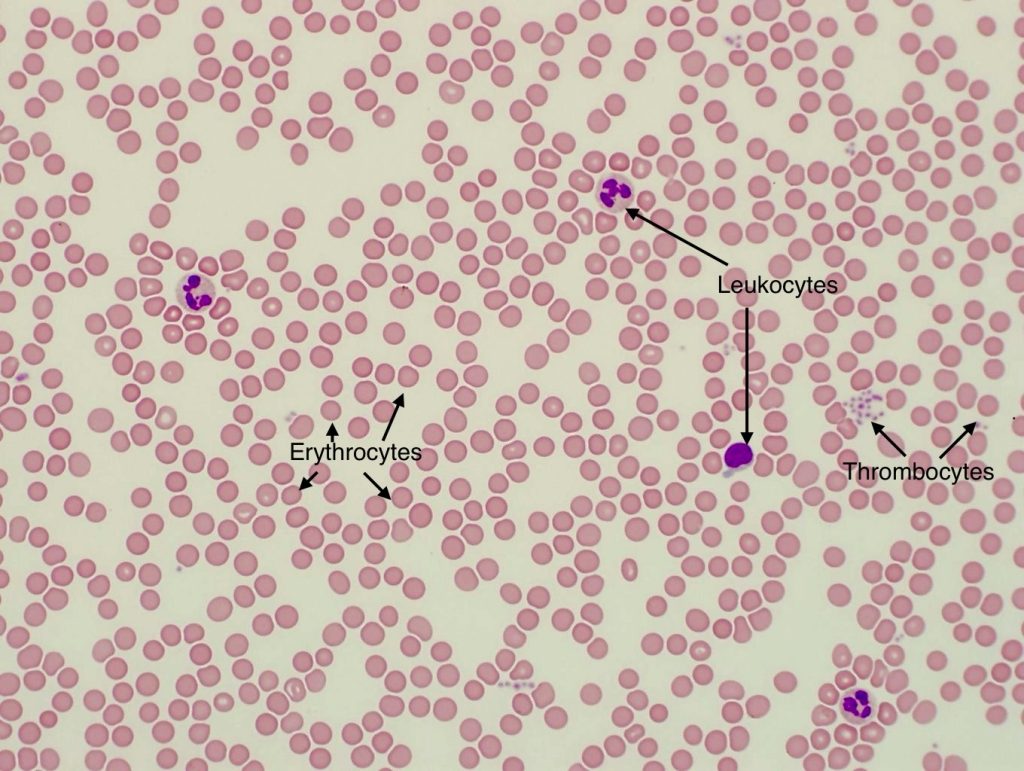
Blood
Characteristic Feature: liquid connective tissue; erythrocytes (red), leukocytes (white), and thrombocytes (platelets); considered connective tissue
Cell shape/Characteristics: circular, beanlike
Function: highway of immune system carries nutrients and waste; primary means of communication between organs
Location: circulatory system
Muscle Tissue Model

Smooth Muscle Model


Muscle Tissue
characterized by the ability to generate a force by converting chemical energy into mechanical energy; they are either smooth or striated (striped)
Distinguishes animals from the other multicellular organisms like plants, fungi, and protists
Affords internally initiated movement and behavior

Skeletal Muscle
Characteristic Feature: skeletal muscle cells; voluntary contraction
Cell shape/Characteristics: long, tubular cells (muscle fibers), vertical stripes
Function: powers walking, lifting, talking, and all other voluntary movements
Location: voluntary muscles
Smooth Muscle
Characteristic Feature: smooth muscle cells; contraction involuntary
Cell shape/Characteristics: thin, horizontally packed; spindle shaped; single nucleus
Function: powers rhythmic, involuntary contractions commanded by the central nervous system
Location: Walls of blood vessels, stomach, and intestines (gut)
Cardiac Muscle
Characteristic Feature: striated; contraction involuntary; intercalated disks (interconnections or dark lines) between adjacent cells organizing into continuous functional fibers
Cell shape/Characteristics: smaller and interconnected; loosely packed, each with its own nucleus (single uninucleate)
Function: highly interconnected cells; promotes rapid spread of signal initiating contraction
Location: walls of heart
Neuron(or nerve cell)Model

Nervous Tissue
consists of neurons, supports cells called glia and Schwann cells which help propagate the nerve impulse and provide nutrients to neurons
Neurons - cells specialized for transmitting nerve impulses; made up of:
Cell body - contains the nucleus and cytoplasmic extensions that conduct nerve impulses
Dendrites - thin, highly branched, short extensions that receive incoming stimulation and conduct electrical impulses to the cell body
Axon - single, long extension of cytoplasm that conducts impulses away from the cell body
It may carry an impulse to a muscle to make it contract or to the dendrites of another neuron
Neurons
Characteristic Feature: nervous tissue; consists of cell body, dendrites, and axon
Function: process and transmit information throughout the body using electrical impulses and chemical signals
Location: throughout the entire human body
Neuron
Neuroglia(glial cells)
Characteristic Feature: they are instrumental in the regulation of neurotransmission
Function: do not conduct impulses; they lend support, aid in nourishment, and provide protection for neurons
Location: through the entire nervous system (central and peripheral)
THE SKELETAL SYSTEM
Axial Skeleton
consists of bones along the longitudinal axis of the body that support the head, neck, and trunk
Cranium
Zygomatic Arch
Maxilla (and Mandible)
4.Cervical
Atlas
Axis
Thoracic
Lumbar
Sacral (Sacrum)
Caudal (Coccyx)
Rib Cage
Sternum
Appendicular Skeleton
RAT DISSECTION