Bontrager Chapter 11 Workbook (positioning cranium)
1/90
There's no tags or description
Looks like no tags are added yet.
Name | Mastery | Learn | Test | Matching | Spaced |
|---|
No study sessions yet.
91 Terms
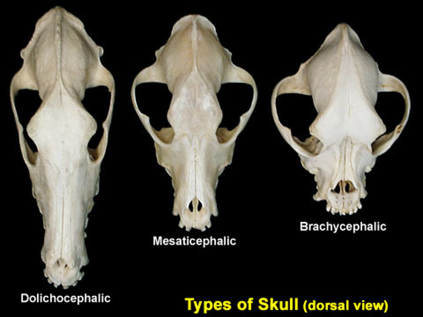
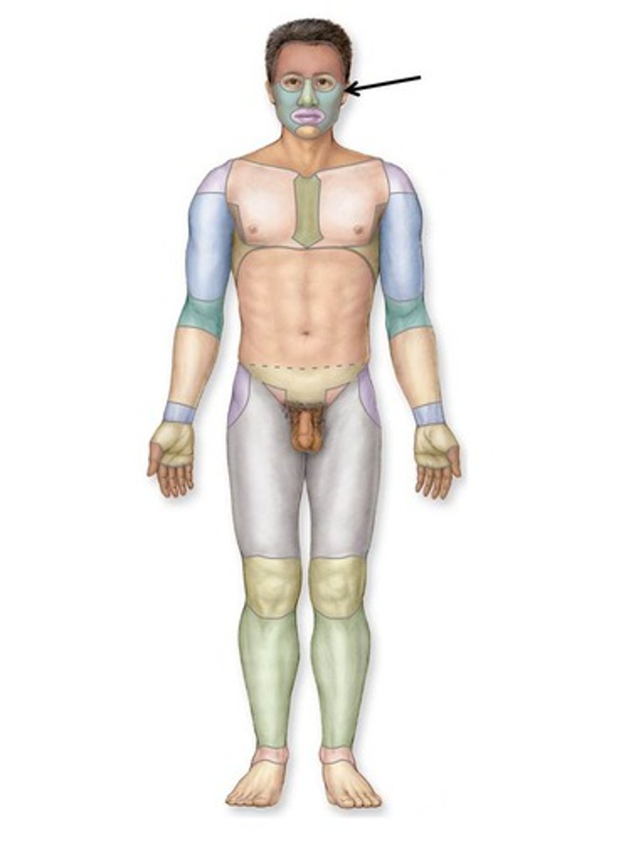
Shape description for the classification of the skull:
Mesocephalic
Width between 75% & 80% of length
(15cm between parietal eminences; 19cm from frontal eminence to external occipital protuberance; 23 cm from vertex to beneath the chin)

Shape description for the classification of the skull:
Brachycephalic
Width ≥80% of length

Shape description for the classification of the skull:
Dolichocephalic
Width <75% of length
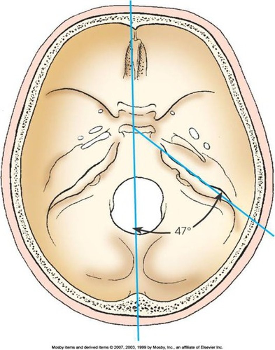
CR angles & degree of rotation stated for basic skull positions are based on the ________ (average) skull, which has an approximate angle of _______ between the midsagittal plane & the long axis of the petrous bone
Mesocephalic, 47 degrees
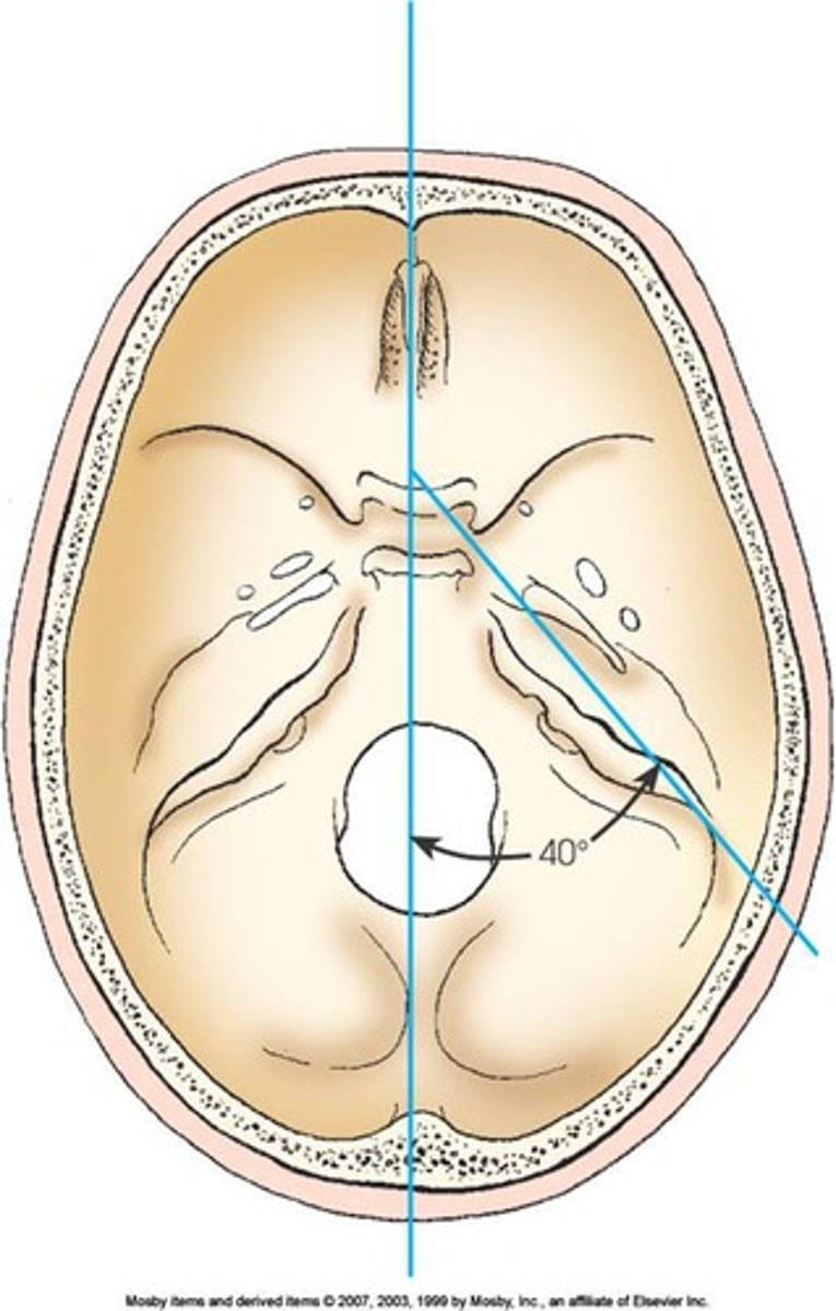
The long, narrow-shaped skull (doliocephalic) has an angle of approximately _______ degrees between the midsagittal plane & the long axis of the petrous bone
+/- 40
True/False: 2 older terms for the orbitomeatal line (OML) are Reid's base line & the anthropologic base line
False (There are other terms for the infraorbitomeatal line)
There is a _____-degree difference between the orbitomeatal & infraorbitomeatal lines, & _____ degrees between the orbitomeatal & glabellomeatal lines
7-8 degrees for both
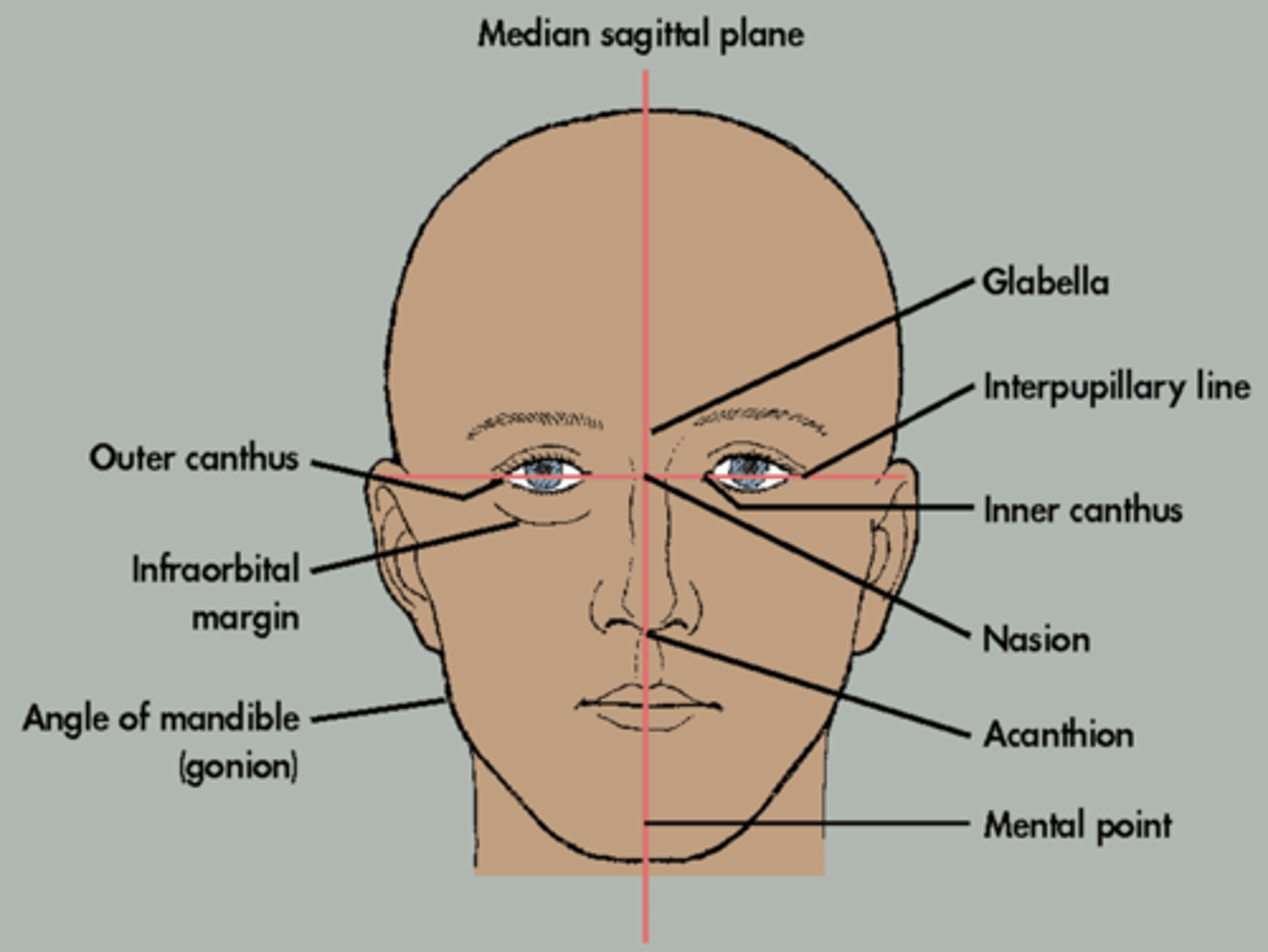
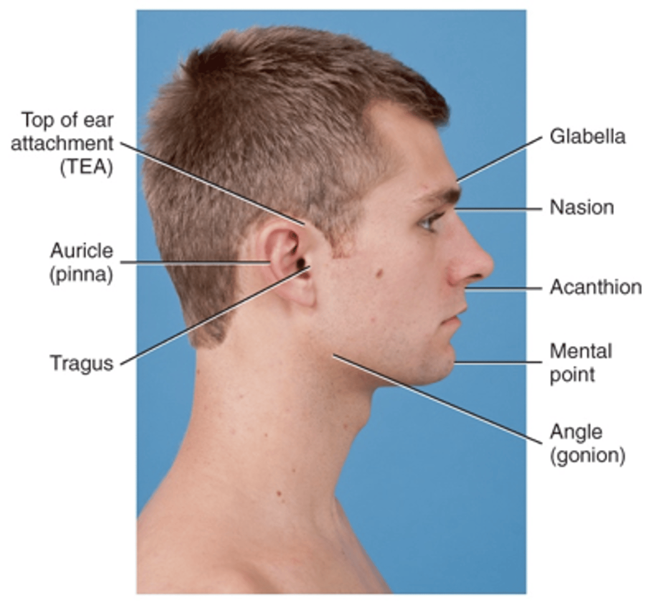
Cranial landmark described:
Lateral junction of the eyelid
Outer canthus
Cranial landmark described:
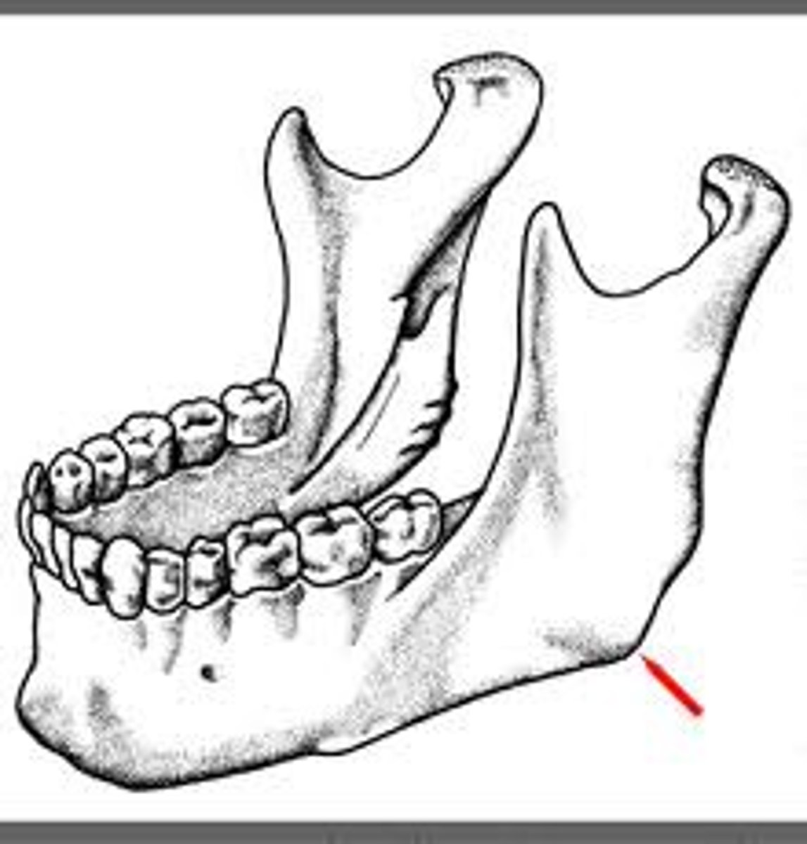
Posterior angle of the jaw
Gonion
Cranial landmark described:
A line between the infraorbital margin & the EAM
IOML
Cranial landmark described:
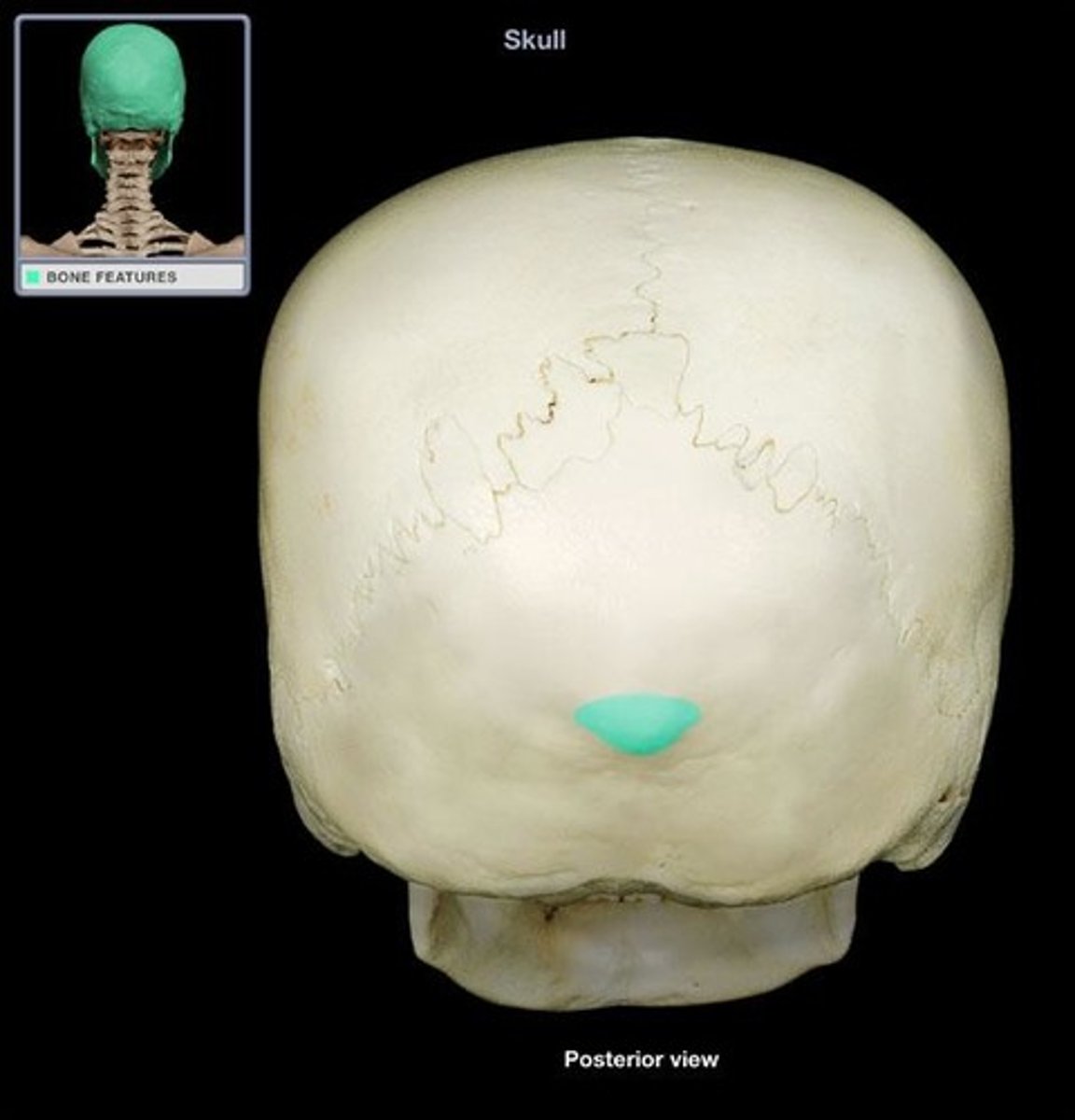
Corresponds to the highest "nuchal" line of the occipital bone
Inion
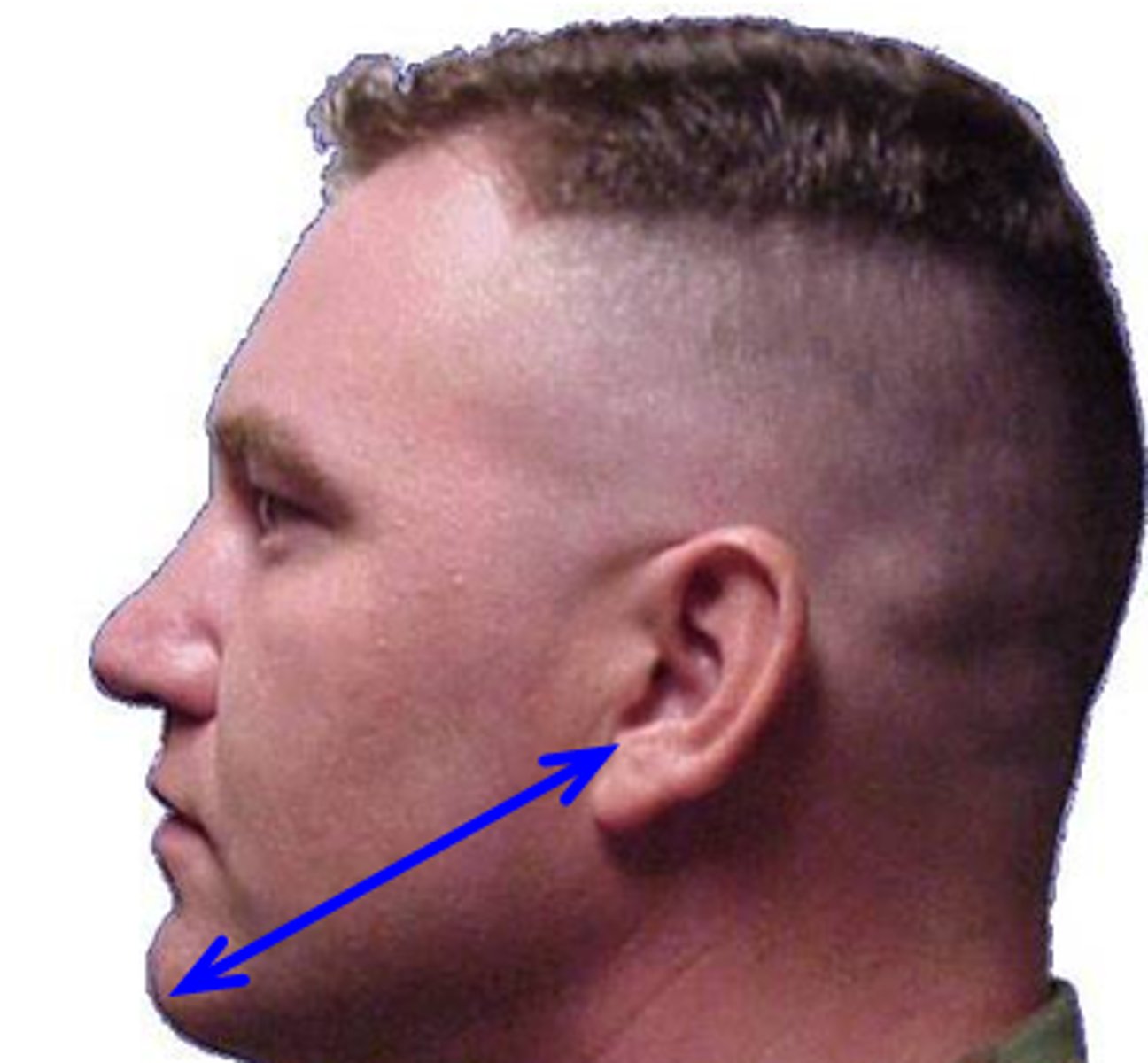
Cranial landmark described:
A line between the mental point & the EAM
Mentomeatal line
Cranial landmark described:
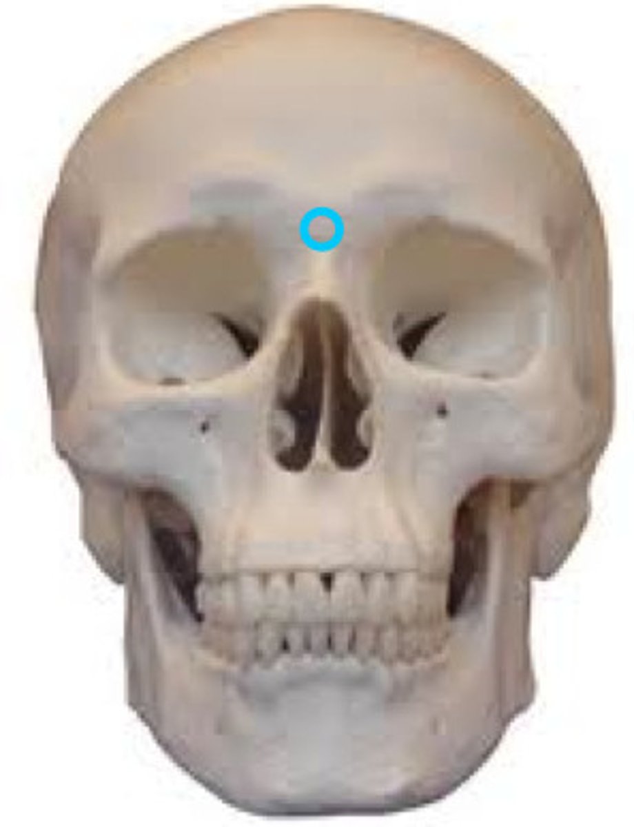
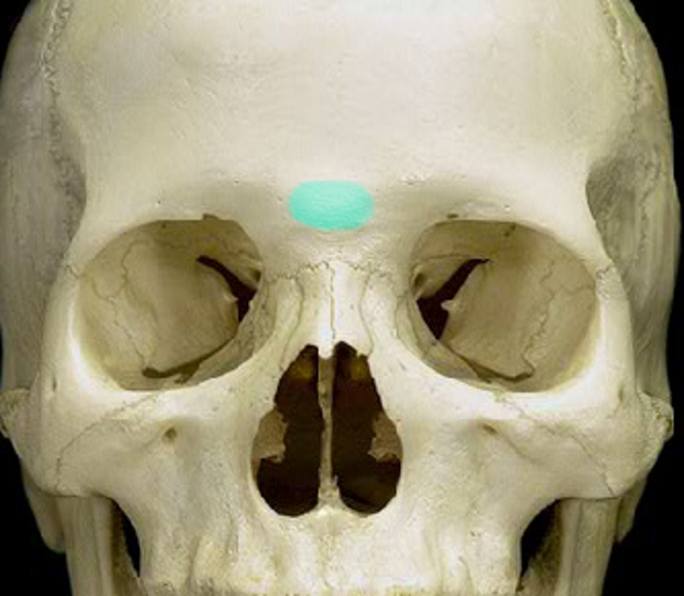
Located at the junction of the 2 nasal bones & the frontal bone
Nasion
Cranial landmark described:
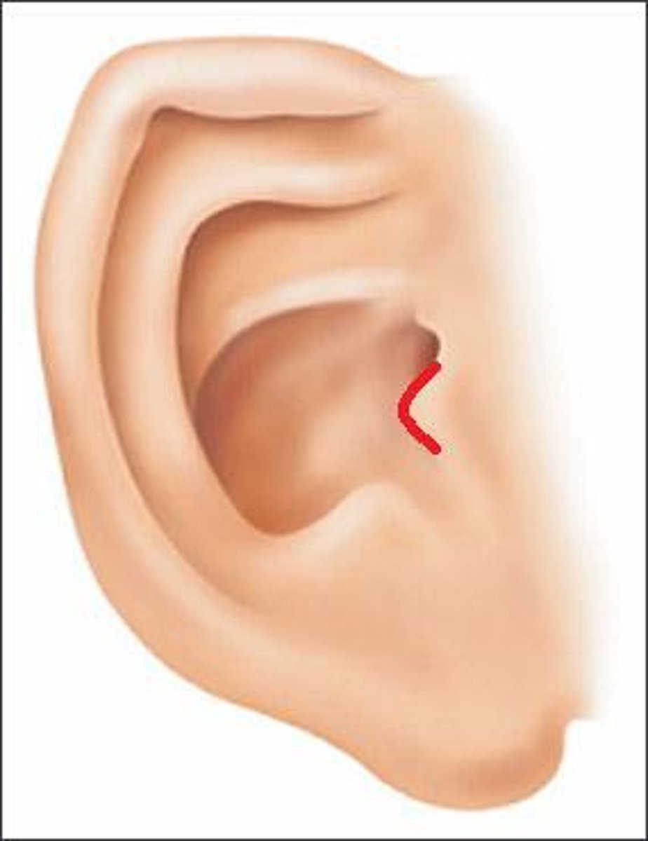
The small cartilaginous flap covering the ear opening
Tragus
Cranial landmark described:
Corresponds to the highest level of the facial bone mass
Supraorbital groove
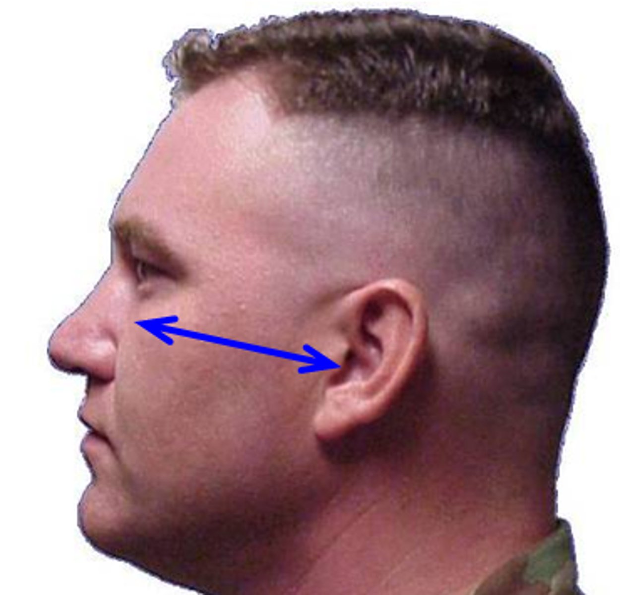
Cranial landmark described:
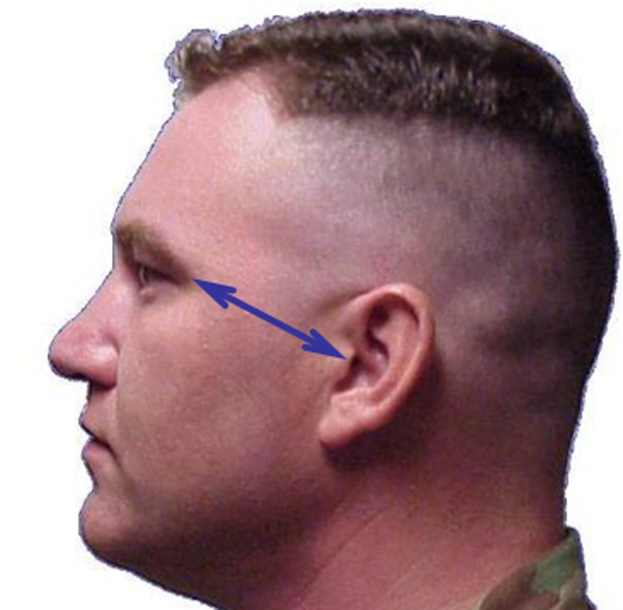
A line between the midlateral orbital margin and the EAM
OML
Cranial landmark described:
The center point of the EAM
Auricular point
Cranial landmark described:
A positioning line that is primarily used for the modified Waters projection
Lips-meatal line
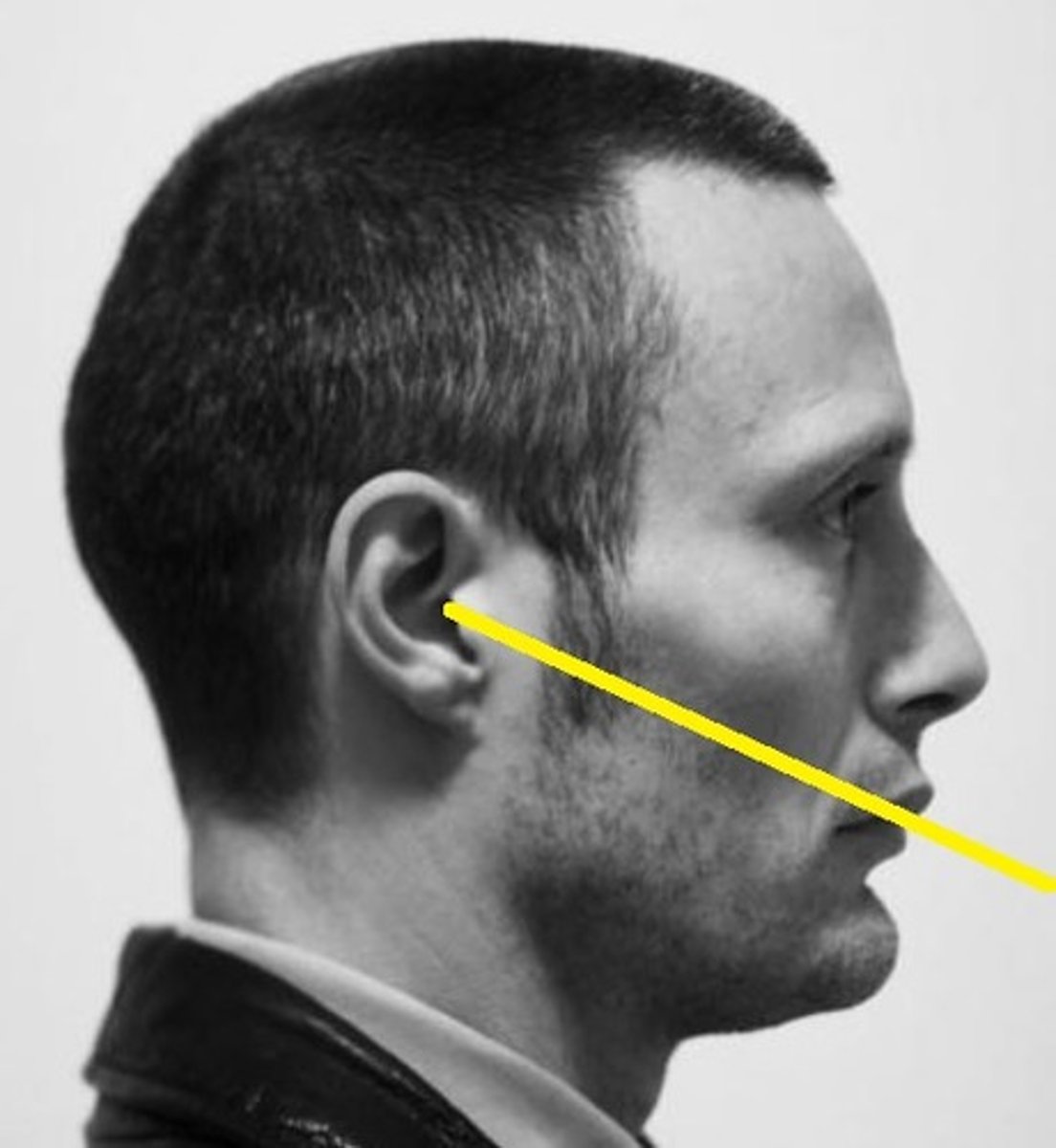
Cranial landmark described:
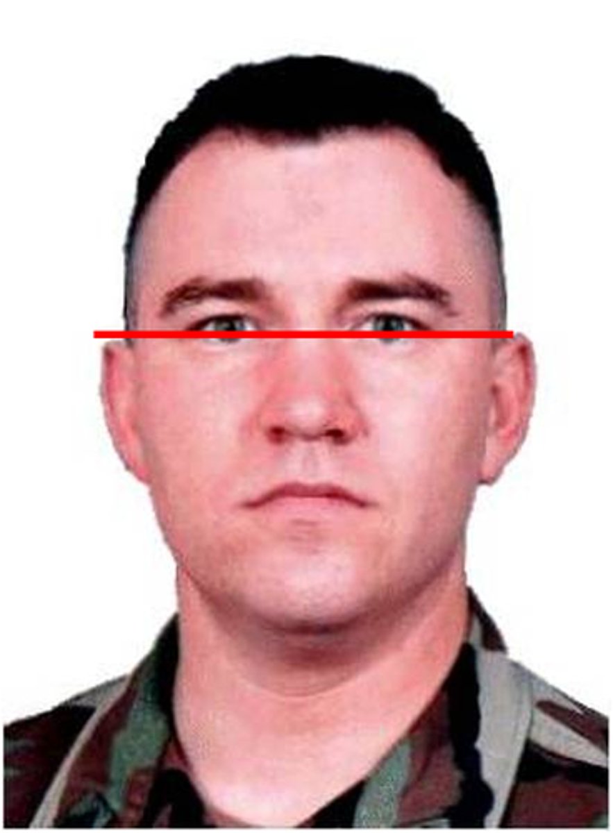
A line used in positioning to ensure that the skull is in a true lateral position
Interpupillary line
Cranial landmark described:
Corresponds to the level of the petrous ridge
TEA
Cranial landmark described:
A smooth, slightly depressed area between the eyebrows
Glabella
What is the average kV range for digital skull imaging?
80-95 kv (75 to 85 analog)
List the 5 common errors made during skull radiography
A. Rotation
B. Tilt
C. Excessive neck flexion
D. Excessive neck extension
E. Incorrect CR angulation
Of the 5 most common error, which 2 are most common?
A. Rotation
B. Tilt
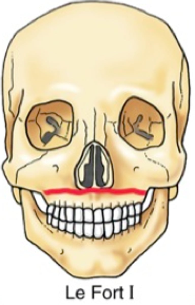
Bilateral horizontal fractures of the maxillae describe a ________ fracture
A. Le Fort
B. Blowout
C. Tripod
D. Contrecoup
A. Le Fort
Which of the following imaging modalities is the most common neuroimaging procedure performed for the cranium?
A. CT
B. Ultrasound
C. MRI
D. Nuclear medicine
A. CT
Which of the following imaging modalities is commonly performed on neonates with a possible intracranial hemorrhage?
A. CT
B. Ultrasound
C. MRI
D. Nuclear medicine
B. Ultrasound
Which of the following imaging modalities is most commonly performed to evaluate patients for Alzheimer disease?
A. CT
B. Ultrasound
C. MRI
D. Nuclear medicine
D. Nuclear medicine
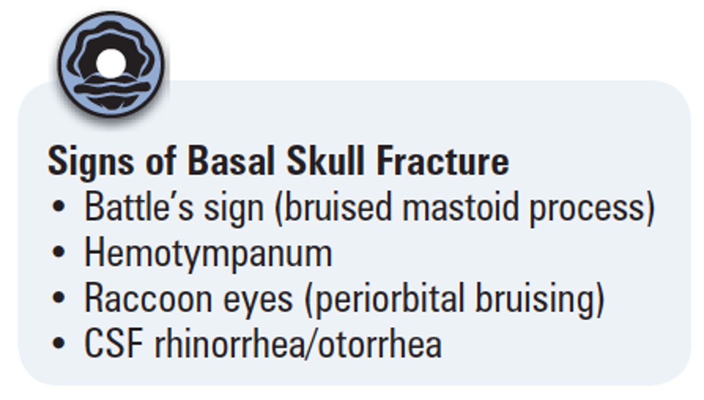
Clinical indication described:
Fracture that may produce an air-fluid level in the sphenoid sinus
Basal skull fracture
Clinical indication described:
Destructive lesion with irregular margins
Osteolytic neoplasm
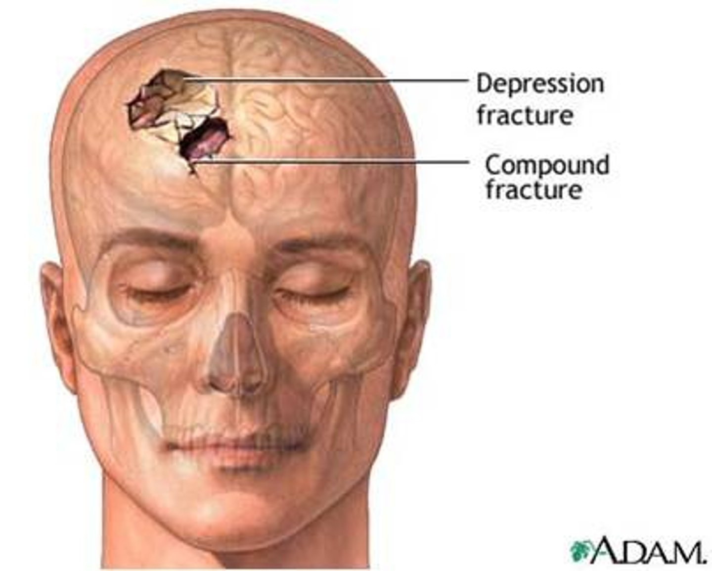
Clinical indication described:
Also called a "ping-pong" fracture
Depressed skull fracture
Clinical indication described:
Proliferative bony lesion of increased density
Osteoblastic neoplasm
Clinical indication described:
A tumor that may produce erosion of the sella turcica
Pituitary adenoma
Clinical indication described:
AKA osteitis deformas
Paget's disease
Clinical indication described:
A bone tumor that originates in the bone marrow
Multiple myeloma
Which of the following clinical indications may require an increase in manual exposure factors?
A. Advanced Paget's disease
B. Metastatic neoplasm
C. Multiple myeloma
D. Basal skull fracture
A. Advanced Paget's disease
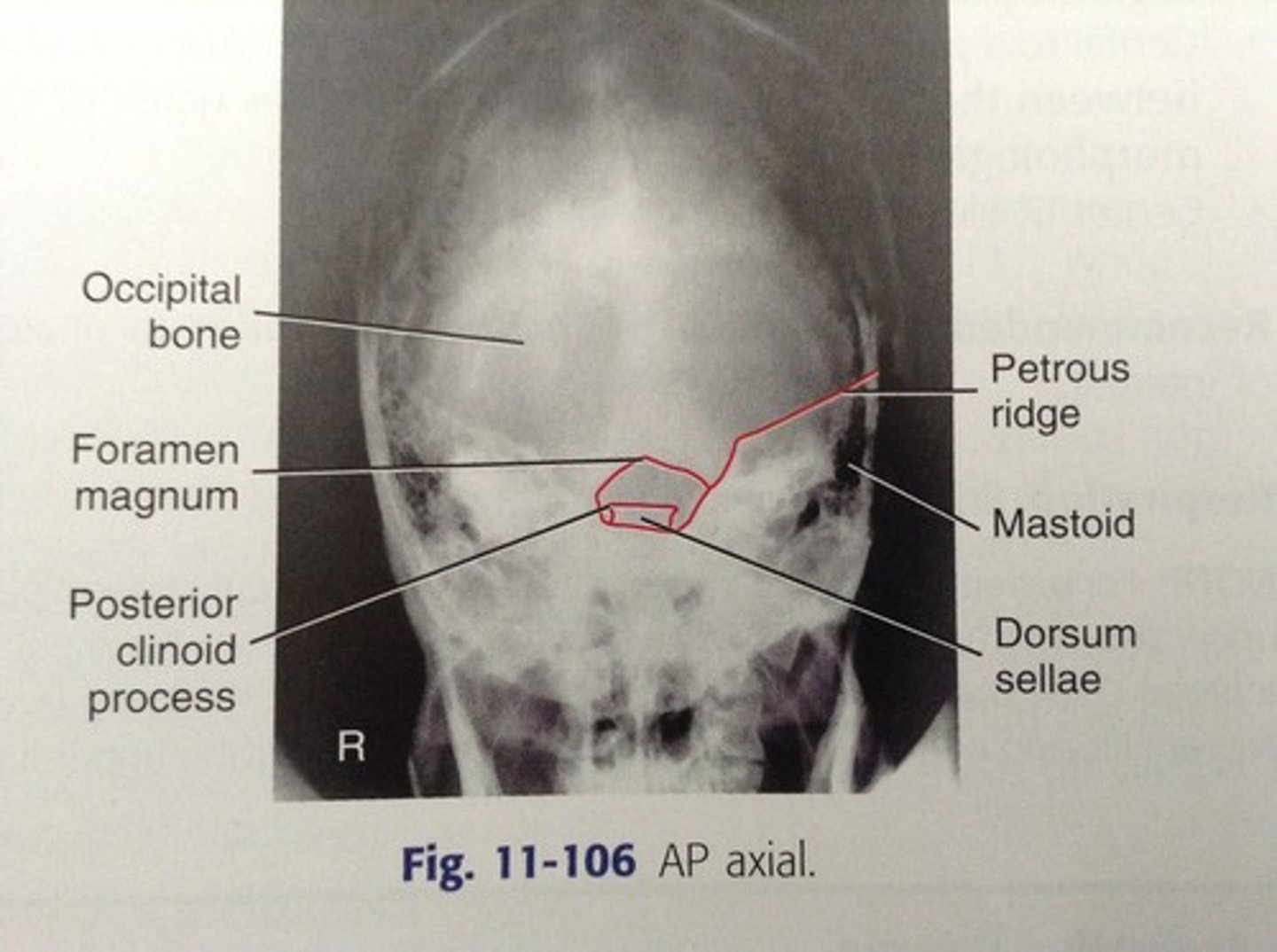
Which cranial bone is best demonstrated with an AP axial (Towne method) projection of the skull?
Occipital
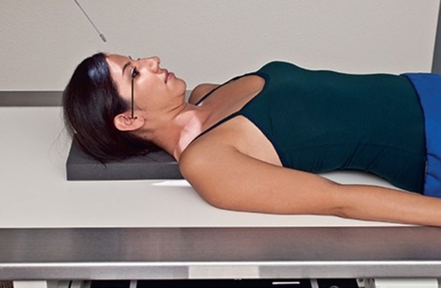
When using a 30 degree caudad angle for the AP axial (Towne method) projection of the skull, which positioning line should be perpendicular to the image receptor?
A. OML
B. IOML
C. GAL
D. AML
A. OML
A properly positioned AP axial (Towne method) projection should place the dorsum sellae into the middle aspect of the:
A. Orbits
B. Clivus
C. Foramen magnum
D. Anterior arch of C1
C. Foramen magnum
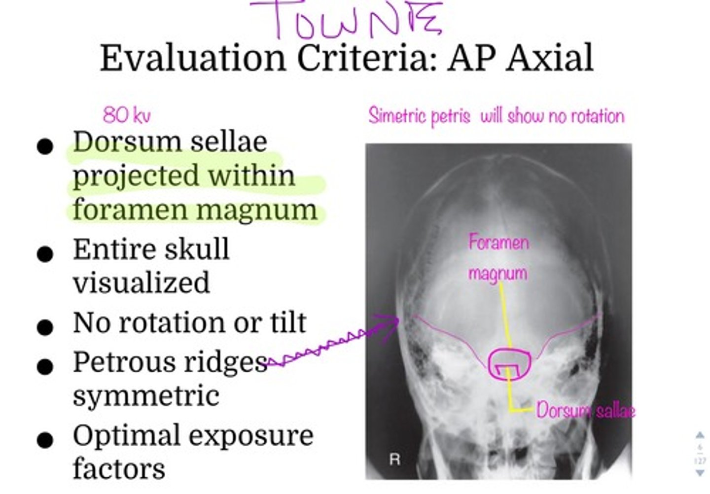
A lack of symmetry of the petrous ridges indicates which of the following problems with a radiograph of an AP axial projection?
A. Tilt
B. CR angle
C. Flexion or extension
D. Rotation
D. Rotation
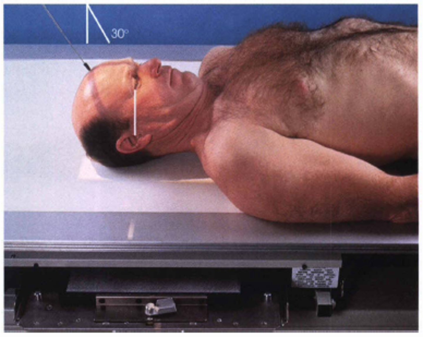
If the patient cannot flex the head adequately for the AP axial (Towne method) projection, the technologist could place the ________ perpendicular to the IR & angle the CR _______ degrees caudad
IOML, 37
What evidence on a an AP axial (Towne method) radiograph indicates whether the correct CR angle & correct flexion were used?
Dorsum sellae & posterior clinoids should be projected into the foramen magnum
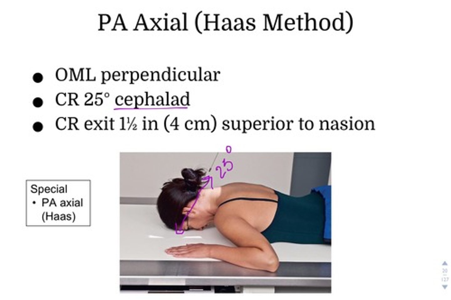
What CR angle should be used for the PA axial (Haas method) projection of the cranium?
25 degrees cephalad
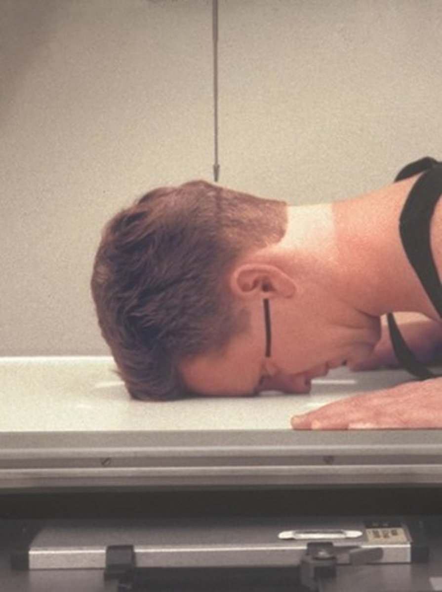
Where is the CR centered for a lateral projection of the skull?
2 inches above the EAM
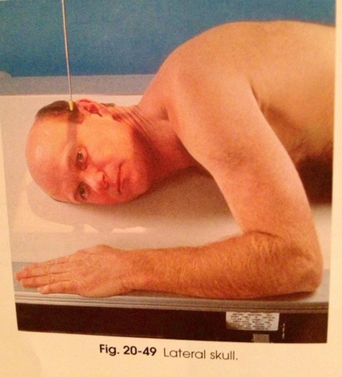
Which specific positioning error is present if the mandibular rami are not superimposed on a lateral skull radiograph?
A. Tilt
B. Rotation
C. Overflexion
D. Incorrect CR angle
B. Rotation
Where will the petrous ridges be projected with a 15-degree PA axial (Caldwell) projection of the cranium?
In the lower one-third of the orbits
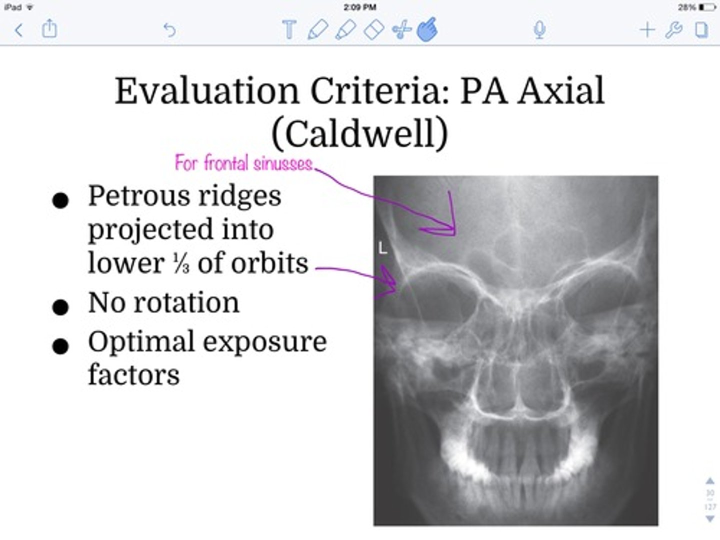
Which positioning error is present if the petrous ridges are projected higher in the orbits than expected for a 15-degree PA axial projection?
excessive flexion or insufficient CR angle
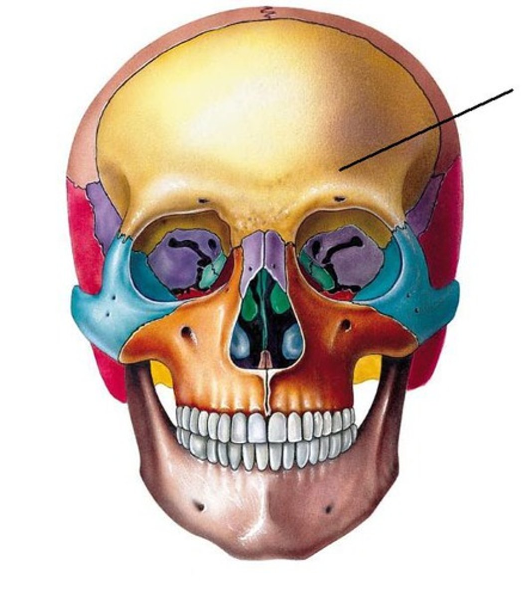
Which projection of the cranium produces an image of the frontal bone with little or no distortion?
0 degree PA
For a patient with possible trauma, what must be determined before performing the SMV projection of the skull?
Rule out any possible cervical fractures or subluxation
What positioning error has been committed if the EAMs are not superimposed with one of them more superior than the other on a lateral projection of the cranium?
Tilt of the skull
Which skull positioning line is placed parallel to the plane of the IR for the SMV projection?
A. OML
B. IOML
C. AML
D. GML
B. IOML
Which of the following projections best demonstrates the sella turcica in profile?
A. AP axial
B. SMV
C. 15-degree PA axial
D. Lateral
D. Lateral
Which of the following projections best demonstrates the foramen rotundum?
A. SMV
B. 25 to 30-degree AP axial
C. 25 to 30-degree PA axial
D. Lateral
C. 25 to 30 degree PA axial
Which of the following projections best demonstrates the clivus in profile?
A. AP axial
B. 15-degree PA
C. Lateral
D. SMV
C. Lateral
Where does the CR exit for a PA axial (Haas method) projection of the skull?
A. 1.5 inches superior to the nasion
B. 3/4 inch anterior to the EAM
C. 2.5 inches above the glabella
D. Level of nasion
A. 1.5 inches superior to the nasion
Which imaging modality is best to differentiate between an epidural & a subdural hemorrhage?
A. CT
B. MRI
C. Nuclear medicine
D. PET
A. CT
A radiograph of an AP axial (Towne method) projection of the cranium shows that the right petrous ridge is wider than the left side. Which specific positioning error is present on this radiograph?
Rotation of skull present; rotation of patient's face toward left
A radiograph of a 15-degree PA axial (Caldwell) projection of the cranium demonstrates that the petrous ridges are projected at the inferior orbital margin. Which positioning error(s) led to this radiographic outcome?
Excessive extension or excessive caudad CR angle - projects the petrous ridges lower than expected (should be in the lower third of the orbit)
A radiograph of a 15-degree PA axial (Caldwell) projection demonstrates that the distance between the right midlateral orbital borders & lateral margin of the skull cortex is greater than the left side. Which positioning error led to this radiographic outcome?
Rotation of the patient's face (skull) to the left
A radiograph of an SMV projection of the skull shows that the mandibular condyles are within the petrous bone. Which specific positioning error led to this problem?
Insufficient extension of the skull, or CR was not perpendicular to IOML
A radiograph of a lateral projection of the skull shows that the orbital plates are not superimposed, one orbital plate is slightly superior to the other. Which specific positioning error led to this image?
Skull tilt
A lateral skull radiograph demonstrates 1 mandibular ramus about 0.5 cm more anterior than the other. Which positioning error occurred?
Skull rotation
An AP axial (Towne method) radiograph for the cranium demonstrates the dorsum sellae projected above, or superior to, the foramen magnum. The foramen magnum is distorted. Which positioning error(s) occurred?
CR angled <37 degrees to IOML, or <30 degrees to OML (would be caused by 30-degree angle to IOML). This issue can be addressed by more flexion of the neck as well.
Situation: A patient comes to the radiology department with a possible tumor of the pituitary gland. Which radiographic projection of the cranium best demonstrates any body involvement of the sella turcica?
Collimated, lateral projection of the sell turcica
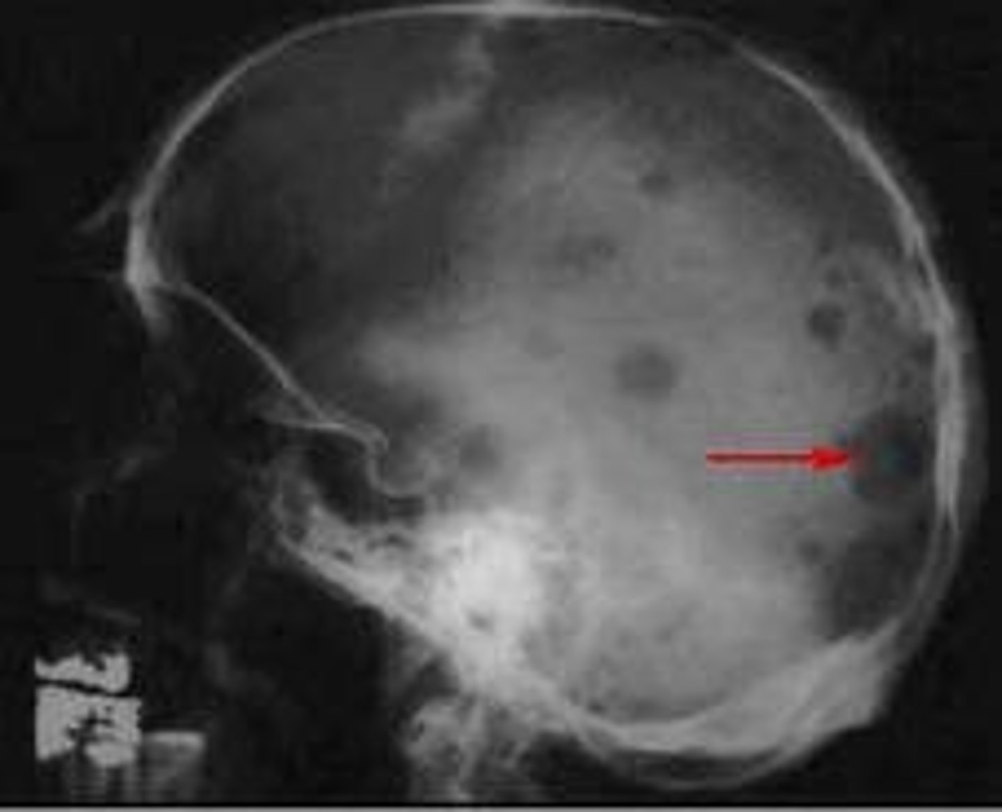
Situation: A patient with a possible linear fracture of the right parietal bone enters the ER. Which single radiographic projection of the skull best demonstrates this fracture?
Right lateral projection of the skull
Situation: A patient comes to the radiology department for a skull series, but the patient cannot assume the correct position for either version of the AP axial (Towne method) projection because of a very short neck & sever spinal kyphosis. What can the technologist do to demonstrate the occipital bone?
Should perform the PA axial projection (Haas method)
Situation: A patient with a possible basal skull fracture enters the ER. No CT scanner is available. Which specific position may provide radiographic evidence of this fracture?
Horizontal beam (dorsal decubitus) lateral position - will demonstrate a possible air-fluid level in the sphenoid sinus
Situation: A neonate has a clinical history of craniosynostosis. Because of the age of the patient, the physician does not order a radiographic procedure of the cranium. What other imaging modality can be performed to evaluate the patient for this condition?
Ultrasound (sonography)
Situation: A patient with a clinical history of acoustic neuroma comes to the radiology department. Which imaging modality or modalities can be performed for this type of pathology?
MRI or CT
A radiograph of an AP axial (Towne method) projection for the cranium shows that the posterior arch of C1 is projected within the foramen magnum. The dorsum sellae is superimposed on the posterior arch as well. What is (are) the positioning error(s)?
Overangulation of the CR or excessive flexion of neck
A radiograph of an AP axial (Towne method) projection for the cranium shows that the mid- to lower mandible is cut off & not demonstrated. What should the technologist do?
No repeat necessary. Because of elongation of the facial mass with the AP axial projection for the skull, cutting off aspects of the mandible is acceptable.
True/False: Facial bone studies should always be performed recumbent whenever possible
False (best to perform erect)
True/False: The common basic PA axial projection for facial bones requires a 15-degree caudad angle of the CR, which projects the dense petrous ridges into the lower one-third of the orbits
True
True/False: An increase in kV of 25-30% (using manual techniques) is often required for the geriatric patient with advanced osteoporosis
False
True/False: CT is ideal for facial bone studies because it allows for the visualization of bony structures as well as related soft tissues of the facial bones
True
True/False: Nuclear medicine is not helpful in diagnosing occult facial bone fractures
False (it is used for this)
True/False: MRI is an excellent imaging modality for the detection of small metal foreign bodies in the eye
False (strong magnets in MRI prohibit this)
What is the name of the fracture that results from a direct blow to the orbit leading to a disruption of the inferior orbital margin?
Blow-out fracture
A "free-floating" zygomatic bone is the frequent result of a ___________ fracture
Tripod
What is the major disadvantage of performing a straight PA projection for facial bones, with no CR angulation or neck extension, as compared with other PA facial bone projections?
Dense petrous pyramids superimpose the orbits, obscuring facial bone structures
Which facial bone structures are best seen with a parietoacanthial projection? (waters method)
Orbits including infraorbital rims, bony nasal septum, maxillae, zygomatic bones, & arches
What CR angle must be used to project the petrous ridges just below the orbital floor with the PA axial (Caldwell method) projection?
A. None. CR is perpendicular
B. 30 degrees
C. 20 degrees
D. 45 degrees
B. 30 degrees
Which structures specifically are better visualized on the modified parietoacanthial (Waters) projection as compared with the basic Waters projection?
Orbital rims & orbital floors
The parietoacanthial (Waters) projection for the facial bones has the _________ line perpendicular to the IR, which places the OML at a _______-degree angle to the tabletop & IR
MML, 37-degree
Where does the CR exit for a parietocanthial (Waters) projection of the facial bones?
Acanthion
For a parietoacanthial (PA Waters) projection, the petrous ridges should be projected directly below the __________ & projected into the lower half of the maxillary sinuses or below the _________ for a modified Waters projection
Maxillary sinuses; inferior orbital rims
Projection that demonstrates:
Floor of orbits (blowout fractures)
Modified Waters method
Projection that demonstrates:
Optic foramen
Parieto-orbital oblique projection
Which paranasal sinuses are best demonstrated with a PA (Caldwell) projection?
Frontal & anterior ethmoid
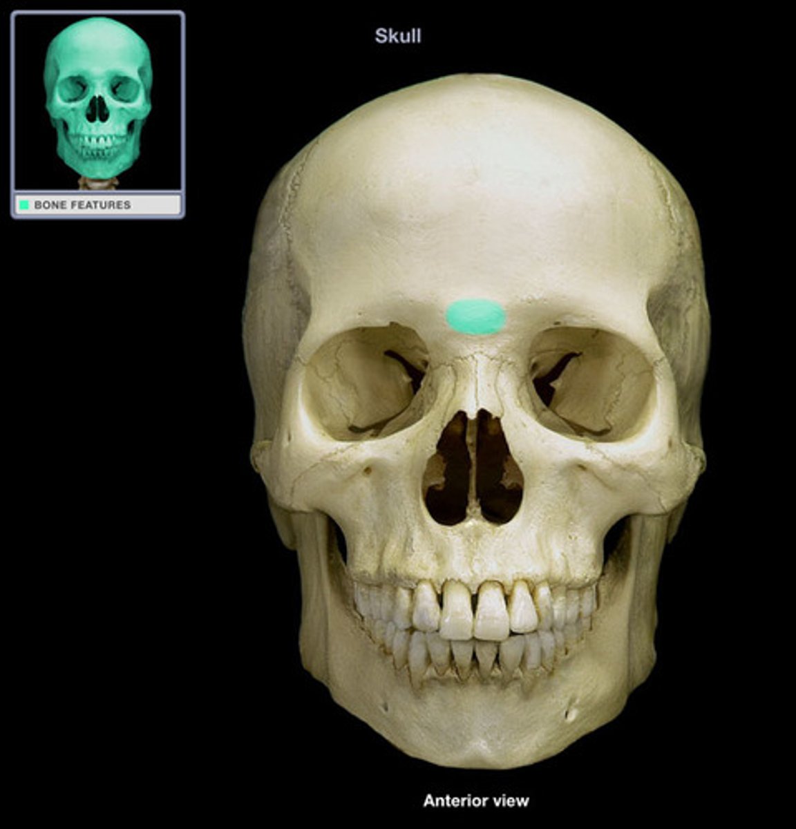
Glabella
Smooth area between the eyes
A line between the glabella and alveolar process of the maxilla
glabellaoalveolar line