Topic 8 - Pancreas and Gallbladder
1/23
There's no tags or description
Looks like no tags are added yet.
Name | Mastery | Learn | Test | Matching | Spaced |
|---|
No study sessions yet.
24 Terms
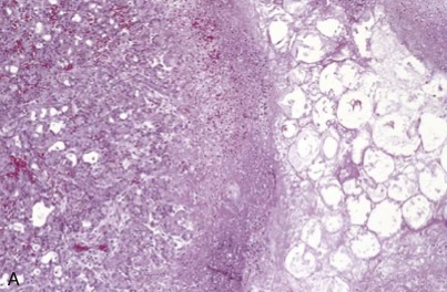
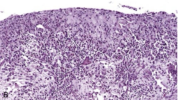
Acute pancreatitis. (A) The microscopic field shows a region of fat necrosis (right) and focal pancreatic parenchymal necrosis (center).
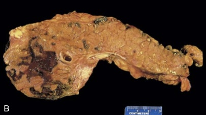
Acute pancreatitis. (B) The pancreas has been sectioned longitudinally to reveal dark areas of hemorrhage in the pancreatic substance and a focal area of pale fat necrosis in the peripancreatic fat (upper left).
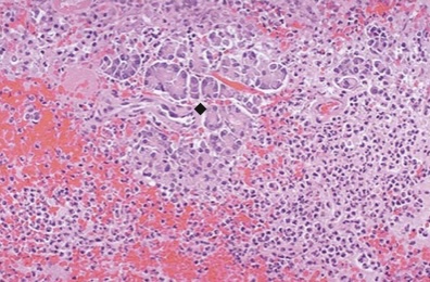
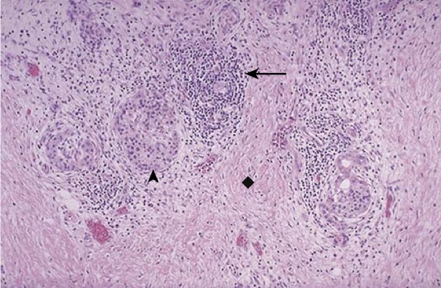
Acute pancreatitis. Inflammation with necrosis and hemorrhage is seen here along with residual pancreatic acini (diamond). The damage involves primarily the acinar cells, but the vasculature is also affected, and if severe and extensive, even the islets of Langerhans may be destroyed.
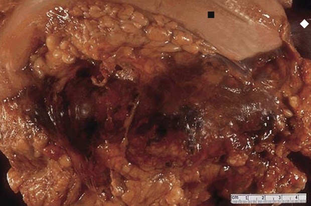
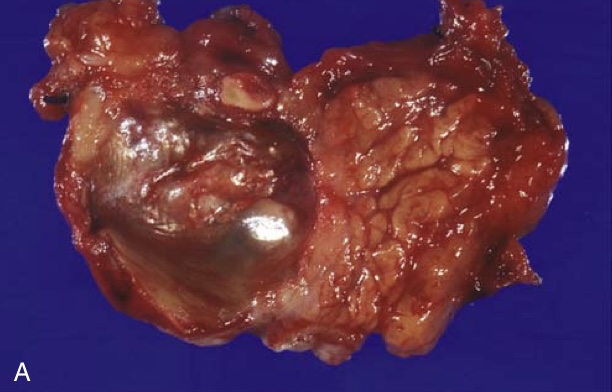
Acute hemorrhagic pancreatitis. At autopsy, the stomach (square) is reflected superiorly, and the spleen (diamond) can be seen at the far upper right. The pancreas is swollen and does not show the typical tan, lobulated architecture. Instead, hemorrhagic necrosis appears as blotchy black to red areas.
Pancreatic pseudocyst. (A) Cross-section revealing a poorly defined cyst with a necrotic brownish wall.
Pancreatic pseudocyst. (B) Histologically, the cyst lacks an epithelial lining and instead is lined by fibrin and granulation tissue, with typical changes of chronic inflammation.
Chronic pancreatitis. Chronic inflammatory cells (arrow) are seen in a collagenous stroma (diamond) that lacks acini and retains a few islets of Langerhans (arrowhead).
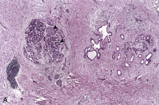
Chronic pancreatitis. (A) Extensive fibrosis and atrophy have left only residual islets (arrowheads) and ducts (right), with a sprinkling of chronic inflammatory cells and acinar tissue.
Chronic pancreatitis. (B) A higher-power view demonstrating a dilated duct with inspissated eosinophilic concretions in a patient with alcohol-related chronic pancreatitis.
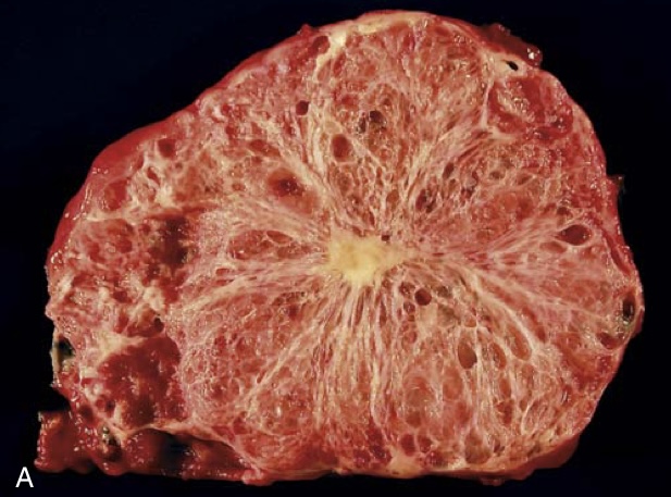
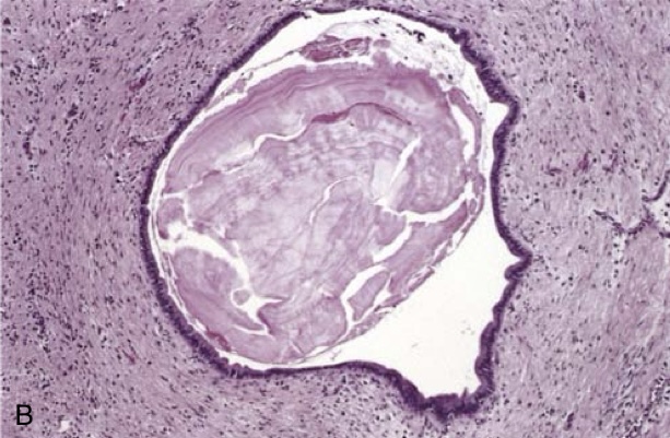
Serous cystadenoma. (A) Cross-section through a serous cystadenoma. The lesion consists of honeycomb of microcystic lesions, only a thin rim of normal pancreatic parenchyma remains. The cysts are relatively small and contain clear, straw-colored fluid.

Serous cystadenoma. (B) The cysts are lined by cuboidal epithelium without atypia.
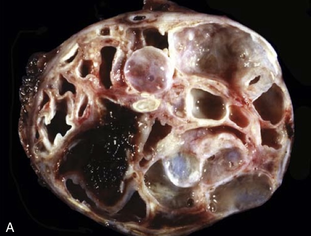
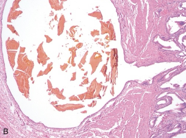
Mucinous cystic neoplasm. (A) Cross-section through a mucinous multiloculated cyst in the tail of the pancreas. The cysts are large and filled with tenacious mucin.
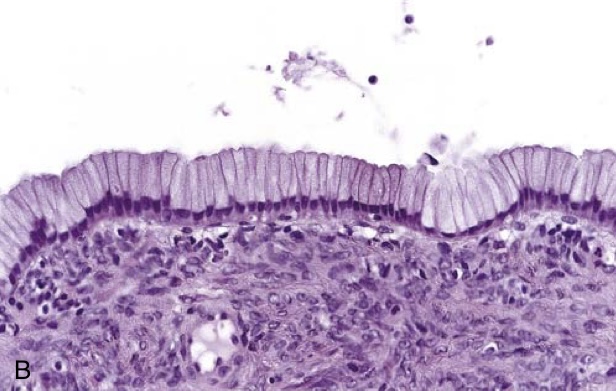
Mucinous cystic neoplasm. (B) The cysts are lined by columnar mucinous epithelium, with a densely cellular“ovarian” stroma.
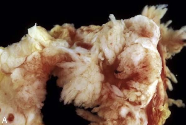
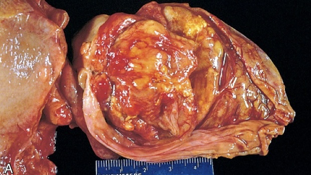
Intraductal papillary mucinous neoplasm. (A) Cross-section through the head of the pancreas showing a prominent papillary neoplasm distending the main pancreatic duct.
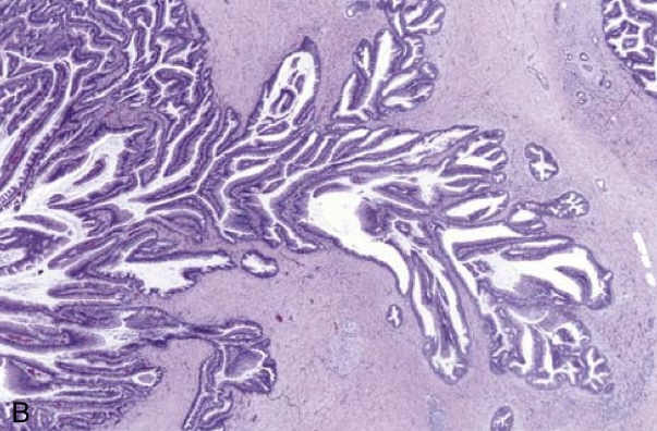
Intraductal papillary mucinous neoplasm. (B) The papillary mucinous neoplasm involves the main pancreatic duct (left) and is extending down into the smaller ducts and ductules (right).
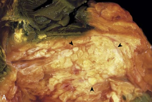
Carcinoma of the pancreas. (A) Cross-section through the head of the pancreas and adjacent common bile duct showing both an ill- defined mass in the pancreatic substance (arrowheads) and the green discoloration of the duct resulting from total obstruction of bile flow.
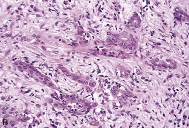
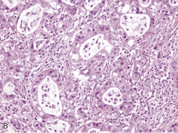
Carcinoma of the pancreas. (B) Poorly formed glands are present in a densely fibrotic (desmoplastic) stroma within the pancreatic substance.
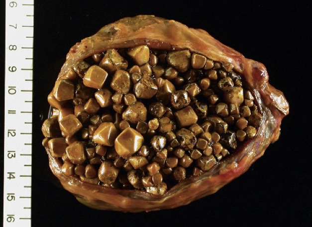
Cholesterol gallstones. The wall of the gallbladder is thickened and fibrotic due to chronic cholecystitis.
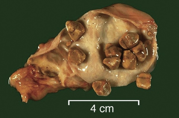
Cholesterol gallstones. Yellow-tan faceted gallstones are present in this gallbladder. The abnormal tan mucosa and pale wall and serosa are the result of scarring from chronic inflammation.
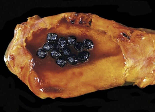
Pigment gallstones. Several faceted black gallstones are present in this otherwise unremarkable gallbladder from a patient with a mechanical mitral valve prosthesis, leading to chronic hemolysis.
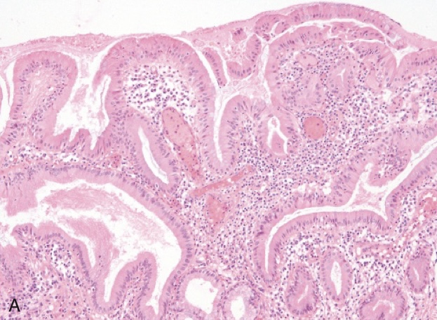
Chronic cholecystitis. (A) The gallbladder mucosa is infiltrated by chronic inflammatory cells.
Chronic cholecystitis. (B) A Rokitansky-Aschoff sinus containing a fragmented bile pigment stone.
Gallbladder adenocarcinoma. (A) The opened gallbladder contains a large, exophytic tumor that virtually fills the lumen.
Gallbladder adenocarcinoma. (B) Microscopically, the tumor shows glandular differentiation along with inflammation.