Medsci 206 Module B
 Call Kai
 Learn
Learn Practice Test
 Spaced Repetition
 Match
Match Flashcards
 Knowt Play
1/110

Earn XP
Description and Tags
rip anki
Name | Mastery | Learn | Test | Matching | Spaced | Call with Kai | Chat |
|---|
No analytics yet
Send a link to your students to track their progress
111 Terms
Sensory systems (main 5 and others)
Vision, hearing, taste, smell, touch, vestibular/balance, proprioceptor/posture and movement of body parts relative to each other, other modalities (ie pain, temperature, itch)
What information (external) do sensory system detence and convey?
Lights, sounds, smells, tastes, touch/pressure
What information (internal) do sensory system detence and convey?
O2, CO2, pH, muscle length, body position, motion and equilibrium
Sensory system - detection and conveying of info about external and internal environment leads to…
Awareness of environment and events
Mosquito - sensory information (antennas, mouth)
Antennas - hearing, vibrating hairs
Mouth - detects chemicals in the air
Sensory hairs - sense movements
Bees!
Use vision, odour (phermone emission), hearing, mechanoreceptor and electrical sense (to sense electrical fields of flowers)
- Hairs are responsible for sensing electrical fields, the greater the movement of hair the greater the neuronal firing
Sensory systems (basis)
Signal
2. Collection
3. Transduction
4. Processing
5. Action
Neural coding of information is conveyed to brain centres - diagram
""
Where does information from the sensory neurons in the head enter the CNS from?
Cranial nerves
Specific pathway of information from sensory neurons entering from below the head:
Sensory neurons below the head enter the spinal cord and pass towards the brain via the spinal nerves
All sensory information goes to the thalamus EXCEPT information from the:
Olfactory bulb - goes straight to the olfactory cortex
""
learn ts
Sensory disorders
Blindness, deaf/impaired hearing, anosmia (loss of smell), ageusia (loss of taste)
How many cone receptors do humans have and what colours are they?
3 cone receptors, red/blue/green
Tapetum
The shiny reflective surface immediately behind the retina of most animals (humans do not have this)
Sensory system basis
Signal (physical stimulus providing information on our surrounding)
2. Collection of signal ie. retinal photoreceptors
3. Transduction of stimulus to nerve signal
4. Processing of information by the brain
5. Generation of resulting action ie an image
(Signal, collection, transduction, processing, action)
Neural coding of information is conveyed to the following brain centres:
Olfactory cortex, auditory cortex, visual cortex, gustatory cortex, somatosensory cortex, vestibular cortex
Attributes of the sensory system
Modality - type/class of stimulus
2. Location - position of stimulus in space
3. Intensity - measure of amount of stimulus
4. Timing - onset, duration and offset of stimulus
5 major classic modalities (human)
Main: vision, hearing, taste, smell, touch
Others: vestibular, proprioception, pain/temp/itch
Receptor specificity - 4 classes
Mechanoreceptors
2. Chemoreceptors
3. Thermoreceptors
4. Photoreceptors
Transduction
Sensory receptors transform signal into electrical energy
Receptor potential
Leads to an electrical reponse/change in membrane potential
Transduction mechanisms
These vary according to the physical stimulus:
1. Direct - ie mechanoreceptors in skeletal muscle
2. Indirect via 2nd messenger systems - ie chemoreceptors in the olfactory epithelium
Mechanical transduction explaination
Mechanoreceptors sense physical deformation of tissue ie pressure on skin/muscles stretching
- Mechanical stimulation causes opening of stretch sensitive ion channels and an increase in Na+ & Ca2+ conductances that depolarise the receptor neurons
- Change in receptor potential triggering an action potential
Example of mechanical transduction: muscle spindle detecting
As muscle fibres stretch channels open, resulting in a change in receptor potential. As more pressure is applied the channels open more frequently.
Transduction via 2nd messenger system - chemoreceptors in the olfactory epithelium
"

Spatial location of receptors:
Provides information on location of stimulus source on body
Enables discrimination of size+shape of object
Enables resolution of fine detail of the stimulus/environment
Receptive field
The area in which a stimuli can activate a sensory neuron
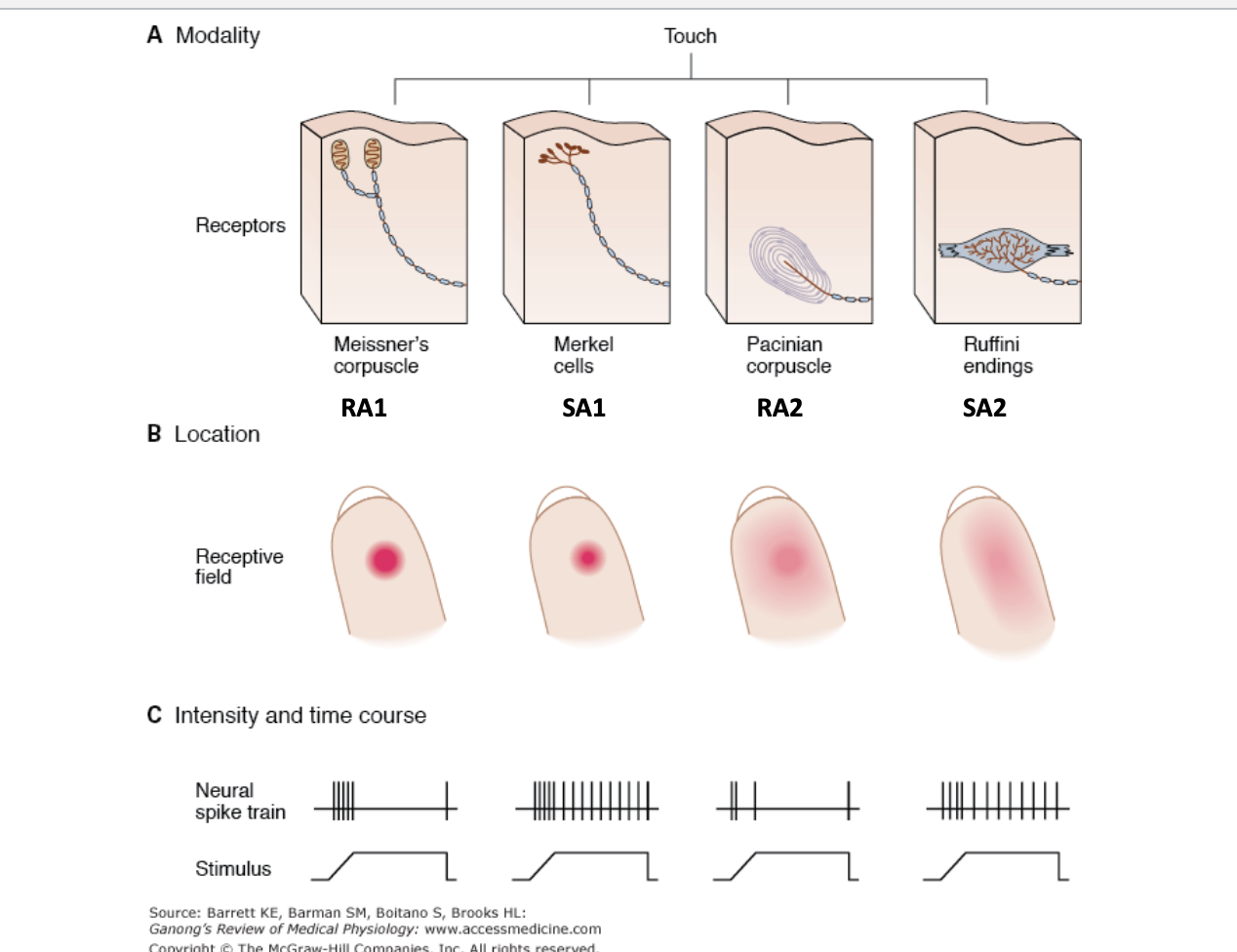
Example of receptive field of touch receptors
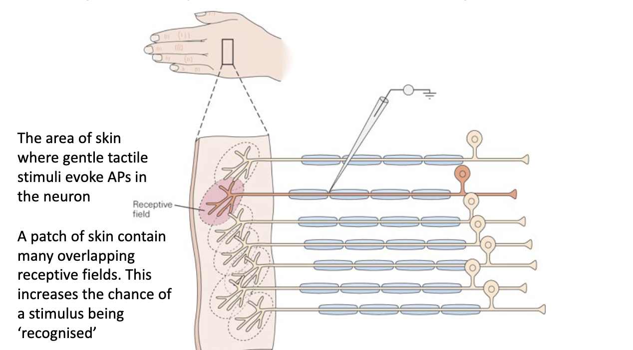
Receptive field sizes
Small receptive fields = high resolution of fine detail
Larger receptive fields = more difficulty in localising stimulus
Ex. receptive fields are small in the fingertips but larger in the palms
The density of receptors define the resolution of the stimulus
Receptor density is not uniform and they may be localised to give a higher definition
Ex. The fovea of the retina has a high density of photoreceptors and small receptive fields
Intensity
Stimulus intensity is encoded by the frequency of action potentials in sensory nerves
Timing
Firing rates of sensory neurons convey information about the stimulus intensity and time course

whatever this is
3 basics parts of vision:
Eyes, optic connections, brain
Basic process of vision
Light passes through the cornea and lens
Light focuses onto retinal photoreceptors
Retina sends information to the brain in the form of electrical signals using the optic nerve
Brain processes the image and allows us to see
Aqueous humor (anterior chamber)
Constantly replenished to ensure there is removal of waste, replenishment of nutrients
Generates intraocular pressure
Is created by the ciliary body
Vitreous humor (between lens and retina)
Clear+transparent to help passage of light to retina
Stagnant (very little exchange/replenishment)
Contains high levels of vitamin C and keep O2 levels low (lens is hypoxic, O2 results in cataract)
Fovea
Region around the middle of the retina that contains cone photoreceptors
Eye Anatomy

Retinal image projection
Lens is convex-shaped causing light rays to converge on the retina. Image is therefore projected upside-down and back-to-front onto the retina
Retina structure from outer retina to inner retina
Pigment epithelium
Photoreceptors
Outer nuclear layer
Outer plexiform layer
Inner nuclear layer
Inner plexiform layer
Ganglion cell layer
Optic nerve layer
Pigment epithelium
Contains melanin to absorb excess light
Provides nutrition to the photoreceptors
Photoreceptors (cones+rods)
Light senstive
Transduces light energy into electrical energy
Outer nuclear layer
Contains cell bodies of rods and cones
Outer plexiform layer
First synaptic interaction between photoreceptors and bipolar cells
Inner nuclear layer
Contains cell bodies of amacrine, horizontal and bipolar cells
Inner plexiform layer
Second synaptic interaction between bipolar and ganglion cells
Ganglion cell layer
Output cells of retina
These are the only retinal cells are capable of firing action potentials
Optic nerve layer
Contains the axons of ganglion cells
In short, photoreceptor cells… while inner retina cells …
Photoreceptor cells detect light, inner retina cells process the information
Responses of retinal cells to darkness (D for darkness, D for depolarise)
ON bipolar cell synapses with ON center ganglion cells
OFF bipolar cell synapses with OFF center ganglion
ON bipolar cell synapses with ON center ganglion cell (retinal cells response to darkness)
mGluR
Presence of glutamate = hyperpolarisation = less glumate
Results in ON center ganglion to decrease firing
OFF bipolar cell synapses with OFF center ganglion (retinal cells response to darkness)
AMPAR
Presence of glutamate = depolarisation = more glutamate
Results in OFF center ganglion cell to increase firing
Responses of retinal cells to light - ON/ON
AMPAR
Presence of glutamate = depolarisation = more glutamate
Results in ON center ganglion cell to increase firing
Responses of retinal cells to light - OFF/OFF
mGluR
Presence of glutamate = hyperpolarisation = less glumate
Results in ON center ganglion to decrease firing
ON/OFF ganglion cells
ON/OFF refers to the response of the ganglion cells to light
Ganglion cell receptive field is defined as:
The area of the retina where stimulation with a small spot of light produces a change in ganglion firing rate.
Area within the receptive field is divided into two regions: center and surround
Receptive field center
Provides direct input from photoreceptors to the bipolar cell
Receptive field surround
Provides indirect input from the photoreceptors to the bipolar cells via horizontal cells

This thing
Turning a spot of light in the centre of an ON-centre ganglion cell receptive field
Produces burst of electrical activity
Turning spot of light on in the centre of an OFF-centre ganglion cell receptive field
Firing decreases
ON-center cell stimulation vs inhibition
Stimulated when center of receptive field is exposed to light
Inhibited when surround of receptive field is exposed to light
Light/no light on receptive field center/surround
Uniform illumination of the visual field is less effective in an ON-center ganglion cell compared to a well-placed spot of light on center of cell receptive field.
This organisation makes ganglion cells sensitive to differences in illumination across the receptive field (luminance contrast)

Receptive fields have overlapping distributions
Several ON/OFF ganglion cells analyse every part of the visual space. Receptive fields convey information about cell firing rates in center and surround - combining information from adjacent receptive fields = brain can construct info about edges/shapes/colours
Visual pathway - transmission of sensation from retina to brain
Monocular information transmitted by optic nerve
Nasal fibres cross at optic chiasm (anatomical crossing point)
Axons synapse at the LGN of the thalamus to large magnocellular areas (magnocellular pathway)
Optic radition
Primary visual cortex (V1)
Visual pathway - steps 1+2
Around 50% of optic nerves cross over to the contralateral side of the brain
Left side of brain receives right visual field information and vice versa
Motion detection
Photoreceptors are not sensitive to an object’s motion direction - motion is rather detected by photoreceptors by special class of ganglion cells that are excited by starburst amacrine cells
Directed selectivity ganglion/M/parasol cells
Neurons that respond differentially to direction of visual stimulus. A moving stimulus can elicit strong spiking of action potentials in one direction but not the opposite
DSGC - motion information
Responds best and has preference to dorsal/ventral/temporal/nasal directions of motion
Inversion of lens = object movement in visual scene corresponding to movement in opposite direction in retinal plane
DSGC - neuron information
Concerned with motion detection and alerting animal to threatening+moving visual imagery
Are large ganglion cells
AKA as M ganglion cells as they terminate in the magnocellular layer of LGN (make up ~5% of total ganglion cell population
Pattern is of open, radiating branches
Fast processing/high-conduction velocity+responds in a transient fashion
Lateral genicular nucleus
Inner two layers (M1 and M2) transmit important information for perception of movement to primary visual cortex
Visual pathway from retina to cortex
Vision for movement —> dorsal stream
Vision for perception —> ventral stream

Summary slide for L2 (vision)

Neural adaptation
Neurons in the brain coding for a particular movement become less sensitive to motion in that direction, therefore looking away from the stimulus results in neurons that detect movement in opposite direction to become more active = appearance of stationary object/image moving. Can see this through the non-adapted eye too due to contralateral crossover of optic nerves
Rods and cones - structure
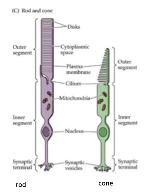
Differences between rods and cones
One type of rod, 3 types of cones
Rods ‘see’ black+white, cones ‘see’ colour
Rods are located in the periphery, cones located in the fovea
Rods 1000x more sensitive to light - can respond to one photon of light while cones require 10-100s of photos to become activated
Rods recover slow, cones recover fast and can rapidly adapt to illumination changes
Scotopic vision (low light conditions)
Only rods are functioning: cones have no light to respond to therefore no colour perception
Photopic vision (daylight conditions)
Only cones are functioning as rods bleach out quickly in bright lights, 3 cone cell types provide wide range of colour perception in bright light
Mesopic vision (moderately low light ie right after sunset)
Both rods and cones are functioning therefore cones still have enough light to provide some colour vision
Processing of colour information is done by parvocellular (P) ganglion cells (LGN - parvocellular pathway)
Compared to M cells, P cells have smaller cell body+dendritic fields, sustained responses, slower conduction velocities, sensitive to light wavelength difference and conveys colour information that way
LGN colour perception
Outer 4 layers (P3-P6) transmit information required for colour perception (esp. red and green) to primary visual cortex
Three anatomical pathways of the LGN
Magnocellular - motion detection
Parvocellular - colour perception
Koniocellular - involved in relaying info from S-cones (blue) with contribution from M and L cones
Trichromatic theory
Based on the fact that all colours can be perceived via a combination of 3 cone receptors
Three types of cone photoreceptors
Red (L-cones): long light wavelengths
Green (M-cones): medium light wavelengths
Blue (S-cones): short light wavelengths
Stimulation of cone photoreceptors
The difference in signals received from the 3 cone types allows the brain to perceive continuous range of of colours.
Ex: yellow-green light stimulates L and M cones equally strongly but only stimulates S cones weakly

Opponent Colour Theory
Certain colours don’t exist in ordinary perception - some colours can be seen simultaneously ie red+yellow (orange) and red+blue (purple) but red+green (reddish-green) and yellow+blue (yellowish-blue) cannot be
Colour opponent channels
3 opponent channels: blue-yellow, red-green and black-white (brightness or luminosity/achromatic channel)
One member of colour pair suppresses the other
P cells comprise of receptive field activated by one colour and receptive field surround activated by another colour
Blue-yellow opponent channel example
Processes information about S cone and combined L+M cone signal differences. If blue light is present S cone is activated and blue+yellow cell receives excitatory input (no L+M cone input) = blue colour perceived
Red-green opponent channel example
Processes information about L+S and M cone firing differences. Red light present results in L cone being activated and red+green cell receives excitatory input (no M cone input) = red colour perceived
Black-white opponent channel example
Transmits info about light intensity
Model for normal colour vision
Trichromatic theory - photoreceptor level
Opponent-process theory - photoreceptor neural interconnection
Protanopia
No red cone cells in retina therefore impairment of long light wavelength perception (reds).
Red-green colour blindness
Deuteranopia
No green cone cells therefore impairment in perceiving medium light wavelengths (greens)
Tritanopia
No blue cone cells in retina (very rare) therefore inability in perceiving short light wavelengths (blues)
Patients confuse greens and blues, yellows may appear pink
Stage theory
Incorporates trichromatic theory and opponent colour theory into two stages: receptor stage (cone cells - blue, green and red) and neural processing stage (colour opponency)
Congenital Colour Vision Defect (CVD) prevalence
8% of males, 0.5% of females. Deutan (green impairment) is most prevalent.
Anomalous trichromats (all three cones are there but have impaired function) is more prevalent than dichromats.
Protan and deutan have severe impact on daily function whereas tritan is relatively uncommon and has less of an impact
Colour vision tests
Ishihara test
Farnsworth D-15
Farnsworth-Munsell
The Anomaloscope
L’anthony Desaturated D-15
City University Test
Lantern test
Four classification of tests:
Pseudoischromatic plates
Arrangement tests
Matching tests
Naming tests