Radiation Protection
 Call Kai
 Learn
Learn Practice Test
 Spaced Repetition
 Match
Match Flashcards
 Knowt Play
1/149

There's no tags or description
Looks like no tags are added yet.
Name | Mastery | Learn | Test | Matching | Spaced | Call with Kai | Chat |
|---|
No analytics yet
Send a link to your students to track their progress
150 Terms
Radiation
Form of energy that travels through space and matter. This energy can move in the form of waves or particles.
Cosmic rays
Terrestrial radiation
Internally deposited radionuclides
Radon (largest source)
Radiation from natural sources
Radon
radioactive gas that comes from the decay of uranium in the earth.
It emits alpha particles, which are not penetrating, and therefore
contributes a radiation dose only to the lung
Medical
Nuclear power generation
Research applications
Industrial sources
Consumer items
Radiation from man-made sources
Radiation protection
tool to protect health against the risks generated by the use of ionizing radiation
Justification
Optimization
Dose Limit
ICRP’s Three Fundamental Principles of Radiation Protection
Prevent deterministic effects
Reduce stochastic effects
Aims of radiation protection
Intensity or the amount of ionization produced by X-rays or gamma rays in air.
What does exposure measure?
Roentgen (R) - Old unit
Air kerma (Gya) - SI unit
C/kg - Coulombs per kilogram (C/kg) - Special Unit
1 R = 2.58 × 10⁻⁴ C/kg
Units for exposure
Exposure
Common in quality control, calibration, and output measurement of X-ray machine.
Energy absorbed per unit mass of tissue
What does absorbed dose measure?
rad (radiation absorbed dose) - Old unit
Gray (Gyt) – SI unit
J/kg = Joules per kilogram - Special Unit
1 rad = 0.01 Gy
1 Gy = 1 J/kg = 100 rad
Units for absorbed dose
Absorbed dose
Used in estimating how much radiation energy is actually absorbed by the patient or material
Overall biological risk of radiation exposure to the entire body, even if only part of the body is irradiated.
What does effective dose measure?
rem (roentgen equivalent man) - Old unit
Sievert (Sv) – SI unit
J/kg = Joules per kilogram - Special Unit
1 Sv = 100 rem
Units for effective dose
Effective dose
Expressing the quantity of radiation received by radiation workers and populations.
Rate of decay of radioactive atoms per unit time
What does radioactivity measure?
Curie (Ci) - Old unit
Becquerel (Bq) - SI unit
d/s - Disintegration per second - Special unit
1 (Bq) = 1 dps
1 (Ci) = 3.7 x 10¹⁰ dps
Units for radioactivity
Radioactivity
Used in nuclear medicine and radiopharmaceuticals
Health physics
Concerned with providing radiation protection and minimizing occupational dose to the public
Health physicist
Radiation scientist concerned with the research, teaching, or operational aspects of radiation safety
Design equipment
Calculate and construct barriers
Develop administrative protocols
Roles of the health physicists
Linear Non-Threshold model,
which addresses the probabilistic risk of cancer, leukemia, and genetic effects even at low radiation doses. Effective implementation requires continuous monitoring of both occupational and patient doses in diagnostic imaging
What model drives the ALARA principle?
Time
Distance
Shielding
Cardinal principles of radiation protection
Directly proportional: when time is doubled, exposure is doubled
Relationship between time and exposure
Exposure = exposure rate x exposure time
Formula for exposure
5-minute reset timer
Reminds the radiologist that a considerable amount of fluoroscopic time has
elapsed. The timer records the amount of x-ray beam on time.
Inversely proportional, computed by the inverse square law
What is the relationship between distance and radiation exposure?
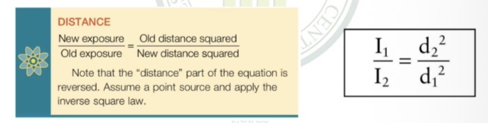
Formula for distance, inverse square law
50 uGya/hr or 5mR/hr
During fluoroscopy, what is the approximate exposure at two steps back?
Because during fluoroscopy, the ptx is an extended source of radiation because of the scattered x-rays generated within the body
Why does the inverse square law not apply to fluoroscopy?
Take two steps back, and take one step to the side and get behind the radiologist
Best step to minimize radiation exposure during fluoroscopy?
Half-value layer (HVL) or
Tenth-value layer (TVL)
The amount that a protective barrier reduces radiation intensity can be estimated if the _____ or _____ of the barrier material is known
One TVL
Is the thickness of absorber that reduces the radiation intensity to one-tenth its original value.
3.3 HVL
1 TVL =
Use of protective apparel
Example of application of shielding in radiology
0.5 mm Pb, equivalent to 2 HVLs, which would reduce occupational exposure to 25%
Lead content of protective aprons and its equivalent, and how much exposure it can reduce
10%
oblique angle
Actual measurements show that such protective aprons reduce exposure to approximately _% because scattered x-rays are incident on the apron at ______ _____.
Protective x-ray tube housing
Every x-ray tube must be contained within protective housing that reduces leakage radiation during use.
100 mR/hr (1 mGya/hr)
1 meter
Leakage radiation must be less than ______ at a distance of _____ from the protective housing.
Control panel
Must indicate the conditions of exposure and must positively indicate when the x-ray tube is energized.
tape measure
A source-to-image receptor distance (SID) indicator must be provided. This can be as simple as a ____ _____ attached to the tube housing, or as advanced as lasers.
2%
The SID indicator must be accurate to within __ of the indicated SID.
Positive Beam Limitation
Automatic, light-localized, variable-aperture collimators automatically adjust the size of the X- ray field to match the size of the image receptor (IR).
Positive Beam Limitation
PBL
2%
The PBL must be accurate to within __ of the SID.
Beam Alignment
In addition to proper collimation, each radiographic tube should be provided with a mechanism to ensure proper alignment of the x-ray beam and the image receptor.
2.5 mm Al
All general purpose diagnostic x-ray beams must have a total filtration (inherent plus added) of at least _____ when operated above 70 kVp.
50 and 70 kVp
Radiographic tubes operated between _____ must have at least 1.5 mm Al.
0.5 mm Al
Below 50 kVp, a minimum of _____ total filtration is required.
Reproducibility
For any given radiographic technique, the output radiation intensity should be constant from one exposure to another.
This is checked by making repeated radiation exposures through the same technique and observing the average variation in radiation intensity.
How is reproducibility checked?
5%
When checking for reproducibility, the variation in x-ray intensity should not exceed ___.
precision radiation dosimeter
three exposures
kVp control
10 exposures
Two methods are available to evaluate exposure reproducibility; both rely on a _____ _____ _____.
First, one can make a series of at least _____ _____ at the same technique factors, having changed technique controls between each exposure. If the result is not reproducible, this is usually the result of error in the ___ _____.
Second, one can select a combination of technique factors and hold them constant for a series of __ _____.
Exposure linearity
The ability of a radiographic unit to produce a constant radiation output for various combinations of mA and exposure time.
10%
The maximum acceptable variation in linearity is ___ from one mA station to an adjacent mA station.
mGya/mAs (mR/mAs)
Radiation intensity is expressed in units of _____.
Operator Shield
It must not be possible to expose an image receptor while the radiologic technologist stands unprotected outside a fixed protective barrier, usually the console booth.
2 meters
The exposure switch of such an imaging system must allow the operator to remain at least __ _____ from the x-ray tube during exposure. The useful beam must be directed away from the radiologic technologist while positioned at this minimum distance.
• 38 cm (15 inches) for stationary fluoroscopic units
• 30 cm (12 inches) for mobile C-arm fluoroscopy
Typical minimum SSD:
• _____ for stationary fluoroscopic units
• _____ for mobile C-arm fluoroscopy
gonadal level
0.25 mm Pb
During fluoroscopy, the Bucky tray is moved to the end of the examination table, leaving an opening in the side of the table approximately 5 cm wide at _____ _____.
This opening should be covered automatically with at least _____ equivalent.
protective curtain
0.25 mm Pb
A _____ _____ or panel of at least _____ equivalent should be positioned between the fluoroscopist and the patient.
cumulative timer
audible signal
A _____ _____ that produces an _____ _____ when the fluoroscopic time has exceeded 5 minutes must be provided.
Dose Are Product (DAP)
measure of the total amount of radiation delivered to the patient
DAP = Radiation dose × Area of the x-ray beam
Formula for DAP
Unit: Gy·cm² (gray × square centimeter)
Unit for DAP
DAP
may be used to monitor radiation output from radiographic and fluoroscopic imaging systems
placed near the x- ray source below the collimator, before the beam enters the patient
Where are DAP meters placed?
… dividing the DAP measurement by the area of the beam at the skin.
The risk for injury to the skin where the beam enters the patient can be derived by …
21 mGya/min (2.1 R/min)
80 kVp
Note:
The intensity of the x-ray beam at the tabletop of a fluoroscope should not exceed _____ for each mA of operation at _____.
100 mGya/min (10 R/min)
200 mGya/min (20 R/min)
If there is no optional high-level control, the intensity must not exceed _____ during fluoroscopy.
If an optional high-level control is provided, the maximum tabletop intensity allowed is _____.
effective radiation dose (E)
The overall stochastic risk to a patient depends on _____ _____ ____, which is related to tissue radiation dose and to the volume of tissue exposed.
Tissue Radiation Dose
Refers to the energy deposited locally
Tissue Radiation Dose
The quantity that best reflects the potential for injury to that tissue (deterministic effect)
If the radiology facility is located on an upper floor
In area survey, when is it necessary to shield floors of radiology rooms as well?
Primary radiation
Secondary Radiation
a. Scatter radiation
b. Leakage radiation
Three types of radiation considered when constructing the protective barrier
Primary Radiation
The most intense radiation and therefore the most hazardous and the most difficult to shield
Scatter radiation
Results when the useful beam intercepts any object, causing some x-rays to be scattered
ptx
During radiography and fluoroscopy, the _____ is the single most important scattering object.
0.1%
The intensity of scatter radiation 1 meter from the patient is approximately ___ of the intensity of the useful beam at the patient.
Leakage radiation
Radiation emitted from the x-ray tube housing in all directions other than that of the useful beam.
1 mGya/hr (100 mR/hr)
If the x-ray tube housing is designed properly, the leakage radiation will never exceed the regulatory limit of _____ at 1m.
Primary protective barrier
designated to the wall to which the useful beam can be directed, and shields from primary radiation
Lead bonded to sheet rock or wood paneling specified for architects and contractors in units of pounds per square foot (lb/ft2 )
Used most often as a primary protective barrier
Concrete, concrete block, or brick
1/16 inch of lead
7 ft high
Primary protective barrier
• _____, _____, or _____ may be used instead of lead.
• As a rule of thumb, 4 inches of masonry is equivalent to _____.
• Radiation protection guidelines have established that primary radiation barriers must be _____.
0.4 mm Pb
conventional gypsum board, glass, or lead acrylic
Often, lead is not required for secondary protective barriers because the computation usually results in less than _____. In such cases, _____ _____, _____, ____ _____ is adequate.
Distance
Occupancy
Control
Workload
Use factor
kVp
Factors that affect barrier thickness
source of radiation and the barrier
The thickness of a barrier naturally depends on the distance between the _____ of radiation and the _____. A wall along which an x-ray imaging system is positioned probably requires more shielding than the other walls of the room
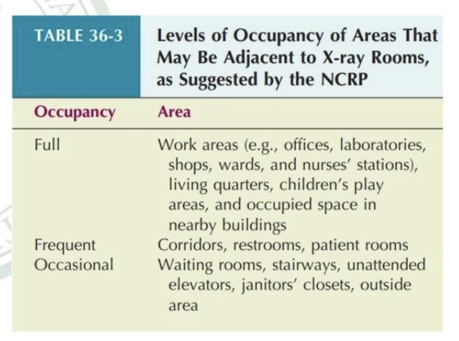
Occupancy
time of occupancy factor (T)
If the area were a rarely occupied closet or storeroom, the required shielding would be less than if it were an office or laboratory that was occupied 40 hours per week.
• This concept reflects the _____.
Controlled area
An area that is occupied primarily by radiology personnel andpatients.
recommended occupational dose limit
less than 1 mSv/wk (100 mrem per week)
The design limits for a controlled area are based on the _____ _____ ____ _____; therefore, the barrier is required to reduce the exposure to a worker in the area to _____.
Uncontrolled area
An area that can be occupied by anyone.
1 mSv/ yr (100 mrem/yr)
20 µSv/wk (2 mrem/wk)
25 µSv (2.5 mrem)
Uncontrolled area:
The maximum exposure rate allowed is based on the recommended dose limit for the public of _____.
• This is equivalent to _____, which is the design limit for an uncontrolled area.
• Furthermore, the protective barrier should ensure that no individual will receive more than _____ in any single hour.
level of radiation activity
workload (W)
units of milliampere minutes per week (mAmin/wk)
The shielding required for an x-ray examination room depends on the _____ of _____ _____ in that room.
• The greater the number of examinations performed each week, the thicker the shielding that is required.
• This characteristic is called _____ and is expressed in _____.
Use Factor
Represents the fraction or percentage of time a specific barrier (like a wall, floor, or ceiling) is directly hit by the primary X-ray beam.
Walls: 1/4
Floor: 1
The NCRP recommends that walls be assigned a use factor of ___ and the floor a use factor of _.
1
secondary radiation barriers
A room designed strictly for chest radiography has one wall with a use factor of _. All others have a use factor of zero for primary radiation and thus would be considered ____ _____ _____.
⬆ use factor = ⬆ shielding
⬇ use factor = ⬇ shielding
⬆ use factor = ___ shielding
⬇ use factor = ___ shielding
kVp
penetrability
150 kVp
75 kVp
100 kVp
30 kVp
For protective barrier calculations, ___ is used as the measure of _____.
• Most modern x-ray imaging systems are designed to operate at up to _____. Most examinations, however, are conducted at an average of _____.
_____ for general radiography, _____ for mammography
75 kVp
The average kVp is usually closer to _____ than to 100 kVp.