Intrapartum Fetal Monitoring, Postdates Pregnancy and Neonatal Resuscitation
 Call Kai
 Learn
Learn Practice Test
 Spaced Repetition
 Match
Match Flashcards
 Knowt Play
1/37

There's no tags or description
Looks like no tags are added yet.
Name | Mastery | Learn | Test | Matching | Spaced | Call with Kai | Chat |
|---|
No analytics yet
Send a link to your students to track their progress
38 Terms
Intra-partum Fetal Monitoring Can be Done
Externally
Doppler (ultrasound transducers)
Tocodynamometer
Internally: when indicated
Fetal scalp electrode
Intra-uterine pressure catheter
Intra-partum Monitoring: Baby
Doppler
Noninvasive ultrasound device that uses sound waves to measure the fetal heart rate
Fetal Scalp Electrode
Internal fetal monitoring device used during labor
Directly measures fetal heart rate
Intra-partum Monitoring: Mom
Tocodynamometer
Pressure sensitive device that measures uterine contractions externally: measures contraction frequency and duration but not intensity
Internal Uterine Pressure Catheter
Rests between uterine wall and the baby
Accurately measures uterine contraction intensity, frequency, and duration
Provides more precise data
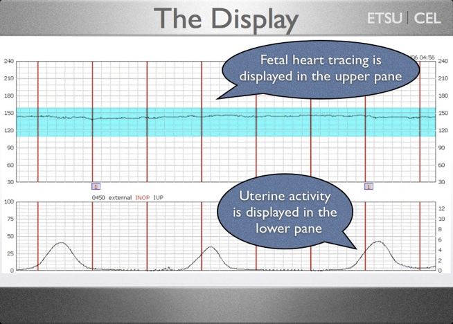
Electronic Fetal Monitor/CTG
Machine that simultaneously records fetal heart rate and uterine contractions
Fetal heart rate → via an ultrasound doppler transducer
Uterine contractions → via tocodynamometer
Top part is baby and bottom is mom
From one red line to another red line is 1 minute
Each box is 10 seconds
Functions of Intrapartum Fetal Monitoring
Assess fetal well being during labor
Detects signs of fetal hypoxia or distress
Monitor fetal heart rate patterns and variability
Evaluate the fetal response to uterine contractions
Identify early indications of acidemia
Guide timely clinical interventions (position change)
Reduce the risk of fetal injury or adverse neonatal outcomes
Provide ongoing data to support labor management decisions
Continuous Fetal Monitoring
Continues monitoring of fetal HR and contractions during labor and delivery using internal or external devices
Indications
Use of Pitocin for induction of augmentation
Epidural or spinal anesthesia
Induced or augmented labor
Non-reassuring fetal HR patterns
Meconium stained amniotic fluid
Maternal HTN or pre-eclampsia
Maternal DM
Maternal fever or suspected infection
Preterm labor
Post-term pregnancy
IUGR
Multiple gestation
Vaginal bleeding during delivery
History of previous C-section or uterine surgery
PROM with risk of infection
Any medical or obstetric complication affecting fetal oxygenation
Has led to an increase in
C-sections
Instrumental vaginal births
Maternal negative impact
Restricts maternal movement
Reduced physical contact with her partner
Intermittent Fetal Monitoring
Fetal HR is checked at scheduled intervals
Recommended in low risk pregnancies
Benefits
Allows more maternal mobility and comfort
Fewer restrictions and less equipment
Endorsed by ACOG and WHO for appropriate candidates
Lower rates of intervention
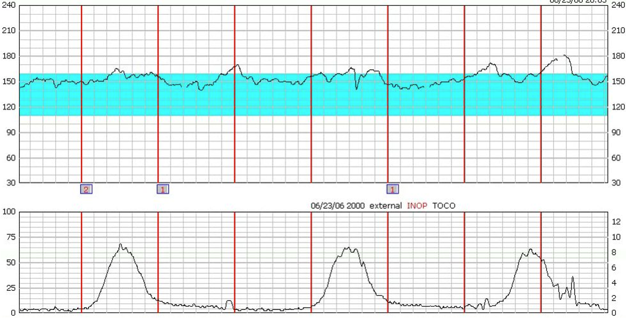
Baseline Fetal Heart Rate
Average heart rate over a 10 minute window
Normal range: 110-160 bpm
Baseline reflects the fetus’s overall:
Oxygenation
Neurological status
Autonomic nervous system
Excludes accelerations and decelerations
Variability
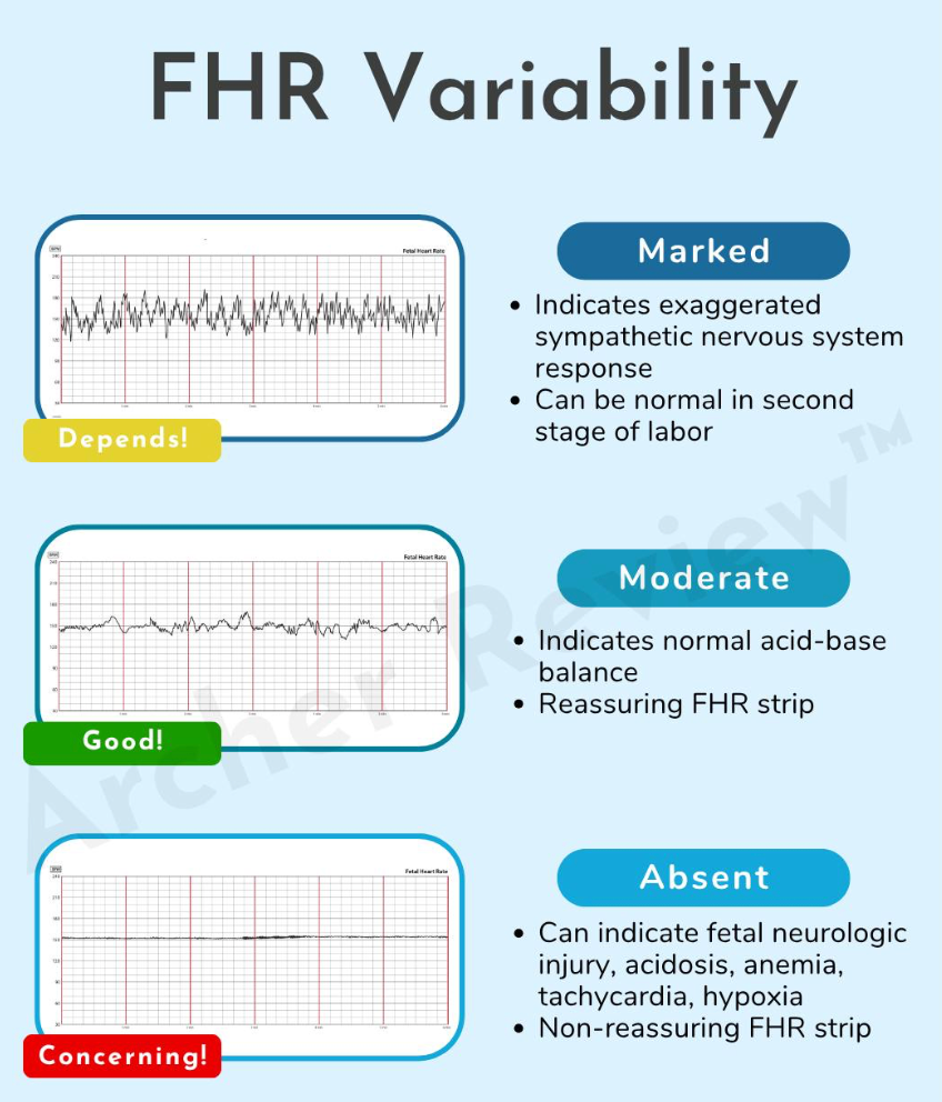
Beat to beat fluctuations in the fetal heart rate
Reflects intact neurologic pathways and adequate oxygenation
If the variability is good → fetus is generally coping well
Types
Absent: no detectible variation (concerning)
Minimal: <=5 bpm (could be sleep, medications, hypoxia)
Moderate: 6-25 bpm (reassuring: oxygenated fetus)
Marked: > 25 bpm (may indicate stress: could be ok and need to assess further)
Accelerations
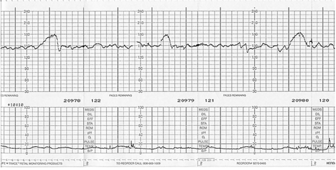
Temporary increase in the fetal heart rate that is reassuring
>= 32 weeks: increase of >= 15 bpm that lasts >= 15 seconds (15 × 15)
< 32 weeks: 10 × 10
Causes
Adequate oxygenation
Responsive autonomic nervous system
Fetal well being
Fetal movement/stimulation
Absence is not abnormal but may require additional monitoring
Decelerations
Periodic decreases in the fetal heart from the baseline
Can indicate:
Normal physiologic responses
Potential fetal distress: depends on timing, shape, and duration
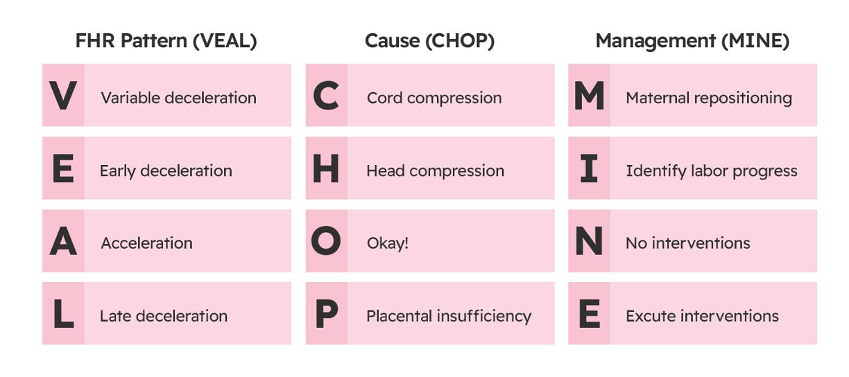
FHR Pattern, Cause, Management
Uterine Contractions
Assess:
Frequency (peak to peak measurement)
Duration
Intensity
Measured by palpating the abdomen
Mild: nose
Moderate: chin
Strong: forehead
IUPC
Mild: < 40 mmHg
Moderate: 40-70 mmHg
Strong: > 70 mmHg
Normal labor pattern
Every 2-5 minutes lasting 60-90 seconds
Rate: number of contractions in 10 minute window
Assessed independently and also in conjunction with fetal heart rate monitoring
Uterine contractions reduce placental blood flow and can cause late decelerations
Category
Overall interpretation and big picture conclusion
Tracings are classified into categories based on their pattern
Category I: normal
Category II: indeterminate
Category III: abnormal
Category II and III tracing interventions focus on:
Improving fetal oxygenation
Reducing uterine stress
Preparing for potential delivery
Category I Criteria
Baseline FHR 110-160 bpm
Variability: moderate (6-25 bpm fluctuations)
No late or variable decelerations
Early decelerations or accelerations may be present
Category II Criteria
Everything’s that not I or III
Requires continued surveillance and reassessment
Intervene, reassess, and continue monitoring
Goal
Improve fetal oxygenation
Correct reversible causes
Interventions
Reposition patient
IV fluids
Stop Pitocin
Give oxygen
Treat maternal hypotension
Amnioinfusion
Fetal scalp stimulation
Close monitoring
Category III Criteria
Absent variability plus
Recurrent late decelerations
Recurrent variable decelerations
Bradycardia
Immediate intervention needed
Goal
Immediate correction or expedited delivery
Interventions
Stop Pitocin immediately
Reposition patient
IV fluids
Administer oxygen
Correct maternal hypotension
Tocolysis if tachysystole present
Amnioinfusion
No improvement → prepare for operative delivery
Fetal Scalp Blood Sampling
Used to directly assess fetal oxygenation and acid base status during labor when there are concerns about fetal distress
Process
Small incision on fetal scalp and tiny blood sample collected
Requires ruptured membranes, cervical dilation, and cephalic presentation
Normal pH → continue monitoring
Low pH → acidemia and expedite immediately
Invasive and used less often today
Contraindications
Infection
Prematurity
Bleeding disorders
Non-stress Test
Non-invasive test to assess fetal well being by monitoring the FHR in response to fetal movement
No uterine contractions are induced and fetus is not stressed during test
Process
Uses electronic fetal monitoring: FHR and contraction measurement
Indicated adequate oxygenation and intact neurologic function
Used outpatient in high risk pregnancies or for reduced fetal movement
Reactive NST
2 or more accelerations in 20 minutes
Reassuring
Non-reactive NST
Fewer than 2 qualifying accelerations in 40 minutes
Non-reassuring but does not automatically mean fetal distress
May indicate
Fetal sleep
Medications
Need for further testing
Can try to give cold water or juice to mom
Contraction Stress Test
Measures the fetal HR after the mother’s uterus is stimulated to contract
Potentially used in rare cases where NSTs are repeatedly nonreactive
Test is done to make sure the fetus can handle contractions during labor and get the oxygen needed from the placenta
Identifies fetal distress before labor begins
Not routinely done
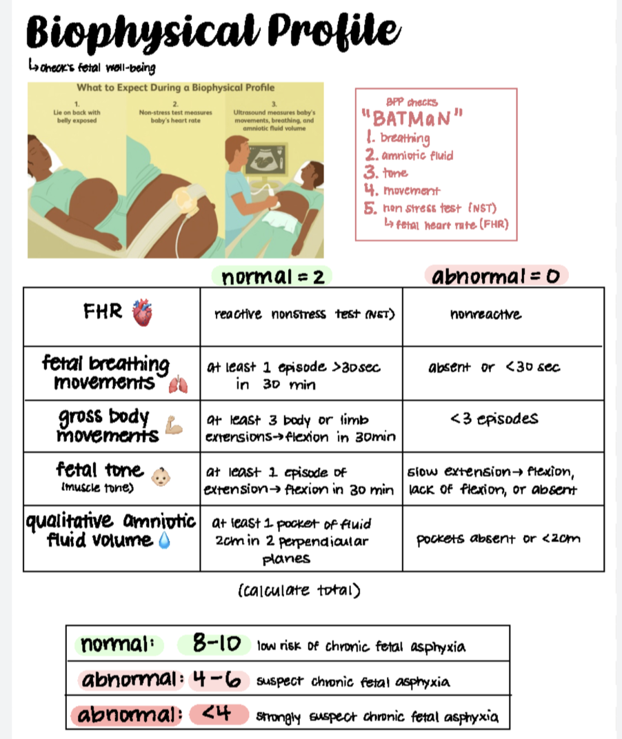
Biophysical Profile
Pre-natal test that combines U/S assessment and FHR monitoring to evaluate fetal well being and oxygenation
Components
NST (non-U/S)
Fetal breathing movements → watch fetal diaphragm or chest wall move
Fetal tone → 1 episode of extension with return to flexion
Gross body movements → minimum of 3 times
Amniotic fluid index
Each component scored 0 or 2 with maximum of 10
Scores
8-10: reassuring
6: equivocal (repeat testing or further evaluation)
4 or less: non-reassuring (consider delivery)
Amniotic fluid is low: score of 8 or 9 is abnormal
NST and BPP
Used for high risk pregnancies for structured monitoring of the fetus at specific intervals
Induction
Intentional initiation of labor using mechanical or medical methods during labor begins on its own, with the goal of achieving a vaginal delivery → ripens the cervix and stimulates uterine contractions
Indications
Postdates pregnancy
PPROM
Maternal conditions (HTN/DM)
Fetal concerns (IUGR)
Elective induction at term
Methods
Pharmacologic
Mechanical
Membrane sweeping (stripping)
Amniotomy (AROM)
Nipple stimulation
Augmentation
Labor has started but needs strengthening
Methods of Induction: Pharmacologic
Prostaglandins:
Soften and dilate the cervix
May stimulate contractions
Used when the cervix is unfavorable (low Bishop score)
Pitocin
Has to be primed and ready with favorable cervix
IV medication
Requires continuous fetal monitoring
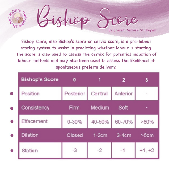
Bishop Score (don’t memorize)
Cervical assessment used to predict the likelihood of a successful labor initiation
5 factors
Cervical dilation
Cervical effacement
Cervical consistency
Cervical position
Fetal station
Score >= 8: cervical favorable and induction likely to succeed
Score < 6: cervix unfavorable and ripening needed first
In between score: gray zone
Methods of Induction: Mechanical
Foley balloon
Balloon inserted into cervix and inflated → applies pressure to promote dilation
Lower risk of tachysystole than medications
Used for cervical ripening
Methods of Induction: Membrane sweeping
Provider manually separates membranes from cervix during vaginal exam → releases natural prostaglandins
Can initiate labor
Not formal induction: just mechanical/natural
Do not use with:
Placenta previa
High risk of bleeding
Vaginal exams are contraindicated
Methods of Induction: Amniotomy
Intentional breaking of amniotic sac
Releases prostaglandins and increases contraction strength
Requires fetal head to be well engaged
Increases risk for: infection and cord prolapse
Big Picture
Unfavorable cervix → prostaglandins or balloon first
Favorable cervix → Pitocin +/- amniotomy
More intervention → more monitoring
Newborn Assessment Immediate Goals
Assess cardiorespiratory status
Establish and maintain ABCs
Identify need for resuscitation
Evaluate adaptation to extra-uterine life
Initial Newborn Assessment
Immediately after birth
Key observations
Respiratory effort (crying, breathing, apnea)
HR
Muscle tone
Color
Gestational age assessment
Presence of anomalies
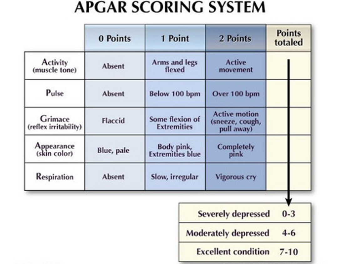
APGAR
Performed at 1 minute and 5 minutes after birth
Interpretations
7-10 → normal adaptation
4-6 → moderate difficulty and may need assistance
0-3 → severe distress and need immediate resuscitation
Does not predict long term outcomes and is not used to decide whether to resuscitate → reflects response to resuscitation
Ongoing Newborn Assessment
Vital signs (HR/RR/temperature)
Physical exam
Skin color and perfusion
Gross malformations, molding, caput, spina bifida
Heart and lung sounds
Abdomen and umbilical cord
Genitalia and anus
Reflexes (Moro, grasp, suck)
Neonatal Resuscitation
Initial questions
Is the infant term
Is the infant breathing or crying
Does the infant have good muscle tone
Yes to all → routine care
No to any → begin resuscitation steps
Initial Resuscitation Steps
Provide warmth
Position airway
Clear secretions if needed
Dry and stimulate
Evaluate breathing and HR
PPV if apnea, gasping, HR < 100 bpm
Chest compressions if
HR < 60 bpm after 30 seconds of effective PPV
Medications
Epinephrine if HR < 60 bpm after ventilation and compressions
Volume expansion if hypovolemia suspected
Pearls
HR is the most important indicator in neonatal resuscitation
Ventilation is the single most effective intervention
APGAR scores assess response, not the need for resuscitation