SNAB Biology - Topic 8 - Grey Matter
1/119
There's no tags or description
Looks like no tags are added yet.
Name | Mastery | Learn | Test | Matching | Spaced | Call with Kai |
|---|
No analytics yet
Send a link to your students to track their progress
120 Terms
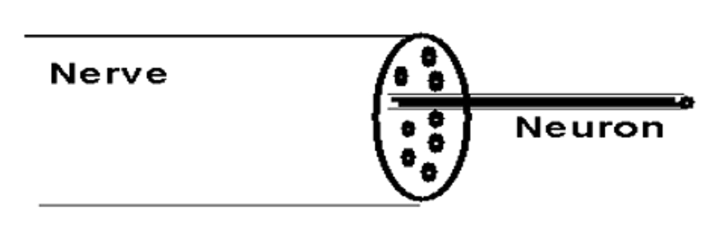
Nerve vs. Neurone
Neuron is a single cell
Nerve is a bundle of neurons surrounded by a protective covering
Different types of neurones
sensory
motor
relay
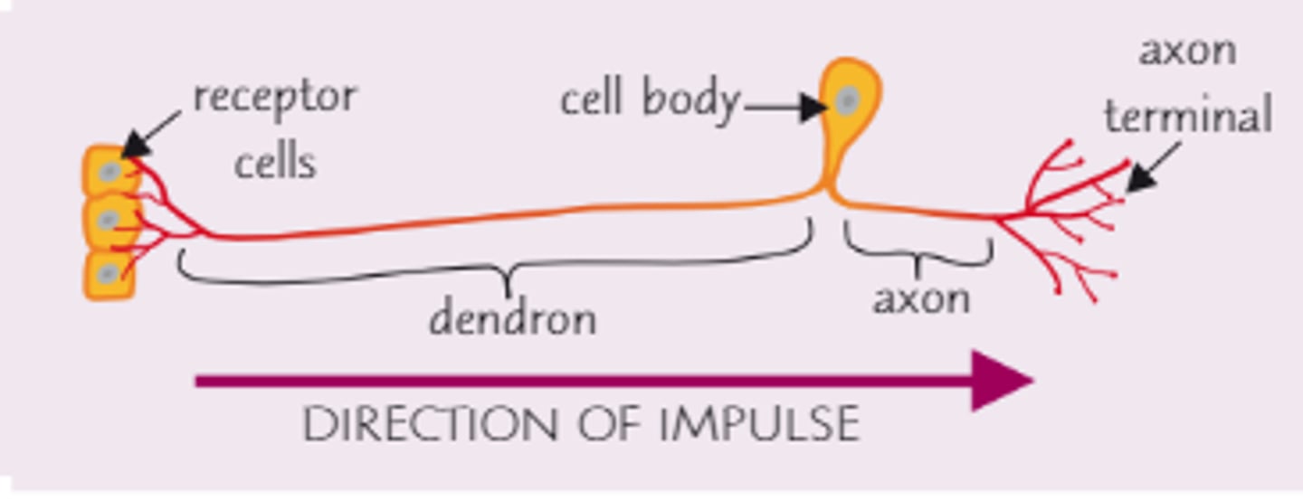
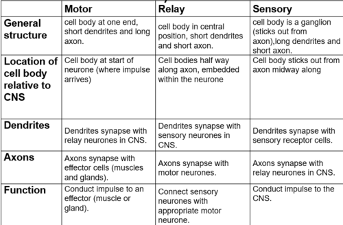
Sensory neurone
One long dendron carries nerve impulses from receptor cells to the cell body which is located in the middle of the neurone.
One short axon carries nerve impulses from the cell body to the CNS
Sensory neurones transmit electrical impulses from receptors to CNS - the brain and spinal cord
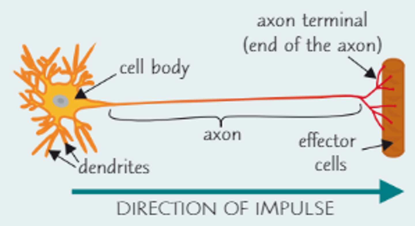
Motor neurone
Many short dendrites carry nerve impulses from the CNS to the cell body
One long axon carries nerve impulses from the cell body to the effector cells
Motor neurones transmit electrical impulses from the CNS to effectors
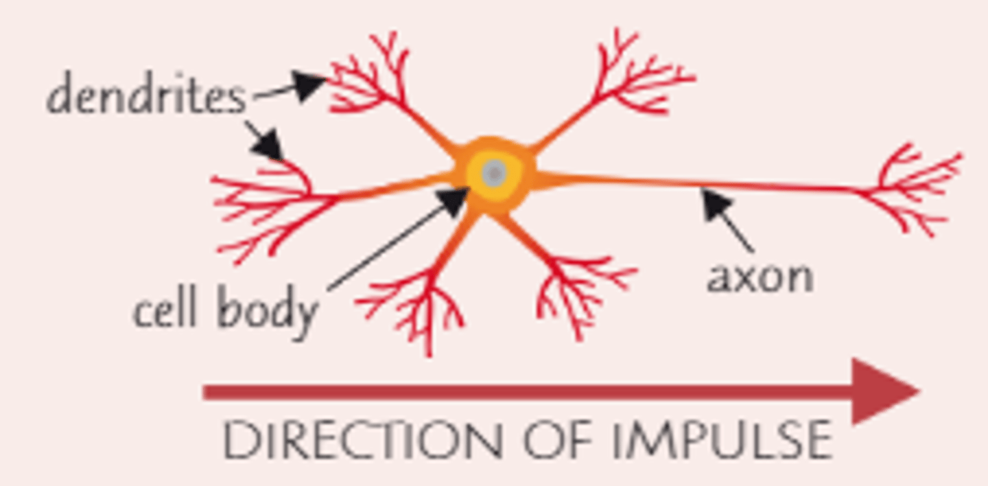
Relay neurone
Many short dendrites carry nerve impulses from sensory neurones to the cell body.
An axon carries nerve impulses form the cell body to motor neurones
Relay neurones are shorter
Relay neurones transmits electrical impulses between sensory neurones and motor neurones
What do all neurons have in common?
•Cell body – nucleus and organelles in cytoplasm
•Dendrites – very fine. Conduct nerve impulses towards the cell body. Forms synapses with other neurones - usually relay neurones
•Axons – single, long, transmits impulses away from the cell body. Conducts impulse
Features of some neurones
•Myelin sheath – fatty insulating layer around the axon. It affects how quickly impulses pass along. Not in every animal
•Schwann cells – produce the the Myelin sheath
Schwann cells have gaps between them. The myelin sheath is not continuous and the gaps between cells are called the ‘Nodes of Ranvier’
Synapse
Site of transmission of electric impulses between neurons or between a neurone and an effector
Receptors
Detect stimuli - they can be cells or proteins on cell surface membranes. There are loads of different types of receptors that detect different stimuli
Receptors communicate with effectors via the nervous system r the hormonal system, or sometimes both.
Effectors
Cells that bring about a response to a stimulus to produce an effect. Effectors include muscle cells and cells found in glands e.g. pancreas
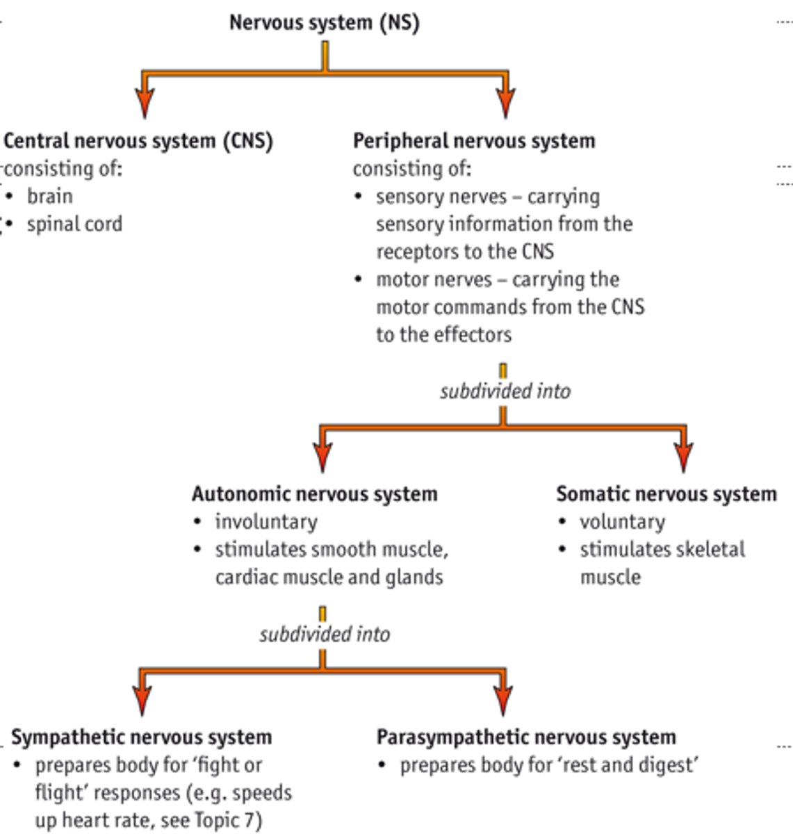
Nervous Systems
CNS and PNS
Eyes in bright light
Circular muscles: contracted
Radial muscles: relaxed
Pupil diameter: small
Controlled by: parasympathetic
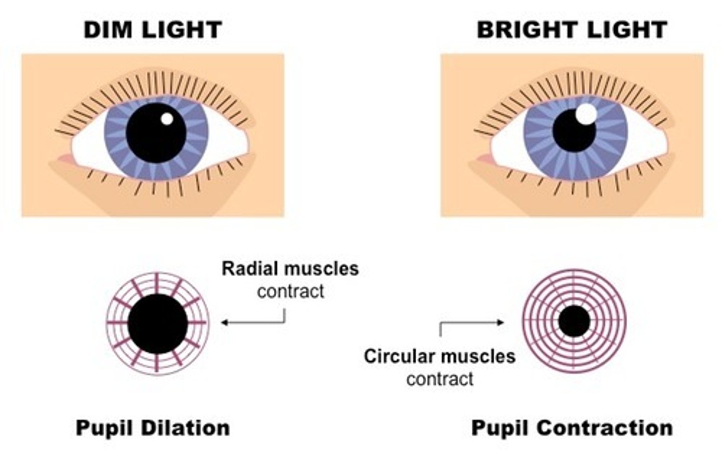
Eyes in dim light
Circular muscles: relaxed
Radial muscles: contracted
Pupil diameter: large
Controlled by: sympathetic
Sympathetic controls radial
contracts to dilate pupil - fight or flight
Parasympathetic controls diameter
contracts to constrict pupil - rest or digest
The hormonal system sends information as chemical signals
The hormonal system is made up of glands and hormones:
• A gland is a group of cells that are specialised to secrete a useful substance, such as a hormone. E.g. the pancreas secretes insulin.
• Hormones are 'chemical messengers'. Many hormones are proteins or peptides, e.g. insulin. Some hormones are steroids, e.g. progesterone.
Hormones are secreted when a gland is stimulated:
• Glands can be stimulated by a change in concentration of a specific substance (sometimes another hormone).
• They can also be stimulated by electrical impulses.
Hormones diffuse directly into the blood, then they're taken around the body by the circulatory system.
They diffuse out of the blood all over the body but each hormone will only bind to specific receptors for that hormone, found on the membranes of some cells (called target cells).
The hormones trigger a response in the target cells (the effectors).
Nervous Communication
Uses electrical impulses.
Faster response — electrical impulses are really fast.
Localised response — neurones carry electrical impulses to specific cells.
Short-lived response — neurotransmitters are removed quickly.
Hormonal Communication
Uses chemicals
Slower response — hormones travel at the 'speed of blood'.
Widespread response — target cells can be all over the body.
Long-lived response — hormones aren't broken down very quickly.
resting state
When a nervous system receptor is in its resting state (not being stimulated), there's a difference in charge between the inside and the outside of the cell. This means there's a voltage across the membrane. The membrane is said to be polarised.
The voltage across the membrane is called the potential difference.
It is generated by ion pumps and ion channels
When a stimulus is detected, the permeability of the cell membrane to ions changes (ions are stopped from moving, or more move in and out of the cell). This changes the potential difference.
If the change in potential difference is big enough it'll trigger an action potential — an electrical impulse along a neurone. An action potential is only triggered if the potential difference reaches a certain level called the threshold level.
Resting potential
Na+/K+ pump creates a conc. gradient across membrane
K+ diffuses out of the cell down the K+ concentration gradient - makes outside of membrane more +ve and inside more -ve, this creates a p.d. - chemical gradient
The p.d. will pull K+ back into the cell - electrical gradient
When electrochemical equilibrium of -70 mV is reached, the 2 gradients (chemical and electrical) counteract each other an there is no net movement of K+ ions.
Action Potential
Has three stages:
1. Depolarisation
•Stimulation causes voltage-dependent Na+ channels to open
•Na+ enters the cell and it becomes positive inside (+40mV)
•Opening of channels causes positive feedback (depolarisation causes more gates to open)
•Action potentials are all-or-nothing
2. Repolarisation
•After 0.5ms, the voltage-dependent Na+ channels close
•Voltage dependent K+ channels now open
•K+ leave the cell down the electrochemical gradient
•This causes the inside of the cell to become negative again
3. Restoring the resting potential
•K+ continues to flow out until the potential difference is more negative than resting potential (hyperpolarised)
•Voltage-gated K+ channels close
•K+ is restored to resting potential (K+ diffuses into the axon via the normal K+ channels)
Refractory period
After an action potential, the neurone cell membrane can't be excited again straight away. This is because the ion channels are recovering and they can't be made to open — sodium ion channels are closed during repolarisation and potassium ion channels are closed during hyperpolarisation. This period of recovery is called the refractory period.
How does the myelin sheath work to increase speed of nerve impulse propagation? (4)
- At nodes of Ranvier there's no myelination
- Nodes are the site of clusters of sodium-gated channels, which open/close when impulse arrives. This allows depolarisation at nodes
- Myelin acts as an electrical insulator
- The impulse/depolarisation jumps to next node. This is saltatory conduction
Synaptic transmission
Incoming action potential causes depolarisation in the synaptic knob. Calcium channels and Ca2+ flood into the synaptic knob
The influx of calcium ions causes synaptic vesicles to fuse with the presynaptic membrane. This release neurotransmitters into the cleft. So calcium ions cause the release of neurotransmitters.
Neurotransmitter (acetylcholine) is released into the synaptic cleft. Acetylcholine binds to the receptor site on the sodium ion channels
The sodium channels on the postsynaptic are normally closed. When the neurotransmitter binds there is a conformational change opening the channel. This allows the sodium ion to flood in
Sodium ion diffuses in (down steep conc. gradient). Postsynaptic neurone becomes depolarised
Depolarisation inside the postsynaptic neurone must be above a threshold value. If the threshold is reached a new action potential is sent along the axon of the post synaptic neurone.
Describe the role of ion channels in the conduction of a nerve impulse (5)
Sodium voltage gated channels open (1)
The sodium ions {diffuse in} (DO NOT ALLOW DIFFUSE INTO MEMBRANE) (1)
Causing depolarisation of the membrane (1)
Sodium ion channels close and K+ channels open (1)
The K+ ions diffuse out (1)
Causes repolarisation (1)
neurotransmitter
A neurotransmitter is a chemical messenger that is released by the presynaptic cell and binds to receptors on the postsynaptic cell, transmitting a signal across the synapse.
Acetylcholinesterase
Acetylcholinesterase at the postsynaptic membrane breaks down the acetylcholine so it can no longer bind to receptors. Some of the breakdown products are then reabsorbed by the presynaptic membrane and reused.
Types of Synapses
Excitatory:
Postsynaptic membrane more permeable to sodium ions
Inhibitory:
Cl- ions diffuse IN
K+ diffuses out
So…INSIDE becomes more negative, therefore harder to depolarise.
Summation
Low frequency action potentials often release insufficient amounts of neurotransmitter to exceed the threshold in the postsynaptic neurone
Summation allows action potentials to be generated
This enables a build up of neurotransmitter in the synapse
Spatial Summation
•A number of different presynaptic neurones share the same synaptic cleft
•Together they can release enough neurotransmitter to create an action potential
•Multiple neurones
Temporal Summation
•A single presynaptic neurone releases neurotransmitter many times over a short period
•
•If the total amount of neurotransmitter exceeds the threshold value an action potential is sent
•
•1 neurone
Eugenol is a drug that inhibits the movement of sodium ions and calcium ions through the cell surface membranes of sensory neurones.
*(ii) Eugenol can be used to reduce pain.
Suggest an explanation for how eugenol affects the movement of calcium ions and reduces pain (6)
{reduced} Ca2+ enters presynaptic membrane
Fewer vesicles {fuses} with presynaptic membrane
Less neurotransmitter {released} into synaptic gap
less neurotransmitters binds to receptors as {postsynaptic membrane}
reduced depolarisation
{threshold intensity} less like to occur
KNOW STRUCTURE OF EYE AND FUNCTIONS OF EACH
CHECK NOTES FOR PPT SLIDES
Role of the ciliary muscle
Near vision:
•Ciliary muscle contracts and pulls forward
•Suspensory ligaments relax
•Lens more spherical (thicker and rounder), focusing on near objects as light is bent more
Distant vision:
Ciliary muscle relaxes and pulls back
Suspensory ligaments pulled tight
Lens thinner, focusing on distant objects, so light doesn't bend much
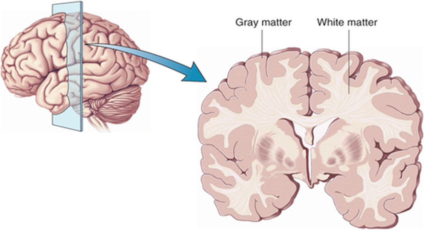
Grey and White Matter
Grey matter
•Outer layer of the brain
•Highly folded
•Consists of nerve cell bodies, synapses and dendrites
White matter
Inner layer of the brain
Consists of axons & myelin sheath
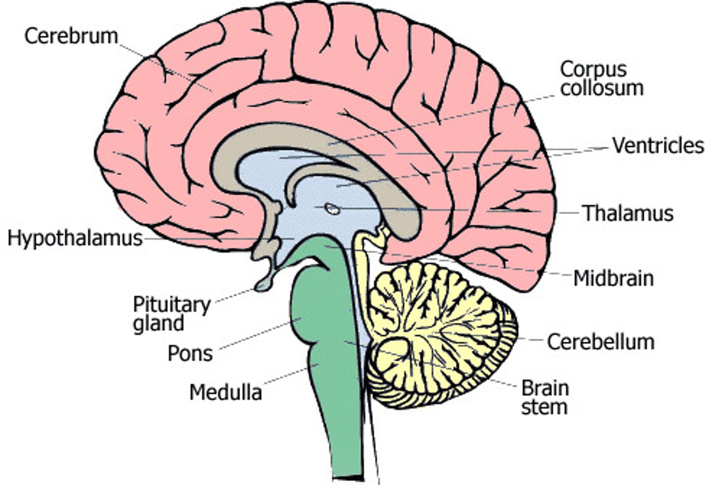
Corpus callosum
Allows communication between the two hemispheres
The Hind brain
1. The cerebrum
1) The cerebrum is the largest part of the brain.
2) It’s divided into two halves called cerebral hemispheres.
3) The cerebrum has a thin outer layer called the cerebral cortex. The cortex has a large surface area so it’s highly folded to fit into the skull.
4) The cerebrum is involved in vision, learning, thinking, emotions and movement.
5) Different parts of the cerebrum are involved in different functions, e.g. the back of the cortex is involved in vision and the front is involved in thinking.
2. The hypothalamus
1) The hypothalamus is found just beneath the middle part of the brain.
2) The hypothalamus automatically maintains body temperature at the normal level (thermoregulation)
3) The hypothalamus produces hormones that control the pituitary gland — a gland just below the hypothalamus
3. The medulla oblongata
1) The medulla oblongata is at the base of the brain, at the top of the spinal cord.
2) It automatically controls breathing rate and heart rate
4. The cerebellum
1) The cerebellum is underneath the cerebrum and it also has a folded cortex.
2) It’s important for coordinating movement and balance.7
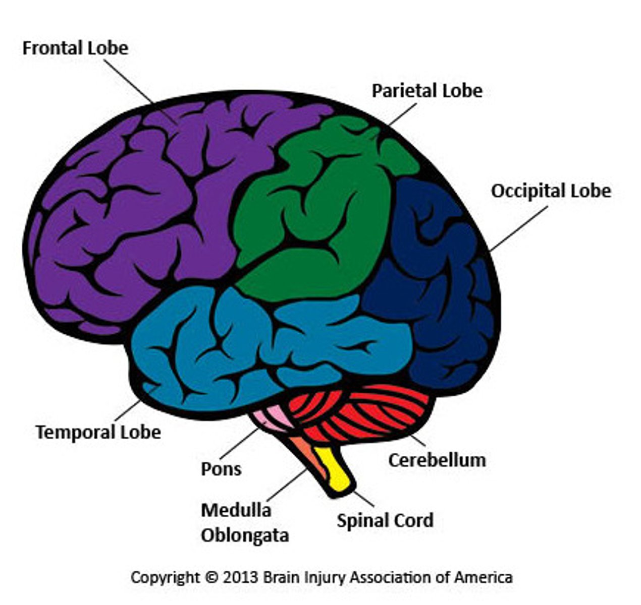
Cerebral Hemispheres
Frontal lobe - thinking, memory, behaviour and movement
Temporal lobe - hearing, learning and feelings
Brain stem - breathing, heart rate, and temperature
Parietal lobe - language and touch
Occipital lobe - sight
Cerebellum - balance and coordination
CT Scans
•Computerised Axial Tomography
•Overcomes limitations of X rays
•Frozen moment pictures
•Looks at structures in brain rather than functions
•Detect brain disease
How it works - CT
•A CT scanner emits uses thousands of narrow beam x-rays as opposed to an x-ray which just uses one radiation beam.
•The final picture is far more detailed than an x-ray
•The x-rays are used to produce an image of a thin slice of the brain on a computer screen in which the different soft tissues of the brain can be distinguished.
Limitations - CT
•Frozen moment pictures.
•They examine structures in the brain rather than functions.
•Used to detect brain disease and to monitor the tissues of the brain over the course of an illness.
•Limited resolution, therefore small structures in the brain cannot be distinguished.
•X-rays can be harmful therefore used less frequently.
•Patient needs to remain still - movement can cause blurring of the image
MRI Scans
•Magnetic Resonance Imaging
•Magnetic field and radio waves
•Detects soft tissue
•Different tissues produce different images
•Better resolution than CT for brain and spinal cord
•Diagnosis of tumours, strokes, brain injuries or infections
How it works - MRI
•Uses a magnetic field and radio waves to detect soft tissue.
Limitation - MRI
-One moment in time
-Structures not functions
-Expensive (more than CT)
-No metal during scan (so not appropriate for pacemakers etc)
-Patient must remain still
fMRI Scans
•Functional Magnetic Resonance Imaging
•Can study brain activity
•Follows the uptake of oxygen in active brain areas
How fMRI works
•Increased neural activity in a brain area results in an increased demand for oxygen.
•This results in an increase in blood flow
•This in turn results in a slight increase in oxygen absorption from the blood.
•Overall there is a large increase in oxyhaemoglobin levels in the enhanced blood flow so..
•Less radio waves are absorbed by oxyhaemoglobin - these areas light up on the screen
Limitations - fMRI
-Expensive
-Difficult to pinpoint exact location in brain hence difficult to interpret
-Not as useful in diagnosing particular conditions as it looks at function not structure
PET Scans
Positron emission tomography
Structure and function studies. Diagnosis: cancers/heart disease/brain disorders. Other uses: plan surgery, monitor and track treatments for diseases e.g. alzheimers/cancer,
Limitations PET Scans
-Limited to 1-2 a year due to safety
-Very expensive
The Visual Cortex is Made Up of Ocular Dominance Columns
1) The visual cortex is an area of the cerebral cortex at the back of your brain.
2) The role of the visual cortex is to receive and process visual information.
3) Neurones in the visual cortex receive information from either your left or right eye.
4) Neurones are grouped together in columns called ocular dominance columns. If they receive information from the right eye they're called right ocular dominance columns, and if they receive information from the left eye they're called left ocular dominance columns.
5) The columns are the same size and they're arranged in an alternating pattern (left, right, left, right) across the visual cortex
Hubel and Wiesel Used Animal Models to Study the Visual Cortex
The structure of the visual cortex was discovered by two scientists called Hubel and Wiesel.
They used animal models to study the electrical activity of neurones in the visual cortex.
They found that the left ocular dominance columns were stimulated when an animal used its left eye, and the right ocular dominance columns were stimulated when it used its right eye.
Hubel and Wiesel (1963) investigated how the visual cortex develops by experimenting on very young kittens:
• They stitched shut one eye of each kitten so they could only see out of their other eye.
• The kittens were kept like this for several months before their eyes were unstitched.
• Hubel and Wiesel found that the kitten's eye that had been stitched up was blind.
• They also found that ocular dominance columns for the stitched up eye were a lot smaller than normal, and the ocular dominance columns for the open eye were a lot bigger than normal.
• The ocular dominance columns for the open eye had expanded to take over the other columns that weren't being stimulated — when this happens, the neurones in the visual cortex are said to have switched dominance.
They then investigated if the same thing happened in an adult cat's brain:
• They stitched shut one eye of each cat, who were kept like this for several months.
• When their eyes were unstitched, Hubel and Wiesel found that these eyes hadn't gone blind.
• The cats fully recovered their vision and their ocular dominance columns remained the same.
They repeated the experiments on young and adult monkeys and saw the same results.
Hubel and Wiesel's experiments showed that the visual cortex only develops into normal left and right ocular dominance columns if both eyes are visually stimulated in the very early stages of life.
What did Hubel and Wiesel discover?
The structure of the visual cortex. They found that the left ocular dominance columns were stimulated when an animal used its left eye, and the right ocular dominance columns were stimulated when it used its right eye.
Ocular dominance - when you use one eye more than the other.
Their Experiments Provide Evidence for a Critical Period in Humans
1) Hubel and Wiesel's experiments on cats show there's a period in early life when it's critical that a kitten is exposed to visual stimuli for its visual cortex to develop properly. This is called the critical period.
2) The human visual cortex is similar to a cat's visual cortex (the human visual cortex has ocular dominance columns too) so Hubel and Wiesel's experiments provide evidence for a critical period in humans.
Visual Stimulation Organises the Neurones During the Critical Period
1) During the critical period of development, synapses that receive visual stimulation and pass nerve impulses into the visual cortex are retained.
2) Synapses that don't receive any visual stimulation and don't pass on any nerve impulses to the visual cortex are removed.
3) This means that if the eyes are not stimulated with visual information during this critical period of development, the visual cortex will not develop properly as many of the synapses will be destroyed.
Arguments AGAINST using animals in medical research
Animals are different from humans, so drugs tested on animals may have different effects in humans.
Experiments can cause pain and distress to animals.
There are alternatives to using animals in research, e.g. using cultures of human cells or using computer models to predict the effects of experiments.
Some people think that animals have the right to not be experimented on, e.g. animal rights activists
Arguments FOR using animals in medical research
Animals are similar to humans, so research has led to loads of medical breakthroughs, e.g. antibiotics, insulin for diabetics and organ transplants.
Animal experiments are only done when it's absolutely necessary and scientists follow strict rules, e.g. animals must be properly looked after, painkillers and anaesthetics must be used to minimise pain.
Using animals is currently the only way to study how a drug affects the whole body — cell cultures and computers aren't a true representation of how cells may react when surrounded by other body tissues. It's also the only way to study behaviour.
Other people think that humans have a greater right to life than animals because we have more complex brains, e.g. compared to rats, fish, fruit flies (which are commonly used in experiments).
Animal Experiments
1) Scientists study the effects of different environments on the brain development of animals of the same species. Individuals of the same species will be genetically similar, so any differences in their brain development are more likely to be due to nurture than nature.
2) To study the effects of different genes, scientists can genetically engineer mice to lack a particular gene and then raise mice with and without the gene in similar environments.
3) Differences between the brain development of the genetically engineered mice and normal mice are more likely to be due to nature than nurture.
Twin Studies
1) If identical twins are raised separately then they'll have identical genes but different environments.
2) Scientists can compare the brain development of separated identical twins — any differences between them are due to nurture not nature, and any similarities between them are due to nature.
3) Scientists can use this comparison to show the relative contribution of environmental and genetic factors to brain development.
4) However, even if they've been raised separately, twins will still have shared the same environment in the womb — so environmental and genetic factors are not completely separated.
7) Identical twins raised together are genetically identical and have similar environments — this means it's hard to tell if any differences between them are due to nature or nurture. So scientists compare them to non-identical twins (who are genetically different but have similar environments) — they act like a control to cancel out the influence of the environment. Any difference in brain development between identical and non-identical twins is more likely to be due to nature than nurture.
Cross-Cultural Studies
1) Children brought up in different cultures have different environmental influences, e.g. beliefs and education.
2) Scientists can study the effects of a different upbringing on brain development by comparing large groups of children who are the same age but from different cultures.
3) Scientists look for major differences in characteristics. Any differences in brain development between different cultures are more likely to be due to nurture than nature. Any similarities in brain development between different cultures are more likely to be due to nature than nurture
Newborn Studies
The brain of a newborn baby hasn't really been affected by the environment.
Scientists study the brains of newborn babies to see what functions they're born with and how developed different parts of the brain are — what they're born with is more likely to be due to nature than nurture.
Brain Damage Studies
1) Damage to an adult's brain can lead to the loss of brain function, e.g. a stroke may cause loss of vision.
2) If an adult's brain is damaged, it can't repair itself so well because it's already fully developed. But a child's brain is still developing — so scientists can study the effects of brain damage on their development.
3) Scientists compare the development of a chosen function in children with and without brain damage.
4) If the characteristic still develops in children who have brain damage, then brain development is more likely to be due to nurture than nature for that characteristic.
5) If it doesn't develop in children who have brain damage, then brain development is more likely to be due to nature than nurture for that characteristic (because nurture isn't having an effect).
Photoreceptors Convert Light into an Electrical Impulse
1) Light enters the eye, hits the photoreceptors and is absorbed by light-sensitive pigments. 2) Light bleaches the pigments, causing a chemical change. 3) This triggers a nerve impulse along a bipolar neurone.
4) Bipolar neurones connect photoreceptors to the optic nerve, which takes impulses to the brain
5) The human eye has two types of photoreceptor — rods and cones.
6) Rods are mainly found in the peripheral parts of the retina, and cones are found packed together in the fovea.
7) Rods only give information in black and white (monochromatic vision), but cones give information in colour (trichromatic vision). There are three types of cones — red-sensitive, green-sensitive and blue-sensitive. They're stimulated in different proportions so you see different colours.
Rod cell in the Dark
•Outer segment: Na+ flows in through non-specific cation (+ve) channels
•Na+ move down concentration gradient to inner segment
•Inner segment: Na+ is actively pumped out
•Cell is slightly depolarised at around -40mV
•This triggers release of neurotransmitter glutamate from rod cells
•Glutamate binds to the bipolar cell stopping it from depolarising
Rod cell in the Light
•Light breaks down rhodopsin to retinal (non-protein) and opsin (protein)
•Opsin triggers chemical reactions resulting in cation (ie Na+) channel closing
•Outer segment: influx of Na+ decreases
•Inner segment: Na+ continues to be pumped out
•Overall, inside of cell is more negative – cell is hyperpolarised
•Glutamate neurotransmitter release stops
•Bipolar cell becomes depolarised – action potential in neurones of optic nerve
What is dark adaptation?
•Occurs when you move from a light room to a dark room
•Initially, blackness is seen because:
-Our cones cease functioning in low intensity light
-Rhodopsin is slow to reform after it is broken down
•Once in the dark, rhodopsin regenerates & the sensitivity of the retina increases over time (can take approximately one hour for rods to adjust).
What is light adaptation?
•Occurs when you move from a dark room to a light room
•Initially all you see is white light because the sensitivity of your receptors is set to dim light
•Rods & cones are both stimulated and large amounts of rhodopsin are broken down
•This produces a flood of signals resulting in the glare
Describe how movement of Na+ ions in a rod cell affects depolarisation in a bipolar neurone? (4)
In the dark Na+ moves {in thru Na+ ions channels}
Na+ ions {removed} at inner segment
Rod cell depolarises
Causing neurotransmitters glutamate to be released
What is a photoreceptor?
A cell which is sensitive to light
Which photoreceptor is found in the eye?
Rod cells
What is the name of the pigment found in the eye in the dark?
Rhodopsin
Which products are formed in the light?
Opsin and retinal
Opsin is the protein component and retinal is the non-protein
Which one triggers the cation channels in the rod cell to close?
Opsin
Some plants open during the day and close at night.
Others only start flowering when the days are long enough
But how do they know????
Animals: Rhodopsin/iodopsin
Plants:
•Phytochromes (red sensitive)
A Tropism is a Plant's Growth Response to an External Stimulus
1) A tropism is the response of a plant to a directional stimulus (a stimulus coming from a particular direction).
2) Plants respond to directional stimuli by regulating their growth.
3) A positive tropism is growth towards the stimulus.
4) A negative tropism is growth away from the stimulus.
Phototropism
• Phototropism is the growth of a plant in response to light.
• Shoots are positively phototropic and grow towards light.
• Roots are negatively phototropic and grow away from light.
Geotropism
• Geotropism is the growth of a plant in response to gravity.
• Shoots are negatively geotropic and grow upwards.
• Roots are positively geotropic and grow downwards
Responses are Brought About by Growth Factors
1) Plants don't have a nervous system so they can't respond using neurones, and they don't have a circulatory system so they can't respond using hormones either.
2) Plants respond to stimuli using growth factors — these are chemicals that speed up or slow down plant growth.
3) Growth factors are produced in the growing regions of the plant (e.g. shoot tips, leaves) and they move to where they're needed in the other parts of the plant.
4) Growth factors called auxins stimulate the growth of shoots by cell elongation — this is where cell walls become loose and stretchy, so the cells get longer.
5) High concentrations of auxins inhibit growth in roots though.
Indoleacetic Acid (IAA) is an Important Auxin
1) Indoleacetic acid (IAA) is an important auxin that's produced in the tips of shoots in flowering plants. When it enters the nucleus of a cell, it's able to regulate the transcription of genes related to cell elongation and growth.
2) IAA is moved around the plant to control tropisms — it moves by diffusion and active transport over short distances, and via the phloem over longer distances.
3) This results in different parts of the plants having different amounts of IAA. The uneven distribution of IAA means there's uneven growth of the plant
IAA distribution for phototropism
Phototropism — IAA moves to the more shaded parts of the shoots and roots, so there's uneven growth.
For shoots: IAA moves to the shaded side — cells elongate and the shoot bends towards the light
For roots: IAA moves to this side — growth is inhibited so the root bends away from the light
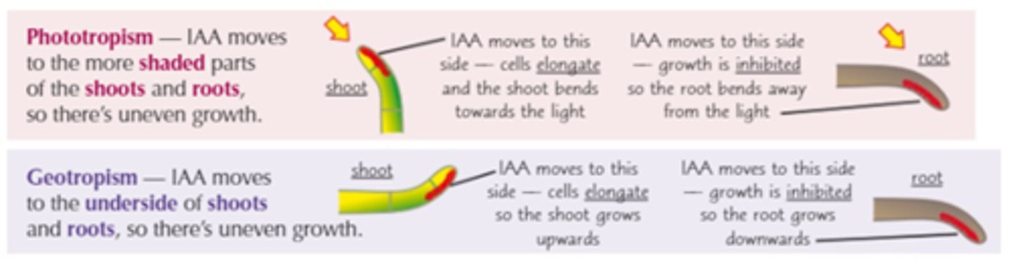
IAA distribution for geotropism
Geotropism — IAA moves to the underside of shoots and roots, so there's uneven growth.
For shoots: IAA moves to this side — cells elongate so the shoot grows upwards
For roots: IAA moves to this side — cells elongate so the shoot grows upwards

What do phytochromes do in plants?
•Trigger germination
•Determine flowering (photoperiods)
•Allow greening
Plants Detect Light Using Photoreceptors
1) Plants detect light using photoreceptors called phytochromes.
2) They're found in many parts of a plant including the leaves, seeds, roots and stem.
3) Phytochromes control a range of responses. For example, plants flower in different seasons depending on how much daylight there is at that time of year, e.g. some plants flower during summer when there are long days.
4) Phytochromes are molecules that absorb light. They exist in two states — the PR state absorbs red light at a wavelength of 660 nm, and the PFR state absorbs far-red light at a wavelength of 730 nm.
5) Phytochromes are converted from one state to another when exposed to light: • PR is quickly converted into PFR when it's exposed to red light. • PFR is quickly converted into PR when it's exposed to far-red light. • PFR is slowly converted into PR when it's in darkness.
6) Daylight contains more red light than far-red light, so more PR is converted into PFR than PFR is converted to PR.
7) So the amount of PR and PFR changes depending on the amount of light, e.g. whether it's day or night, or summer or winter.
8) The differing amounts of PR and PFR control the responses to light by regulating the transcription of genes involved in these responses. E.g. flowering — in some plants, high levels of PFR stimulates flowering. When nights are short in the summer, there's not much time for PFR to be converted back into PR, so PFR builds up and genes involved in flowering are transcribed. This means the plants flower in summer
What is habituation?
A decrease in the strength or occurrence of a behavior after repeated exposure to the stimulus that produces the behaviour.
Habituation allows animals to avoid wasting energy by responding to repetitive harmless stimuli.
How is habituation acheived?
With repeated stimulation, Ca2+ channels become less responsive so less Ca2+ crosses the presynaptic membrane
Less neurotransmitter is released
There is less depolarisation of the postsynaptic membrane so no action potential is triggered in the motor neurone.
Sensitisation
•Sensitisation refers to the process by which a synapse becomes more efficient in its response to a stimulus (e.g. increased startle to loud noise).
Serotonin (neurotransmitter)
•Made from the amino acid tryptophan and there are specific serotonin post-synaptic receptors
•Secreted by neurones in the brain stem and the axons extend to many areas of the brain (Incl. cerebral cortex, cerebellum, spinal cord)
•Serotonin affects mood, emotion, sleep, appetite
Depression
•Lack of serotonin linked to depression
•Causes not understood – genetic or environment?
•Multifactorial condition – what does this mean?
Traditionally thought of as a serotonin deficiency....
Drug treatments for depression
•First generation antidepressants:
•Monoamine oxidase inhibitors (MAOIs) - break down neurotransmitters but can have adverse side effects
•Second generation antidepressants:
•Selective serotonin uptake inhibitors (SSRIs)
How can drugs affect synaptic transmission?
Neurotransmitter synthesis and storage
Neurotransmitter release (Ca2+ channels)
Neurotransmitter receptor binding (cation channel opening)
Neurotransmitter reuptake
Neurotransmitter breakdown (enzymes are responsible for this)
MDMA
Ecstasy binds to the reuptake molecule for serotonin
It prevents re-uptake at the pre-synaptic membrane which means it continues to bind to receptors on the post synaptic membrane
The concentration of serotonin is therefore increased
May have a similar effect on dopamine transporters too
Leads to feelings of well being
Side effects
Anxiety
Dehydration
Aggression
Sweating
Lack of appetite
Sadness
Dopamine
Dopamine is a neurotransmitter that helps control the brain's reward and pleasure centres. Dopamine also helps regulate movement and emotional responses
Why are brain disorders particularly hard to treat?
The blood–brain barrier is the separation of circulating blood and the brain extracellular fluid in the central nervous system (CNS)
Prevents infection of the brain
It does this by having endothelial cells which restrict the diffusion of microscopic objects which are large and hydrophilic
Small hydrophobic molecules, such as oxygen, CO2 or hormones can diffuse through easily
Unfortunately, because antibodies and antibiotics are too large to cross the blood–brain barrier, infections and diseases of the brain that do occur are often very serious and difficult to treat.
How can we treat Parkinson's?
Dopamine cannot be given as it is as it cannot cross the blood-brain barrier. For each drug that has been trialled, can you work out how it might work?
1.Selegiline inhibits certain enzymes in the brain that would normally break down dopamine
2.L-dopa is an intermediate stage in the formation of dopamine which can cross the blood-brain barrier and be used to make dopamine.
3.Dopamine agonists are the same shape as dopamine so activate dopamine receptors directly.
Selegeline
•MAO inhibitor
•Inhibits the enzyme MAO which breaks down dopamine in the brain
•This increases dopamine levels
L-dopa
•Dopamine cannot pass across the blood brain barrier, but the precursor L-dopa can
•In the brain, L-dopa is converted to dopamine
•This increases dopamine levels
Dopamine agonists
•Activate the dopamine receptor directly
•Trigger action potentials at the post-synaptic neurone
•They mimic the role of dopamine in the brain
Human Genome Project
1) The Human Genome Project (HGP) was a 13 year long project that identified all of the genes found in human DNA (the human genome).
2) The information obtained from the HGP is stored in databases.
3) Scientists use the databases to identify genes, and so proteins, that are involved in disease.
4) Scientists are using this information to create new drugs that target the identified proteins, e.g. scientists have identified an enzyme that helps cancer cells to spread around the body — a drug that inhibits this enzyme is being developed.
5) The HGP has also highlighted common genetic variations between people.
6) It's known that some of these variations make some drugs less effective, e.g. some asthma drugs are less effective for people with a particular mutation.
7) Drug companies can use this knowledge to design new drugs that are tailored to people with these variations — these are called personalised medicines.
8) Doctors can also personalise a patient's treatment by using their genetic information to predict how well they will respond to different drugs and only prescribe the ones that will be most effective.