BME 311 Lecture 2
 Call Kai
 Learn
Learn Practice Test
 Spaced Repetition
 Match
Match Flashcards
 Knowt Play
1/41

There's no tags or description
Looks like no tags are added yet.
Name | Mastery | Learn | Test | Matching | Spaced | Call with Kai | Chat |
|---|
No analytics yet
Send a link to your students to track their progress
42 Terms
imaging depth in the body
-confocal microscopy: 100um Resolution: 1um
-optical coherence tomography (OCT): 1 mm Resolution: 2-10um
-ultrasound: 1 cm Resolution: 150um
-high-res computed tomography (CT): 10cm Resolution: 300um
-magnetic resonance imaging: entire body Resolution: 1 mm
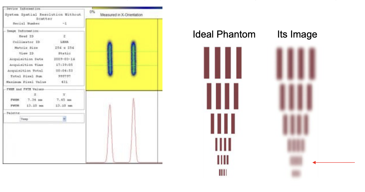
what is spatial resolution
-the size in the image of a “point” source of contrast
-PSF = point spread function
-measured in each dimension, can differ in x,y,z

spatial resolution provides the measurement of
how well the imaging system can distinguish small objects
imaging volume is discretized into
-pixels (2D), often square but not always
-voxels (3D), often cubic (but often called “pixel” too)
smaller resolution value is better
and we call this higher resolution
sometimes defined by pixel size, sometimes larger than pixel size
-pixel size closely related to resolution (MRI)
-pixel size is arbitrary in PET/SPECT (smaller than resolution)
-pixel size chosen should not cause loss of info in the data, but not too many to waste memory
resampling
changing voxel size and/or alignment
-done to save disk space of match with other images
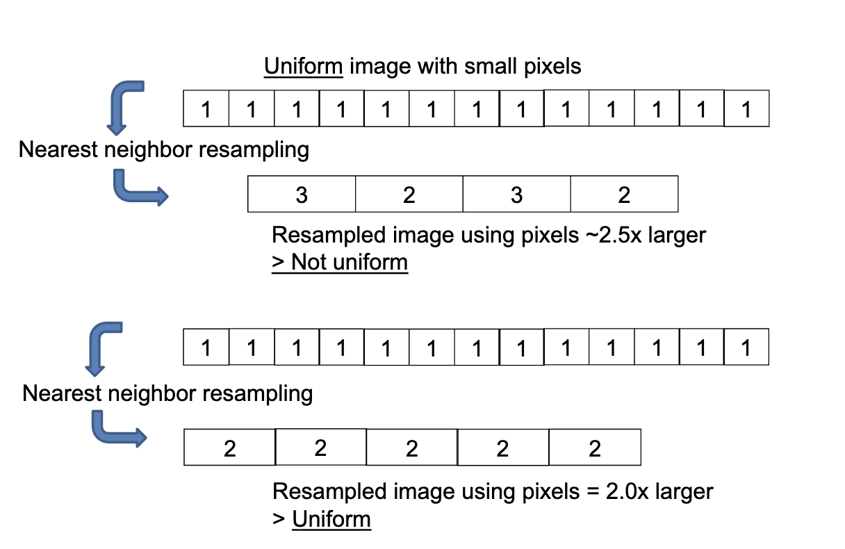
-simple resampling to finer grid can create impression of better resolution
-simplistic resampling (eg. nearest neighbor) by non-integer factors can create artifacts
-easiest using integer factors = 2x, 3x
-best using interpolation (bilinear or other)
misinterpreting shape-resolution
pixel size or image appearance does not always reflect resolution

discretization issues
artifact generation: Nearest-neighbor resampling with non-integer scaling creates artificial patterns
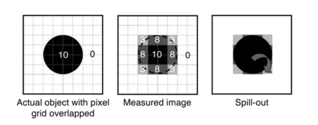
partial volume effect
-voxel/pixel contains >1 type of tissue or signal value
-so pixel has intermediate value

what is temporal (time) resolution
-how fast an image can be taken
-how fast the imaging system creates an image
-how fast a change in the object can be detected

time resolution subtleties: meaning on multiple levels
-detection of raw signal itself (<1 nanosecond for PET)
-creation if image (variable for PET, defined by pulse sequence for MRI and ultrasound)
-acquisition of enough data to answer clinical/research question - better quality takes more time
-varies even within modality depending on pulse sequence, radiotracer
time resolution subtleties: changes during acquisition are averaged or worse
-patient motion in PET causes blurring but in MRI can cause major artifacts
-biological variation is typically averaged
sensitivity
-change in image/change in actual parameter
-higher sensitivity allows visualization of smaller differences
-for a given noise level, better contrast
-specific definition varies depending on imaging modality
what is contrast
-ability to see differences between different signals
-subject contrast (biology/chemistry)
-detector contrast (computer)
-affected by both background and noise
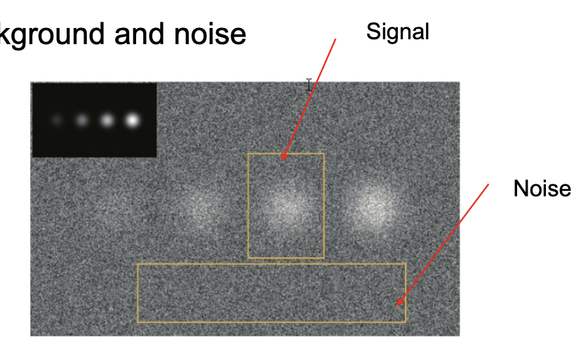
effects of noise
lose details

computers in bioimaging
originally: designed to perform mathematical computations and data processing quickly
now: information display, storage, networking-info transfer and communication
file size
number of voxels x memory per voxel + metadata
bit- 0 or 1
bytes-8 bits»256 different values
kilobyte(kB)-1024 bytes
megabyte(MB)-1024 kB
gigabyte(GB)-1024 MB
terabyte(TB)-1024 GB
data type of voxel value is important
-integer can be 2/4/8 bytes
-integers can be signed or unsigned
-2 bytes=65,537 values
-float can be 4/8 bytes, these are decimal numbers with exponent, and there are different formats (bit/little endian)
-no 2 byte float-sometimes float data is stored as integer*2 with an overall multiplier and offset
analog and digital data
all data originates as analog signal, must be digitized

analog transmission
-all detector signal start out as analog, but distortions possible
-inaccuracies when signal amplifies
-attenuation losses along signal wires
-electronic noise
digital transmission advantage
-immune to accumulation of errors due to signal distortion
-distortions do not have sifficient amplitude to cause 0 to be mistaken as 1
-most digital circuits do not amplify incoming data, make a fresh copy of it
-other safeguards to main information integrity-can have additional redundant information to detect errors
-eg. parity bits-additonal bit with each byte which changes if any bit changes
conversion of continuous analog data to digital (ADCs)
-only certain time points of the continuous analog signal converted=sampling
-conversion to digital=digitization or quantization
characteristics of ADC: sampling rate-number of times per second sample and digitize signal

nyquist theorem
to represent a signal of frequency, you need to sample at 2f. ex. if the sampling interval deltat is 2ms (500 Hz) then the highest frequency you can represent is fN=1/(2×0.002)=250Hz
also applies to spatial information=signal changing over distance
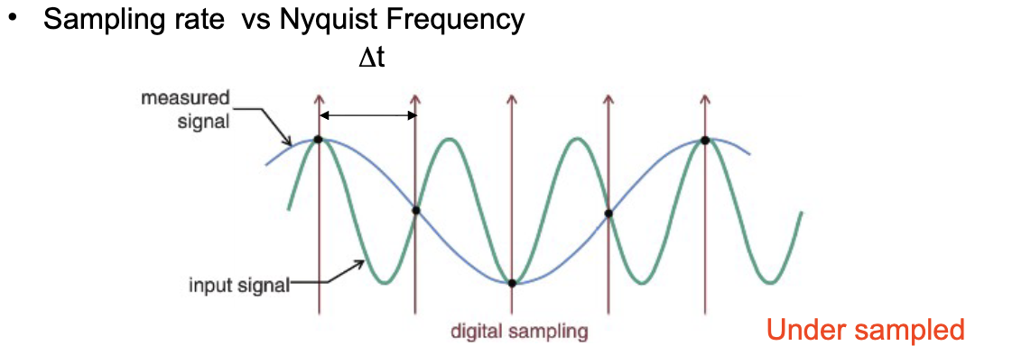
to display digital images from imaging modality onto video monitor
digital-to-analog converter (DAC)

disadvantages of digitization
-loss if information due to both sampling and quantization
-values of analog signal between sampling times is lost
-minimum sampling rate needed-nyquist limit
-analog signal continuous value-digital signal finite values
-N-bit signal restricted to 2N values


storage of digital images
-digital image, usually square or rectangular array of numbers -a matrix
-pixel size=field of view (FOV) of the image/number of pixels across the image
eg. if CT has 23 cm x 23 cm FOV and images aquired in 512 pixel by 512 pixel format, pixel size is 23cm/512=0.45 mm in x and y directions
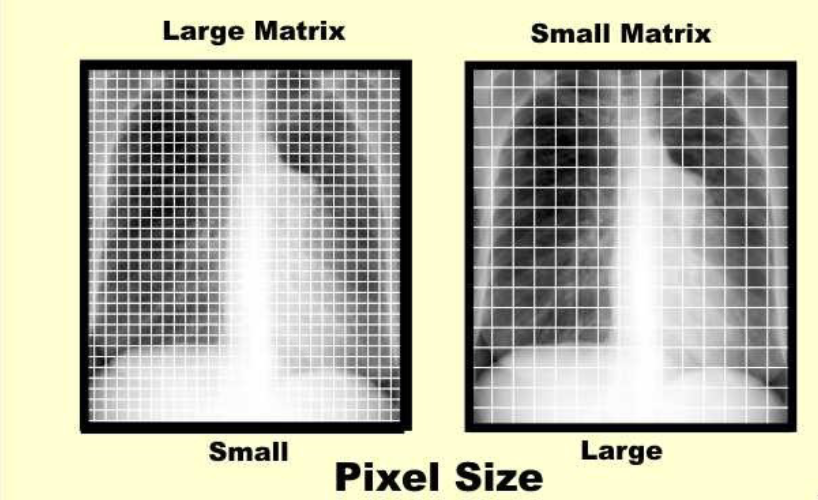
digital contrast resolution
-largest number stored in single pixel is determined by number of bits or bytes used for each pixel (i.e. gray levels)
-1 byte used = maximal number is 28-1=255(8 bits)
-amount of contrast resolution provided by an imaging modality determines the number of bits per pixel needed
-ultrasound limited contrast resolution-6 or 7 bits enough - 8 bits used
-x ray CT high contrast resolution -12 bits required to represent full range of CT numbers

visual interpretation of image data
-image data is just numbers, needs to be converted to visual representation
-the way this conversion is done affects observed contrast and ability to diagnose
-human eye can detect only a limited range of brightness
-select mapping that optimizes display of important features-contrast enhancement
how?
-translation table selection
-windowing


translation table
-lookup table produces digital value indicating video intensity to the DAC
-video intensity can also be represented as different colors

windowing
-can use entire range of intensity to display portion of total range of pixel values
-windowing can be combined in translation table

processing of digital images
-image reconstruction to get initial image from raw data
-many “post-processing” techniques to apply to images
-post-processing of image generally does not unequivocally improve image quality-eg cannot resolve objects less than limits of the modality
-but can make useful tradeoffs in image quality features or combine
common processing techniques
-addition or subtraction of digital images
images need to be in the same format, pixel size, etc
resultant image is in the same format
image subtraction common in angiography to remove effects of anatomic structures
spacial filtering=smoothing
-statistical nature of acquisition process can give grainy appearance-noise
-reduced by spatial filtering operation=smoothing
-each value of pixel in smoothed image is weighted average of the corresponding pixel in the unprocessed image with its neighbors
-reduces noise but also worsens spatial resolution-tradeoff but sometimes improves things like logic detectibility
quantification (quantitation)
-physical and biological meanings
-physics-how well absolute voxel value represents true value-often estimated with phantoms-can vary within each modality
-biology-values derived from raw images that represent functional processes (glucose metabolic rate)-sometimes measure reliability with test-retest” study
registration (coregistration, realignment, reslicing, spatial normalization)
-aligning 2 images of the same region of body
-equalize pixel sizes and rotate/shoft/scale
-”rigid body” if images are of same person and organ shape does not change (eg brain)
-”non rigid”, “deformable”,”warping” etc if not (etc to template)
translational methods
-readily allow transition from lab to clinic
-eg PET tracers tested in animals, then humans
for single parameter, p-value is used
-assuming groups do not differ (“null hypothesis" is true), this is the probability that your result is by chancw
MRI
-usually good resolution
-anatomy is obvious
-good contrast between soft tissue types
-low signal in cortical bone (like skull)
-usually gray scale
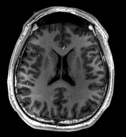
CT
-great resolution
-bone signal very strong, soft tissue weaker with poor contrast between soft tissue types (unless contrast agent used eg in blood)
-usually gray scale
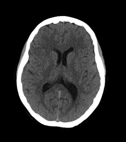
PET/nuclear
-anatomy can be hard to discern
-resolution low, noisy appearance
-be careful-often fised (overlaid) over CT or MRI for anatomy
-usually colored scale
