Biol 260 - Exam 2 - Muscles and Tissues
 Call Kai
 Learn
Learn Practice Test
 Spaced Repetition
 Match
Match Flashcards
 Knowt Play
1/97

There's no tags or description
Looks like no tags are added yet.
Name | Mastery | Learn | Test | Matching | Spaced | Call with Kai | Chat |
|---|
No analytics yet
Send a link to your students to track their progress
98 Terms
Connective tissue consists of what two basic elements:
Cells
Extracellular matrix
Characteristics of Cells and Tissues
Cells: do not have any free surfaces
immature cells have names that end in -blast
mature cells have names that end in -cyte
Tissue: highly vascularized and has a nerve supply
except tendons and cartilage
What do these connective tissues do:
Mature
Fibrous
Supporting
Liquid
Mature: connective tissue present at birth and persists through life
Fibrous: flexible and contains viscous ground substance and many fibers
Supporting: protects and supports soft tissues of the body
Liquid: extracellular matrix is liquid
Examples of mature CT?
Bone
Dense irregular
Hyaline
What makes up the supportive ct?
Chondroblasts and chondrocytes
Osteoblasts and osteocytes
What makes up the liquid ct?
Formed elements: cellular components of blood
Erythrocytes
Leukocytes
Platelets (thrombocytes)
Ground substance
Aka ECF = fluid/gel component of EM
contains water, gases, nutrients, minerals, wastes, hormones, and other chemicals
The three classes of large molecules that make up the ground substance
Glycosaminoglycan (GAG)
Proteoglycans
Adhesive glycoproteins
Glucosaminoglycan (GAG)
Long polysaccharide w/ unusual composition
Negatively charged, attracts sodium and potassium ions, making GAG absorb and retain water
Regulate water and electrolyte balance
Proteoglycans
Giant molecule shaped like a bottle brush w/ central core protein and bristles of GAGs
Attach to hyaluronic acid to make enormous complex
Slows spread of pathogens and helps hold tissues together
Form thick colloids like gravy, gelatin, or glue
Adhesive glycoproteins
Protein/carbohydrate complexes
Bind plasma membrane proteins to extracellular collagen and proteoglycans
Bind components of tissue together
Mark paths to guide migrating embryonic cells to their destination tissue
Characteristics of Epithelial tissue
Closely packed cells with little extracellular material
Cells sit on basement membrane
Avascular
Good nerve supply
Rapid cell division
Function of the basement membrane is to
Anchor cells
Guide for cell migration during development
Restricts passage between epithelial and connective tissues
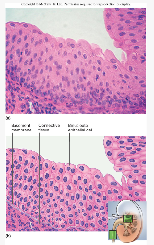
Urothelium aka transitional epithelium
Appearance:
Somewhat resembles stratified squamous
epithelium but w/ rounded surface cells that
bulge at the surfaceTypically 5-6 cells thick when relaxed, only 2-3 cell
layers thick when stretchedSome cells have two nuclei
Locations:
Urinary tract – kidney, bladder, ureter, urethra
Functions
stretching to allow filling of urinary tract
protects underlying tissues from damage by urine
4 major types of tissues
Epithelial
Connective
Muscular
Nervous
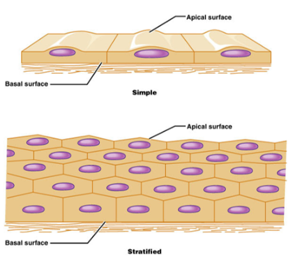
Simple vs Stratified
Simple: one layer
Stratified: multiple layers of cells
Label the apical vs basal surface in both simple and stratified epithelia
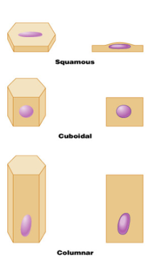
Classification of epithelia are?
Squamous, cuboidal, or columnar
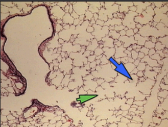
Simple Squamous Epithelial
Function: filtration (kidneys) and rapid diffusion (air sacs of lungs)
Location: kidney glomeruli, alveoli of lungs, lining of heart, kidney corpuscles, and blood and lymphatic vessels
Keratinized Stratified Squamous Epithelia
Function: protects underlying tissue in areas subject to abrasion and penetration, retards water loss through skin
Location: epidermis, palms and soles (heavily keratinized)
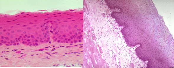
For stratified epithelia, why do we name based off cells on apical surface?
All cells connected to basal membrane will either be cuboidal or columnar
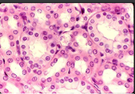
Simple Cuboidal
Description: Single layer of cubelike cells with large, spherical central nuclei
Function: Secretion and absorption, production of mucous coat, movement of respiratory mucus
Location: Kidney tubules, ducts and secretory portions of small glands, and liver
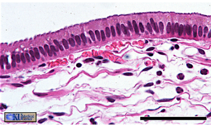
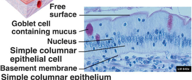
Simple Columnar
Description: Single layer of tall, column-like cells with oval nuclei
Function: Protection, secretion of mucus, absorption
Location: Lining of stomach, intestine, gallbladder, uterine tube, collecting ducts of kidneys
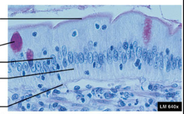
Label the simple columnar
free surface
goblet cell containing mucus
nucleus
basement membrane
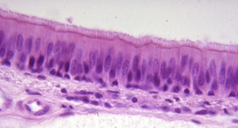
Ciliated Pseudostratified Columnar
Function: Secretion and propulsion of mucus
Location: Lining of ducts, ciliated: lines trachea and most of upper respiratory tract
Characteristics:
Has goblet cells that secret mucus & is ciliated
Looks layered, but only has a single layer of cells
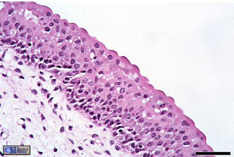
Transitional
Description: Multiple layers of cells that can change shape depending on the degree of stretching.
Function: Stretches readily and permits distension of urinary organ by contained urine
Location: Lines the ureters, bladder, and part of the urethra
Connective tissue
Found throughout the body; most abundant and widely distributed of all the tissues
Four main types:
connective tissue proper
cartilage
bone
blood
Characteristics: Tissues with large matrix inside of it, rich supply of blood vessels, composed of many types of cells
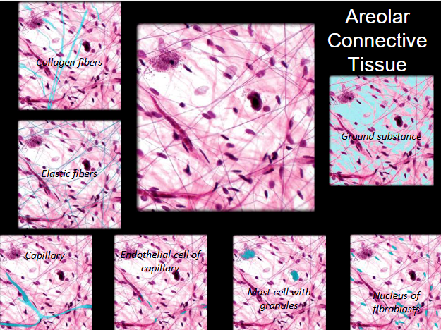
Loose Areolar CT
Appearance: loose arrangement of collagenous and elastic fibers
CT Type: CT proper
Fibers: Collagen, elastic
Function: Wraps and cushions all internal organs - attached to all epithelial tissue (connects skin to deep tissues)
Location: in serous membranes, between muscles, passageways for nerves and blood. underlies all epithelia
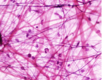
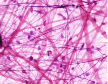
Label the Areolar CT
Dense Regular Tissue
CT Type: CT Proper
Fibers: Collagen fibers tightly packed together
Function: Withstand force in one direction - resist stress
Location: In tendons and some ligaments
Characteristics: Parallel collagen fibers in one direction and compressed fibroblast nuclei
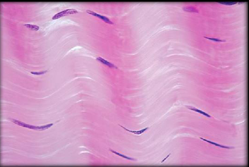
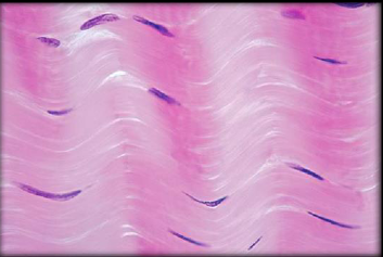
Label the Dense Regular CT
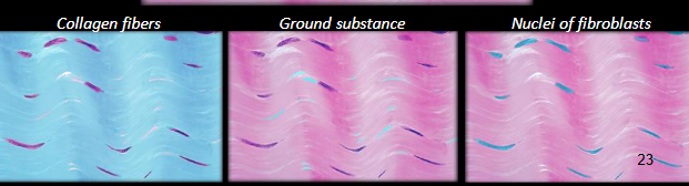
Tendons do what? Ligaments do what?
Tendons attach muscles to bones and ligaments hold bones together
Adipose CT
CT Type: CT Proper
Function: Insulation, cushions and protects organs, and reserve fuel
Location: Under skin in the abdomen, around kidneys & eyeballs, in breasts
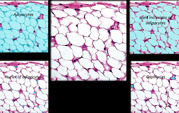
Characteristics: Nucleus pressed against cell membrane
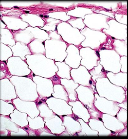
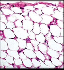
Label the Adipose CT
Reticular CT
CT Type: CT Proper
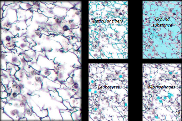
Function: Provides a framework for white blood cells, mast cells, and macrophages - all a part of the immune system
Location: Lymphoid organs: spleen, lymph nodes, and bone marrow
Characteristics: Mesh of reticular fibers and fibroblasts
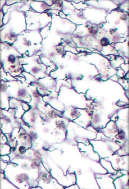
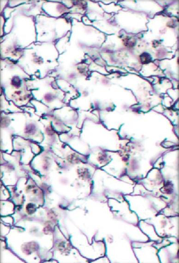
Label the Reticular CT
Bone (osseous tissue) CT
Function: Structural support, give body its shape, act as attachment points for muscles
Location: Bones
Characteristics: Arranged in cylinders that surround central (haversian or osteonic) canals that run longitudinally through shafts of long bones
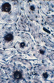
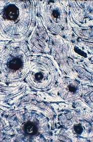
Label the Compact Bone
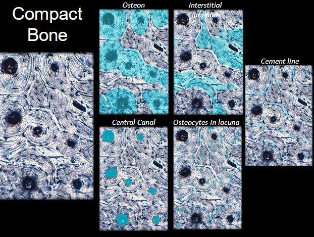
Haversian Canal
A series of tubes around narrow channels formed by lamellae
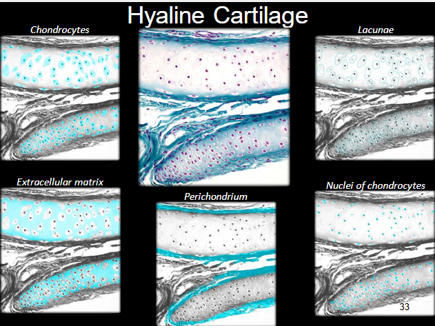
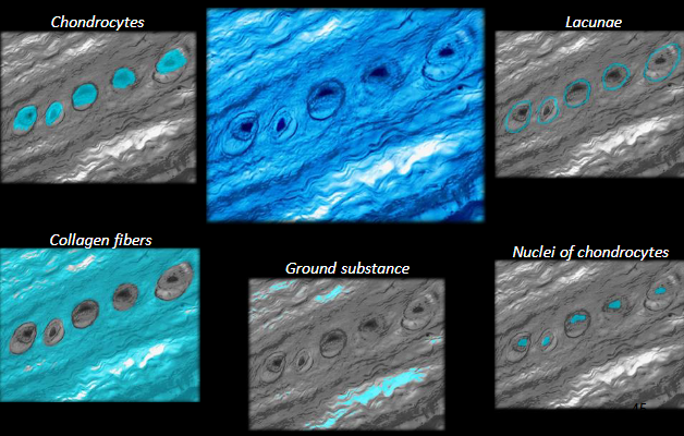
Hyaline Cartilage
Function: Supports and reinforces structures (like bone), but can bend - eases joint movement
Location: Ends of long bones, connects ribs to sternum, found at end of nose, in trachea and larynx
Characteristics: Pairs of lacunae, clear, glassy microscopic appearance. Covered by perichondrium
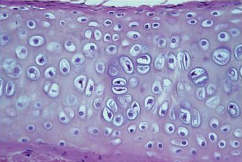
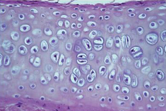
Label the Hyaline Cartilage
Blood CT
Function: transport respiratory gases and dissolve matter from place to place
Location: in blood vessels
Characteristics: Formed in the bone marrow and fluid connective tissue
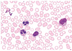
Plasma
Blood’s liquid ground substance
Formed elements
Cells and cell fragments
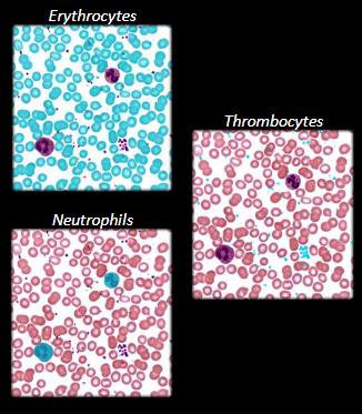
Erythrocytes
Red blood cells: transport O2 and CO2
Leukocytes
White blood cells: defense against infection and other diseases
Thrombocytes
Cell fragments involved in clotting and other mechanisms
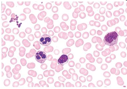
Label the blood CT
Smooth Muscle tissue
Function: involuntary control - propels substances along redetermined pathways
Location: digestive, respiratory, and urinary tract - visceral muscles
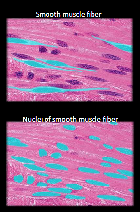
Characteristics: lines hollow structures, smooth and no striations
relatively short, fusiform cells
One centrally located nucleus
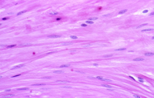
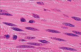
Label the Smooth Muscle
Cardiac Muscle
Function: Propels blood into circulation as it contract
Location: only in the heart
Characteristics:
involuntary control
one nucleus surrounded by light-staining glycogen
shorter, branched, and blunted at ends
intercalated discs allow the cardiac muscle to act as a unit
striated
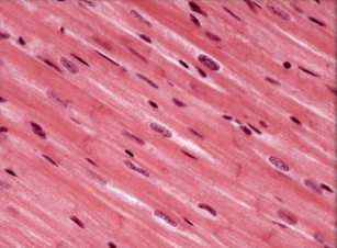
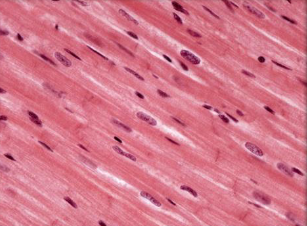
Label the Cardiac Muscle
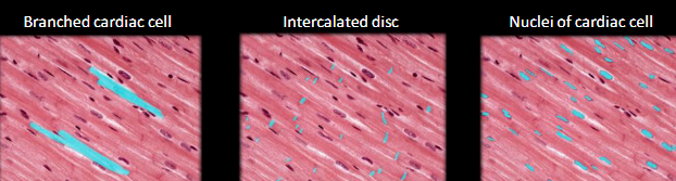
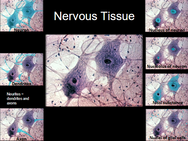
2 main cell types
Neuroglia: functionally support, protect and insulate neurons and bind them together
Neurons: respond to stimuli and conduct impulses to and from all body organs
Nervous tissue
Function: specialized for communication by electrical and chemical signals
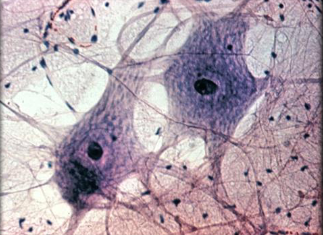
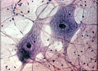
Label the Nervous tissue
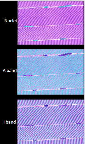
Skeletal Muscle
Functions: voluntary control
Location: in skeletal muscles attached to bones or occasionally to skin
Characteristics:
Cells are long
multi-nucleated
striated
threadlike cells
contains multiple nuclei
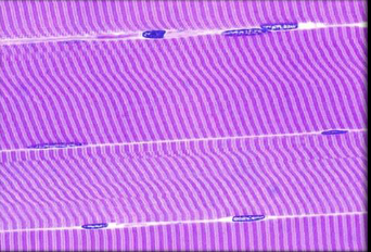
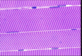
Label the skeletal muscle
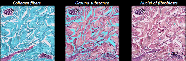
Dense irregular CT
Function: provides strength and elasticity to the skin and underlying structures - withstands unpredictable stresses
Location: found in the dermis of the skin, joint capsules, and organ capsules - fibrous sheaths
Characteristics:
densely packed
randomly arranged
collagen fibers
few visible cells
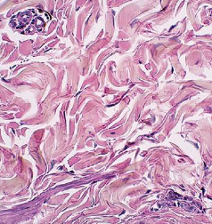
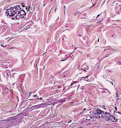
Label the Dense Irregular CT
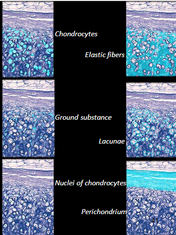
Elastic Cartilage CT
Function: provides flexible, elastic support
Location: external ear and epiglottis
Characteristics:
covered with perichondrium
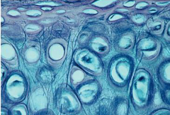
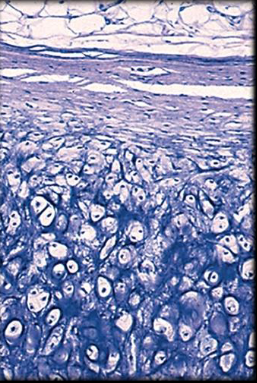
Label the Elastic Cartilage
Fibrocartilage CT
Function: resists compression and absorbs shock
Location: pubic symphysis, menisci, and intervertebral discs
Characteristics:
cartilage containing large, coarse bundles of collagen fibers
never has perichondrium
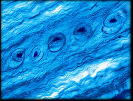
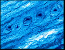
Label the fibrocartilage
2 main layers of the skin
Epidermis: stratified into 4 structural layers (or 5 layers in palms and soles)
Dermis: consists of 2 layers
Two types of sweat glands
Eccrine
Apocrine
Eccrine Sweat Glands
simple, coiled tubular glands
more common than apocrine
regulate body temperature through perspiration/evaporation
Apocrine sweat glands
Found: skin of axilla, groin, anal regions, areolae areas of the breasts, and bearded areas of men
Secretions are more viscous (thicker) than eccrine secretions
Epithelial
Tissue composed of layers of closely spaced cells
Function:
Protection
Secretion
Excretion
Absorption
Filtration
Sensation
Locations:
Epidermis
Inner lining of digestive tract
Liver and other glands
Connective Tissue
Tissue with usually more matrix than cell volume, often specialized to support and protect organs and to bind other tissues and organs to each other
Locations:
Tendons
Ligaments
Cartilage
Bone
Blood
Nervous Tissue
Tissue containing excitable cells specialized for rapid intermission of coded information to other cells
Locations:
Brain
Spinal cord
Nerves
Muscular Tissue
Tissue composed of elongated, excitable muscle cells specialized for contraction
Locations:
Skeletal muscles
Heart
Walls of viscera
What’s the difference between longitudinal, cross section and oblique?
Longitudinal sections are sliced along the length of the structure
Cross sections are cut horizontally across it
Oblique sections are cut at an angle, allowing visualization of different cellular features.
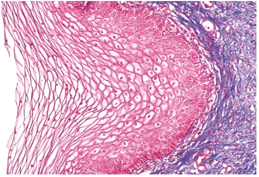
Stratified Squamous Epithelium - nonkeratinized
Appearance: Same as keratinized but without the surface of dead cells
Function: Resists abrasion and penetration by pathogenic organisms
Locations: Tongue, oral mucosa, esophagus, anal canal, vagina
The cells of fibrous CT include?
Fibroblasts
Macrophages
Leukocytes
Plasma cells
Mast cells
Adipocytes
Three types of protein fibers characteristics?
Collagenous fibers:
made of collagen
tough and flexible
resist stretching
most abundant protein
glistening white fibers
makes up tendons, ligaments, and the dermis of the skin
Reticular fibers:
thin collagen fibers
coated with glycoprotein
form a spongelike framework for spleen and lymph nodes
Elastic fibers:
thinner than collagen fibers
branch and rejoin each other
made of elastin
coasted with glycoprotein
stretch and recoil
Characteristics of muscle
Excitability = respond to stimuli with electrical changes across the plasma membrane
Conductivity = electrical signal travels rapidly along the cell
Contractility = cell shortens when stimulated
Extensibility = able to stretch beyond normal resting length without damage
Elasticity = shortening of a muscle after it is stretched and released; ability of muscle to return to shape after stretching or contracting
Fascia
Fibrous tissue covering of bones and muscles
holds muscles w/ similar functions together
Epimysium
Outer layer covering muscle; made of dense irregular connective tissue
Perimysium
Surrounds groups of 10-100 muscle fibers creating bundles called “muscle fascicles”
made of dense irregular ct
Endomysium
Penetrates interior of each fascicle and separates fibers from one another
mostly reticular fibers
What forms a tendon?
Three layers of fascia which extend beyond muscle fibers and intertwine with the collagen
Aponeurosis
When layers fuse in a broad sheet