Staining: Carbohydrates & Staining
 Call Kai
 Learn
Learn Practice Test
 Spaced Repetition
 Match
Match Flashcards
 Knowt Play
1/80

There's no tags or description
Looks like no tags are added yet.
Name | Mastery | Learn | Test | Matching | Spaced | Call with Kai | Chat |
|---|
No analytics yet
Send a link to your students to track their progress
81 Terms
Microscopic examination of a control section of small intestine stained with colloidal iron shows marked staining of the lamina propria. This could most likely be corrected by:
ensuring the use of a potassium ferricyanide-hydrochloric acid solution
rinsing slides with 3% acetic acid before and after the colloidal iron solution
using diastase digestion prior to colloidal iron staining
carefully preparing a new colloidal iron sollution
carefully preparing a new colloidal iron sollution
Feedback:
Colloidal iron solutions must be carefully prepared according to the instructions, or poor results will be ontained. Potassium ferrocyanide is used, not potassium ferricyanide in the Prussian blue reaction, slides should be rinsed with 12% acetic acid, and diastase digestion would have no effect.
H&E stained sections of a gastrointestinal (GI) biopsy show red blood cells, collagen, and muscle all staining the same shade of pink-orange. Which of the following may be the cause?
More than on answer may be correct
leaving slides in xylene too long
staining in eosin too long
improper bluing of the hematoxylin
differentiating in alcohols is too brief
staining in eosin too long
differentiating in alcohols is too brief
Feedback:
Differentiation of the eosin staining occurs in the 95% alcohol anf subsequent dehydration alcohols. If differentiation is too brief, all eosinophillic elements will have the same shade of pink-orange.
Leaving the slides in xylene too long and improper bluring of the hematoxylin would not have an affect on the differentiation of the eosin.
Leaving the slides in eosin too long will produce an intense cytiplasmic stain that will also make differentiation between red blood cells, collagen, and muscle difficult.
Chromaffin cell tumors of the adrenal gland are known as:
adenomyomas
carcinoids
sarcomas
pheochromocytomas
pheochromocytomas
Feedback:
Pheochromocytomas arise in the chromaffin tissue portion of the adrenal medulla.
Giemsa is an example of a metachromatic stain.
True
False
False
Feedback:
Giemsa is not an example of a metachromatic stain. Metachromatic stains use a single dye that displays a color shift. An example is toluidine blue, where the mast cells stain violet rather than blue. Giemsa is a polychromatic stain.
Pigment that is formed following a reaction of ferrous ions with potassium ferricyanide is known as:
Prussian blue
Turnbull blue
Argyrophil
Anthracotic
Turnball blue
Feedback:
Turnbull blue is the result of ferrous ions reacting with potassium ferricyanide.
Alcian blue-stained (pH 2.5) sections reveal very weak staining, and the dye solution needs to be replaced; however no alcian blue is immediately available. A method that could be substituted with almost identical results is:
Congo red
colloidal iron
PAS
aldehyde fuchsin
colloidal iron
Feedback:
Colloidal iron will stain acidic mucosubstances (carboxylated and sulfated) just as alcian blue will. Congo red is a stain for amyloid; the PAS stains only neutral mucosubstances; aldehyde fuchsin stains only sulfated mucosubstances, but not carboxylated.
*In the colloidal iron method, ferric ions are adsorbed by:
neutral mucosubstances
both sulfated and carboxylated mucosubstances
carboxylated mucosubstances only
sulfated mucosubstances only
both sulfated and carboxylated mucosubstances
Feedback:
Ferric ions are adsorbed by both sulfated and carboxylated mucosubstances. This is a more sensitive method for acid mucosubstances than the alcian blue, but it is not as specific. Neutral mucosubstances will not be stained.
Very weak PAS staining is noted on a section of kidney. One problem-solving action that could be tried is to:
shorten the time in periodic acid
decrease the water wash after the Schiff reagent
verify that the Schiff reagent is stored at room temperature
check the Schiff reagent with formaldehyde
check the Schiff reagent with formaldehyde
Feedback
The viability of Schiff reagent can be checked by adding a few drops of Schiff reagent to 10 mL of concentrated formaldehyde; it should immediately turn reddish-purple if good. Periodic acid times should not be decreased for good oxidation to occur; the water wash after Schiff reagent is necessary to help the full development of color; and Schiff reagent should be stored in the refrigerator.
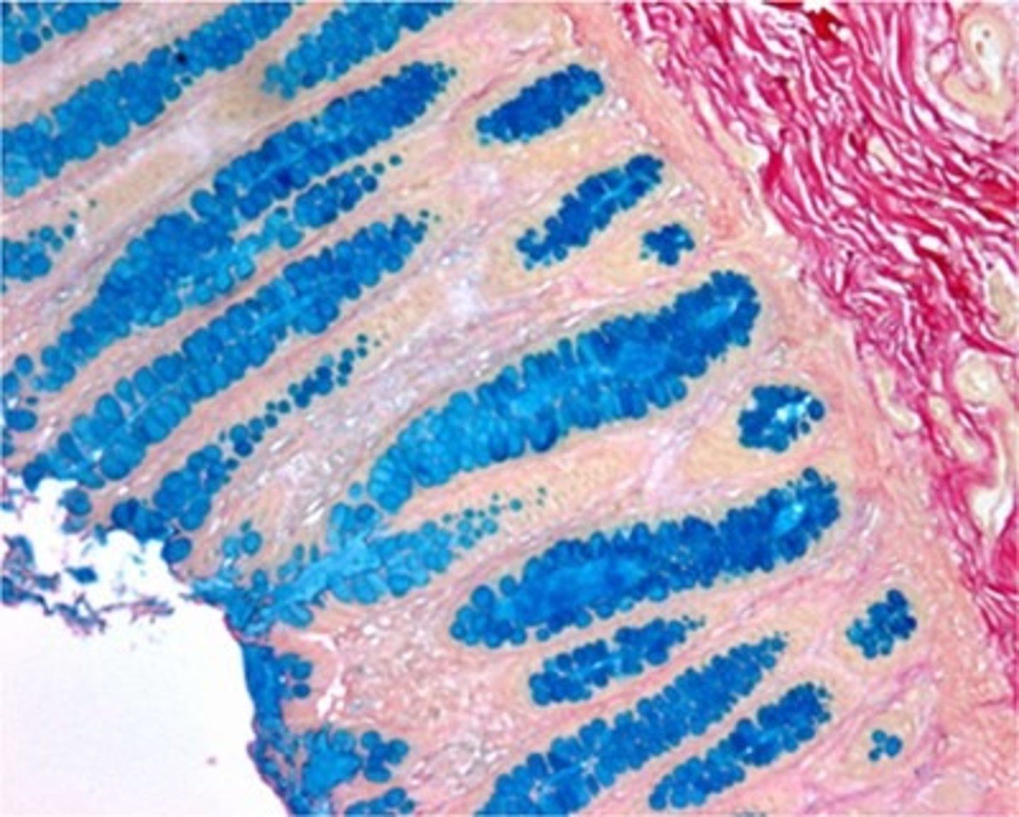
The staining technique used in this section is most likely:
aldehyde fuchsin
colloidal iron
toluidine blue
methylene blue
colloidal iron
Feedback:
This section has been stained with the colloidal iron procedure. Aldehyde fuchsin would stain the mucin purple; toluidine blue is a met not stain mucin.
All of the statements about bile and Hall's technique are true EXCEPT:
Biliverdin is the product of reduced bilirubin in the liver
Fouchet reagent oxidizes bilirubin into biliverdin
Bile is the normal end product of red blood cell degradation
Hemeglobin breaks down into bile
Biliverdin is the product of reduced bilirubin in the liver
Feedback:
Bilirubin is the product of reduced biliverdin.
Individual virus particles cannot be seen by light microscopy, but can be visualized by:
phase microscopy
polarizing microscopy
electron microscopy
fluorescence microscopy
electron microscopy
Feedback:
Individual virus particles are too small to be examined by light microscopy, but can be demonstrated will with electron microscopy.
The problem shown in this image could most likely be corrected by:
insuring that the stain is done at 370C
cutting slightly thicker sections
preparing new stock solution
adjusting the pH
preparing new stock solution
Feedback:
The mucicarmine stock solution has either been improperly prepared or has deteriorated. A new solution should be prepared or obtai thicker sections would not help the weak mucicarmine staining; and if the stock and working solutions are properly prepared, no pH adjustment is necessary.
*The preferred fixative for pituitary tissue sections prior to staining with Gomori's chrome-alum-hematoxylin is:
Zenker•s
Helly's
Bouin•s
Carnoy's
Bouin's
Feedback:
Picric acid in Bouin•s fluid gives this tissue a strong affinity for acid dyes.
Amyloids are identified by an abbreviation of their originating protein preceded by a/an:
A
D
L
H
A
Feedback:
Amyloids are identified by an A followed by an abbreviation of their originating protein. AA is "amyloid associated"; it is a nonimmunoglobulin protein made in the liver. AL is and amyloid which is immunoglobulin light chain derived; AH is immunoglobulin heavy chain derived; there is no AD.
Reagent problems are most apparent if the control used for the PAS reaction is:
kidney
lymph node
liver
umbilical cord
kidney
Feedback:
Reagent problems are more readily apparent if a section of kidney is used as a routine control for substances other than glycogen. A small piece of kidney may also be included on the glycogen control to check for reagent problems. Liver containing large amounts of glycogen may show a weak, but still very apparent reaction, with deteriorating solutions; weak reactions may lead to false-negative results on components that normally do not yield strong reaction. Lymph node is not a control for polysaccharides and mucosubstances, and the umbilical cord is the control for determining the presence of hyaluronic acid.
Proteolytic emzymes used in enzyme-induced epitope retrieval (EIER) protocols can ONLY be used at 37-400 C.
True
False
False
Feedback:
Select proteolytic emzymes can be used in environments ranging from room temperature to 37-400 C.
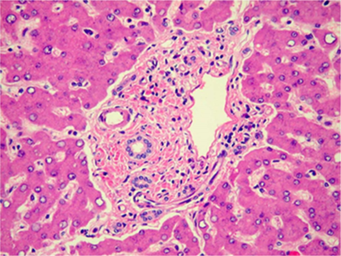
The structure in the middle of this section is a/an:
portal triad
endocrine gland
glomerulus
lymph nodule
portal triad
Feedback:
This is a portal triad in a section of liver. It is so named because it contains branches of three different structures: hepatic portal vein, h You answered the question correctly.
The PAS reaction is used for the demonstration of:
hyaluronic acid
dermatan sulfate
chondroitin sulfate B
neutral mucopolysaccharides
neutral mucopolysaccharides
Feedback:
The PAS reaction is a reliable stain for the demonstration of neutral mucopolysaccharides. Hyaluronic acid and dermatan sulfate (same as chondroitin sulfate B) are acid mucopolysaccharides; they are PAS-negative, but may be demonstrated with alcian blue.
Combining alcian blue at a pH of 2.5 with hyaluronidase digestion will differentiate:
sulfated and carboxylated mucins
acidic and neutral mucins
stromal and epithelial mucins
carcinomas and lymphomas
stromal and epithelial mucins
Feedback:
Stromal mucins (hyaluronic acid) will be digested by hyaluronidase, but epithelial mucins will not. Sulfated and carboxylated mucins can be differentiated by staining with alcian blue at pH 2.5 and pH 1.0. Neutral mucins will not stain with alcian blue; only acidic mucins will stain. Carcinomas will be unaffected and lymphomas do not produce mucin.
The metal found in the structural formula of alcian blue is:
aluminum
iron
copper
calcium
copper
Feedback:
Alcian blue is a copper phthalocyanin basic water soluble dye colored blue because of the copper content. None of the other metals is found in alcian blue.
All of the following reagents demonstrate lipids EXCEPT:
Osmium tetroxide
Oil Red O
Alizarin Red
Sudan Black
Alizarin Red
Feedback
All of the reagents dye lipids except for Alizarin Red, which demonstrates calcium deposits in tissue.
A rapid bile identification technique that involves destruction of tissue sections by acid is the:
Gmelin test
Ralph method
Hall technique
Gomori reaction
Gmelin test
Feedback
In Gmelin's test for bile, a coverslip is placed over deparaffinized and hydrated sections; nitric acid is allowed to diffuse beneath the coverslip, destroying the tissue. The presence of bile pigment is indicated by color changes from yellow to green to blue to purplered. This method is somewhat unreliable and should be repeated several times to confirm the results.
Which of the following chemicals is added to Congo red solution in order to suppress background staining?
sodium acetate
potassium phosphate
potassium sulfate
sodium chloride
Feedback
Sodium chloride and a high alcohol content will result in less background staining. The other chemicals are not used in Congo red solutions.
sodium chloride
Feedback:
Sodium chloride and a high alcohol content will result in less background staining. The other chemicals are not used in Congo red solutions.
For optimum apple green birefringence, sections for Congo red staining should be cut at:
3-5 pm
6-7 pm
8-10 pm
12-14 pm
8-10 pm
Feedback:
The thickness of sections for Congo red staining is critical, with 8-10 pm optimal for apple green birefringence. Sections that are too thin will show faint red birefringence and those that are too thick will show yellow birefringence.
Why would zinc salts be used in fixatives in immunohistochemical (IHC) staining methods?
To provide contrast to tissues
To enhance protein cross-linking
To retain the epitopes in a satisfactory manner
To prevent background staining
To retain the epitopes in a satisfactory manner
Feedback:
Zinc is a substitute for mercury and is a less toxic metal. It is used in fixatives to prevent excessive crosslinking from formaldehyde fixation, thus preserving (classified as a preservative) and not masking the antigenicity of the tissue.
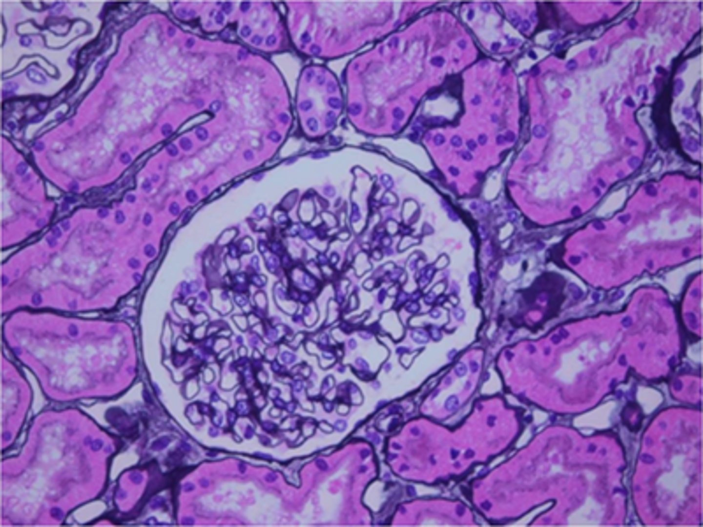
The component stained black in this image is:
reticulin
calcium
basement membrane
elastin
basement membrane
Feedback:
The basement membrane is stained black in this Jones-stained sections of kidney.
Which of the following protocols is used for the purpose of demonstrating nissl and myelin, or the absence of either?
Holzer's crystal violet Staining
Hematoxylin and eosin staining
Bielschowsky silver staining
Luxol fast blue (LFB) with cresyl echt violet(CEV) staining
Luxol fast blue (LFB) with cresyl echt violet(CEV) staining
Feedback:
The luxol fast blue and cresyl echt violet staining procedures combined allow pathologists to demonstrate both Nissl substance and myelin, or their absence.
Hematoxylin and eosin, Holzer's crystal violet, and Bielschowsky silver staining protocols are NOT used for this purpose as they do not demonstrate myelin or nissl.
Since argentaffin and argyrophil granules are found in similar tissues, two stains (ie. Fontana Masson and Grimelius) must be done on same tissue to differentiate between carcinoid tumors.
True
False
True
Feedback:
The intestine and central nervous tissue have both argentaffin and argyrophil granules. To differentiate between tumors, two stains must be done. Grimelius and Churukian-Schenk stains will demonstrate both argyrophil and argentaffin granules, therefore, Fontana Masson must be performed as well to demonstrate only the argentaffin granules!
Prolonged formalin fixation increases tissue basophilia.
True
False
True
Feedback:
Formalin fixation of tissue increases basic dye (such as hematoxylin) uptake.
Connective tissue fibers are either classified as loose or dense.
True
False
True
Feedback:
Connective tissue fibers are either classified as loose or dense.
Connective tissue is the most abundantly found tissue in the human body. The connective tissue in the human body has three major functions which are protection, support, and binding.
The substance stained orange-red in this image is most likely:
collagen
amyloid
glycogen
epithelial mucin
amyloid
Feedback:
Amyloid has been stained orange-red by the Congo red technique. Collagen unstained in the lower right corner of the image.
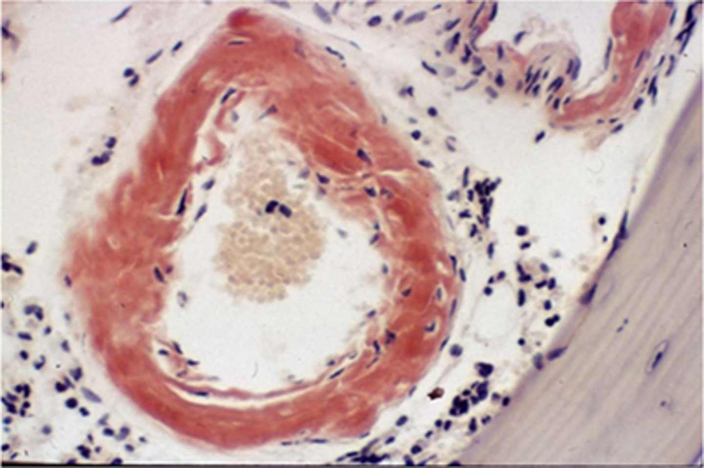
Choose the three components of Gordon and Sweets working ammoniacal silver solution for reticular fibers.
Silver nitrate
Ammonium hydroxide
Methenamine
Sodium hydroxide
Silver nitrate
Ammonium hydroxide
Sodium hydroxide
Feedback:
Gordon and Sweets ammoniacal silver solution contains silver nitrate, sodium hydroxide, and ammonium hydroxide. Methenamine is typically used in PAMS (periodic acid methenamine silver) procedures for basement membranes.
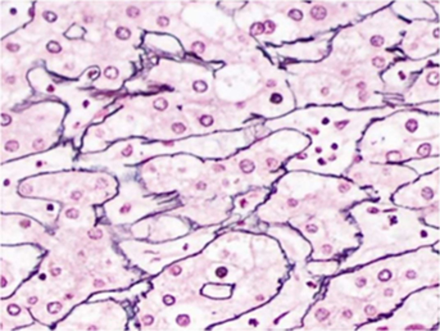
Microscopic review of a PAS-stained section of kidney reveals a lack of staining of the glomerular basement membrane. Which of the following is the most likely cause of the problem?
inadequate oxidation
thickening of the basement membrane
lack of treatment with sodium metabisulfite
prolonged time in Schiff reagent
inadequate oxidation
Feedback:
Inadequate oxidation would cause a lack of staining of the glomerular basement membrane. Basement membranes would show a more intense stain if thickened, or if sections were given a prolonged time in Schiff reagent. Many procedures omit the sodium metabisulfite; if omitted, there may be more non-specific staining, not lack of staining.
A suspected bacterial infection on the skin would most likely be stained with which of the following reagents/dyes?
Crystal violet and Gram's iodine
Carbol-fuchsin and methylene blue
Schiff reagent and light green
Methenamine-Silver nitrate and light green
Crystal violet and Gram's iodine
Carbol-fuchsin and methylene blue
Feedback:
Bacteria are reliably demonstrated by Acid-fast stains (Carbol-fuchsin and methylene blue) and Gram stains (crystal violet and Gram's iodine).
Fungi are demonstrated by PAS reaction and GMS (methenamine silver) stain.
When performing alkaline phosphatase (AP) immunohistochemistry (IHC), the blocking of endogenous peroxidase activity is required.
True
False
False
Feedback:
An alkaline phosphatase (AP) enzyme label is NOT affected by peroxidase activity.
While examining a GMS-stained section microscopically it is noted that elastin, crenated red cells, and mucin have stained black, confusing interpretation. This is most likely caused by:
overexposure to hot silver nitrate
underexposure to gold chloride
underexposed to sodium thiosulfate
performing the procedure in a microwave oven
overexposure to hot silver nitrate
Feedback:
The most common infraction committed when performing this procedure is that of leaving the section in hot silver nitrate too long. It is necessary in isolated instances, such as with the organisms of sporotrichosis, to leave the section in silver nitrate until many components are black, however, this in not ordinarily desirable.
Which of the following fixatives would give the best demonstration of glycogen with the PAS reaction?
alcoholic formalin
glutaraldehyde
Helly solution
Zenker solution
alcoholic formalin
Feedback:
Of the fixatives listed, alcoholic formalin is preferred for the demonstration of glycogen. Glutaraldehyde is a dialdehyde, and may leave free aldehyde groups in the tissue to react with Schiff reagent. Mercuric chloride-containing fixatives, such as Helly and Zenker solutions produce uneven staining of glycogen.
Which of the following can be substituted for diastase in the PAS procedure to specifically remove glycogen?
sialidase
a-amylase
hyaluronidase
proteinase
a-amylase
Feedback:
Glycogen will be depolymerized by a-amylase into smaller sugar units (maltose and glucose) which may be easily washed out of the sections. Sialidase removes sialidase acid, hyaluronidase removes hyaluronic acid, and proteinase downgrades proteins.
The only member of the polysaccharide group that normally can be demonstrated in human tissue sections is:
cellulose
glycogen
pectins
dextrin
glycogen
Feedback:
Glycogen is the only polysaccharide that normally can be demonstrated in human tissue sections. Pectins are found in plants; dextrin is formed by the hydrolysis of starch, a plant component; cellulose is also found in plants. Starch may be present in human tissue sections as a contaminant from surgical glove powder, or in pulmonary alveolar walls in cases where drug addicts have self-injected starch-containing barbiturates into the blood-stream. Cellulose (undigested food particles) may be seen in sections from the gastrointestinal tract, or in sections of skin from wood splinters. Both starch and cellulose are PAS-positive and are birefringent.
Melanin pigment is best described as:
Birefringent
Argentaffin
Argyrophil
Exogenous
argentaffin
Feedback:
Melanin is an argentaffin pigment that reduces silver to its metallic form.
A myeloma can be described as a malignant neoplasm of this origin.
Skin
Plasma cell of the bone marrow
Connective tissue
Gland
plasma cell of the bone marrow
Feedback:
A myeloma can be described as a malignant neoplasm of plasma cell in the bone marrow origin.
Carcinomas can be described as a malignant neoplasms of epithelial origin.
Sarcomas can be described as a malignant neoplasm of connective tissue origin.
Adenomas can be described as a malignant neoplasm of glandular tissue origin.
Carcinoma is defined as a malignancy of this tissue type.
Nervous
Epithelial
Connective
Muscle
epithelial
Feedback:
Carcinoma is the term used to define the family of malignant tumors found in epithelial tissue.
Rhabdomyosarcoma can be described as a malignant neoplasm of this tissue origin.
Plasma cell
Gland
Skin
Muscle
Muscle
Feedback:
Rhabdomyosarcoma can be described as a malignant neoplasm of muscle origin.
Adenocarcinomas can be described as a malignant neoplasm of this glandular origin.
Carcinomas can be described as a malignant neoplasms of epithelial origin.
Sarcomas can be described as a malignant neoplasm of connective tissue origin.
Myelomas can be described as a malignant neoplasm of plasma cell of the bone marrow origin.
What type of fluorescence in situ hybridization (FISH) is performed on formalin-fixed, paraffin-embedded (FFPE) tissue?
Metaphase FISH
Interphase FISH
Interphase FISH
Feedback:
When we perform FISH in FFPE tissue, it is considered to be interphase FISH. Interphase is the portion of the cell cycle in which the DNA is unwound. Human tissue is normally fixed in 10% neutral buffered formalin (NBF) and embedded in a paraffin wax. Cells within the tissues are undergoing various phases of the cell cycle at different times. Many cells will be in the interphase portion of the cell cycle.
In traditional cytogenetics laboratories, cultured cells from blood and amniotic fluids are harvested so that many of the cells will be in "metaphase." Metaphase is a phase of the cell cycle in which the chromosomes are lined up on the metaphase plate, just before segregating into daughter cells.
The alcian blue stain will demonstrate only sulfated mucosubstances if performed:
at pH 5.6
at pH 2.5
at pH 1.0
with hyaluronidase digestion
at pH 1.0
Feedback:
Alcian blue performed at pH 1.0 will demonstrate only sulfated mucosubstances, as the carboxyl (acidic) groups are not ionized and therefore cannot electrostatically attract the cationic (basic) dye. If performed at pH 2.5 or pH 5.6, both carboxylated and sulfated mucosubstances will be ionized and will be demonstrated. Hyaluronic acid does not contain sulfated mucosubstances.
*Which of the following staining procedures relies on the oxidation-reduction chemical reaction for dye binding?
Toluidine blue stain
Masson•s trichrome stain
Phosphotungstic acid-hematoxylin (PTAH) stain
Gordon and Sweets silver stain
Gordon and Sweets silver stain
Feedback:
The Gordon and Sweets staining method relies on the oxidation-reduction chemical reaction for dye binding.
Toluidine blue, Masson's trichrome and PTAH staining do not rely on the oxidation-reduction chemical reaction for dye binding.
Microscopic review of PAS-stained sections of kidney reveals very weak staining of the glomerular basement membrane. All of the following are possible corrective actions with the exception of ensuring that the:
Schiff reagent is stored in the refrigerator
Schiff reagent is not overused
sections are washed well following Schiff reagent
periodic acid is the correct concentration
sections are washed well following Schiff reagent
Feedback:
If sections are not washed well following the Schiff reagent, the result will most likely be non-specific staining due to reoxidation of any loosely adsorbed Schiff reagent; this would give a more intense color, not a weaker stain. All other answers are possible corrective actions.
Amyloid control slides are staining very pale with the Congo red method. They have stained satisfactorily in the past. One cause of this problem might be:
slides have been cut and stored for a prolonged time
the Congo red solution was too freshly prepared
high salt content in the Congo red solution
an alkaline pH
slides have been cut and stored for a prolonged time
Feedback:
Staining will decrease if cut sections are stored for a prolonged period before use. Congo red solutions should be freshly prepared, have a high salt content, and have an alkaline pH.
*A procedure for demonstrating cytoplasmic secretory granules of the juxtaglomerular complex is:
Bowie's
Von Kossa
Mayer's mucicarmine
Verhoeff-Van Gieson
Bowie's
Feedback:
Bowie's stain is an excellent method for demonstrating these cells, but requires fixation in Helly's fluid and overnight exposure of the sections to Bowie's solution.
Human parainfluenza (HPIV) demonstrates as a nuclear staining pattern localized in the cytoplasm of infected cells.
True
False
False
Feedback:
Cytoplasmic localization is granular.
Included in the acid mucopolysaccharides are hyaluronic acid, chondroitin sulfates and:
heparin
pepsin
dextrin
gastrin
heparin
Feedback:
Heparin is an acid mucopolysaccharide. Pepsin is an enzyme, dextrin is a breakdown product of starch (a neutral polysaccharide), and gastrin is a hormone; none of these is an acid mucopolysaccharide.
The stain shown in this image is:
PAS
Congo red
muc•carmme
aldehyde fuchsin
PAS
Feedback:
The stain shown in this image is the PAS. It has stained basement membranes rose. The ather stains listed would not stain basement membranes. Congo red demonstrates amyloid; mucicarmine and aldehyde fuchsin stain acidic mucins.
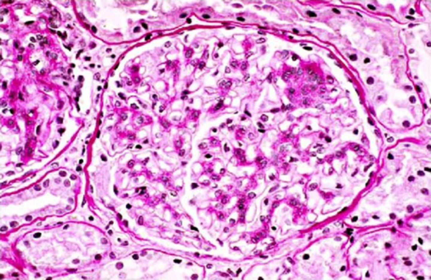
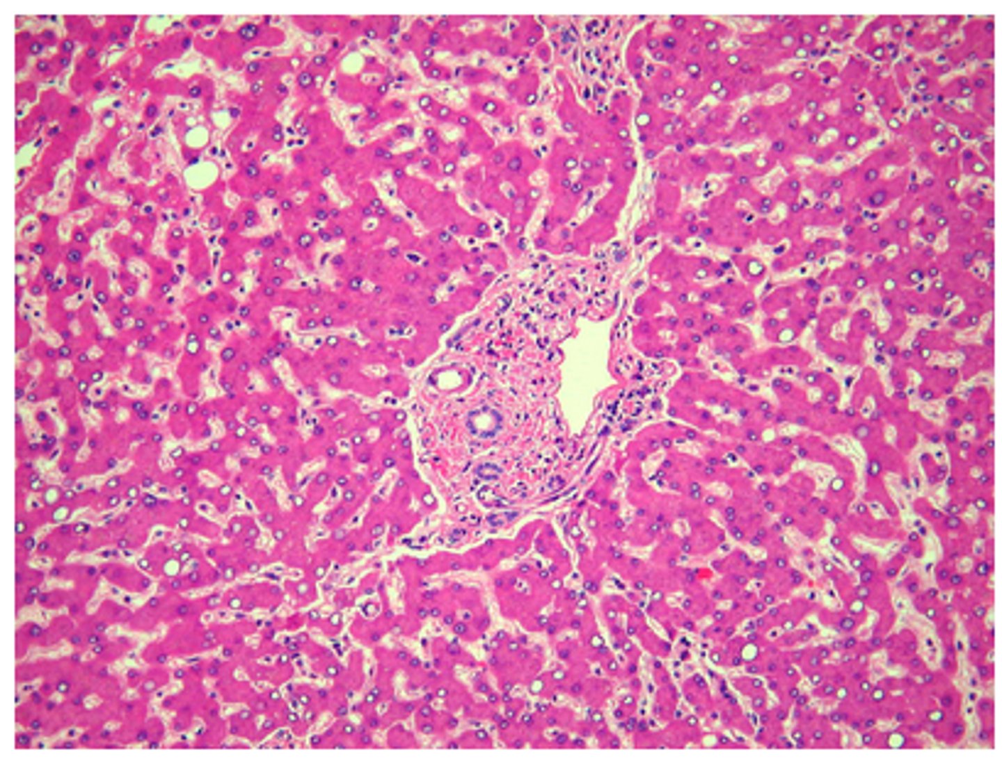
The tissue shown in this image is:
kidney
pancreas
liver
salivary gland
liver
Feedback:
This tissue can be identified as liver by the cords of hepatocytes and by the portal triad in the center of the image.
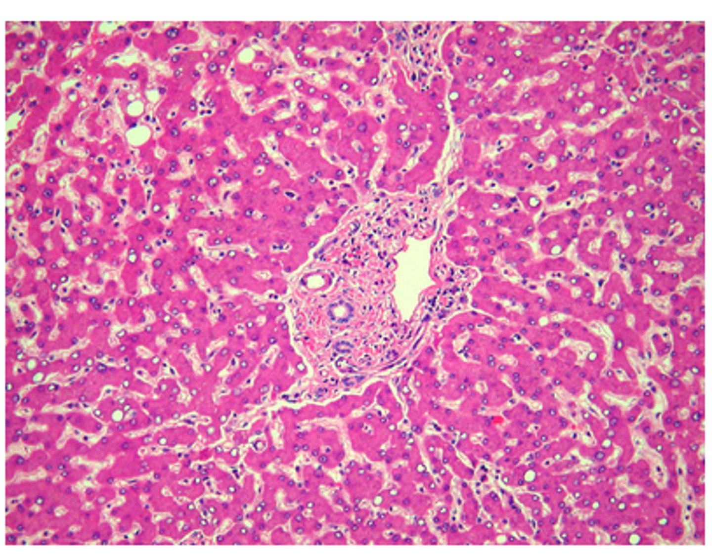
Select the differentiating solution in the Verhoeff-Van Gieson procedure for elastic fibers:
Weigert iodine solution
5% sodium thiosulfate
2% ferric chloride
acid alcohol
2% ferric chloride
Feedback:
In the Verhoeff-Van Gieson procedure ferric chloride serves first as a mordant, then as an oxidizer, and lastly as a differentiator.
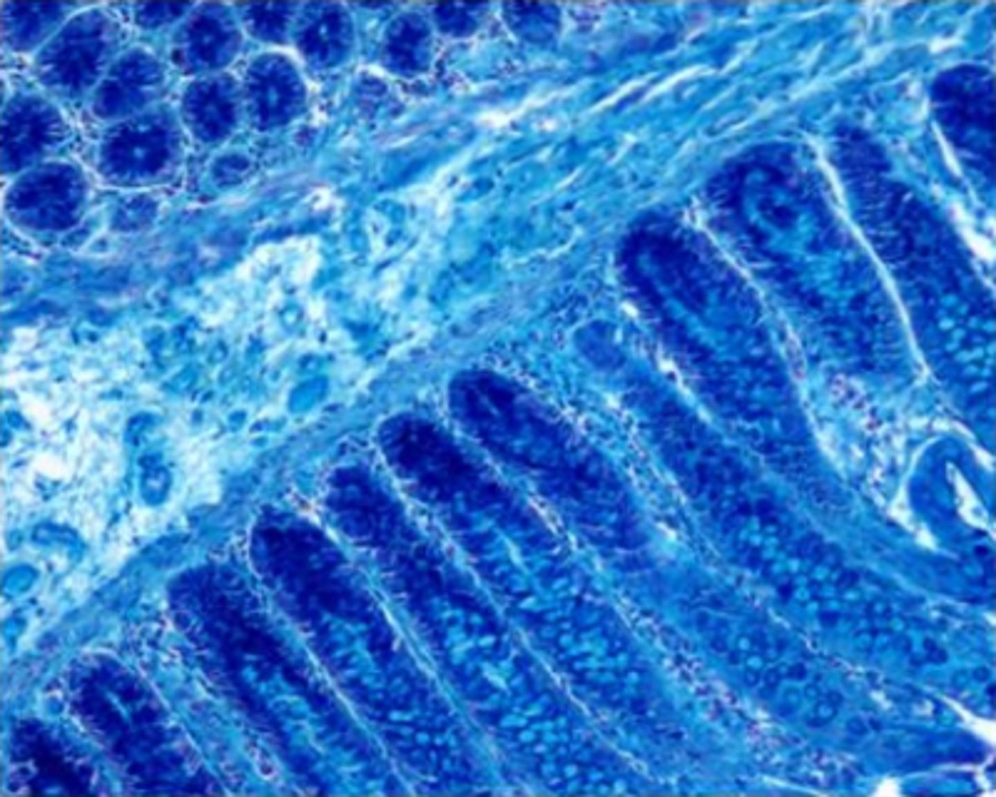
The problem seen in this colloidal iron-stained section is:
lack of counterstaining
marked staining of the submucosa
shortened time of differentiation
solution pH was between 1.5 and 1.9
marked staining of the submucosa
Feedback:
There is marked staining of the submucosa and also the lamina propria. This is improper staining. The counterstain is not the problem; this stain does not require differentiation, and a pH between 1.5 and 1.9 is proper for the colloidal iron stain.
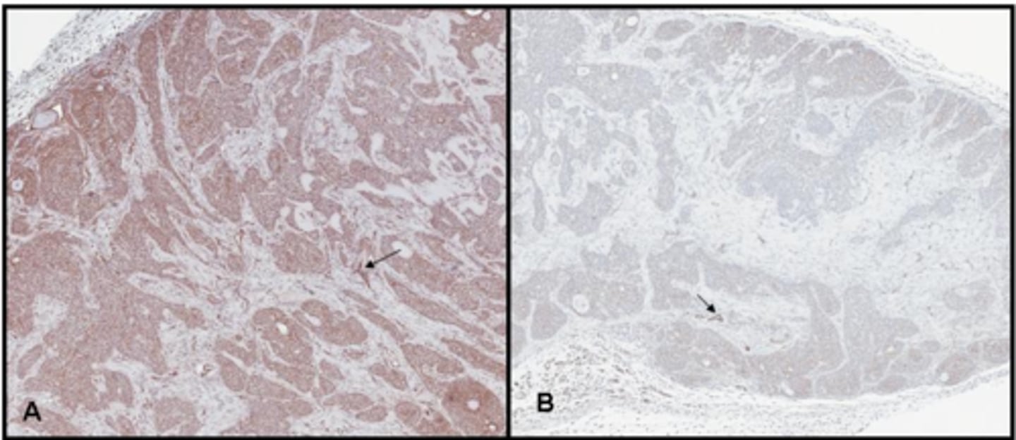
Which of the tissue section images below demonstrates an example of a good quality stain?
A
B
B
Feedback:
Notice that image A has excessive background staining which masks the crisp clean staining of the blood vessels as shown in image B. Excessive background distracts from the true positive and diagnosis can be compromised A good technician is able to recognize background fram true positive staining and is able to troubleshoot and correct the problem.
In Cajal•s method for astrocytes, sections of brain are fixed in formalin that contains:
sodium acetate
ammonium bromide
mercuric chloride
calcium chloride
ammonium bromide
Feedback:
In Cajal•s method for astrocytes, sections of brain are fixed for no less than 2 days and no more than 25 days in formalin ammonium bromide.
This term is used to describe proteins that are neutral but capable of carrying either positive (basic) or negative (acidic) charges which is dependent upon the pH of the solution.
Anionic
Cationic
Amphoteric
Synthetic
Amphoteric
Feedback:
Amphoteric is the term used to describe proteins that are neutral but capable of carrying either positive (basic) or negative (acidic) charges which is dependent upon the pH of the solution.
Cationic refers to a basic dye, or staining properties.
Anionic refers to an acidic dye, or staining properties.
Synthetic referes to a dye that does not occur naturally.
An adenocarcinoma can be described as a malignant neoplasm of this origin.
Glands
Epithelium
Connective tissue
Plasma cells
Glands
Feedback:
An adenocarcinoma can be described as a malignant neoplasm of this glandular origin.
Carcinomas can be described as a malignant neoplasms of epithelial origin.
Sarcomas can be described as a malignant neoplasm of connective tissue origin.
Myelomas can be described as a malignant neoplasm cf plasma cell of the bone marrow origin.
Which of the following is the most likely cause of the problem seen in this colloidal iron-stained section?
lack of properly acidified solution
failure to dialyze the colloidal iron solution
the solution was not properly ripened
use of an incorrect counterstain
lack of properly acidified solution
Feedback:
The colloidal iron solution must be acidified to a pH if between 1.5-1.9. The Mueller colloidal iron solution does not require dialyzing and is the solution most frequently used today.
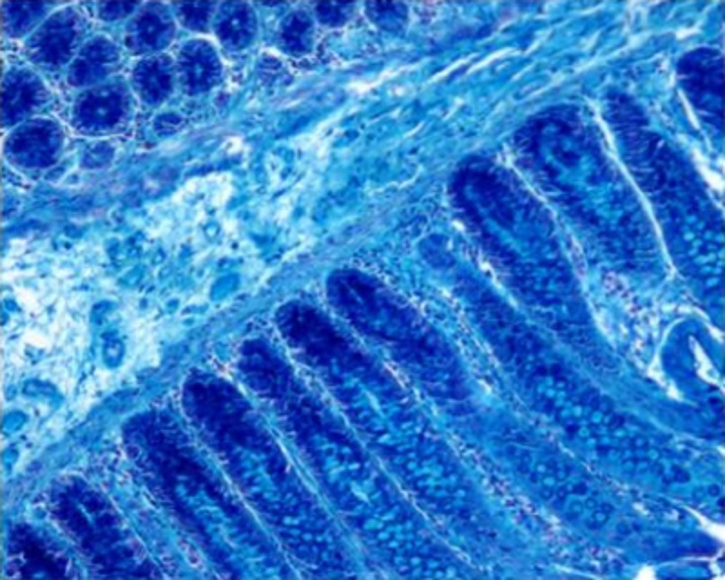
*The tissue shown in this image would most likely be chosen as a control for:
amyloid
mucicarmine
PAS with and without digestion
alcian blue with and without hyaluronidase
PAS with and without digestion
Feedback:
This is a section of liver and is frequently chosen as a control for glycogen demonstration (PAS with and without diastase). It would not be a control tissue for the other techniques; there is no amyloid or mucin present.
Two main categories of cytological specimens are:
FNA and gynecol ogical
Cervical and abdominal
Gynecological and non-gynecological
Fresh and fixed
Gynecological and non-gynecological
Feedback:
Cytology specimens are categorized into gynecological and non-gynecological specimen groups.
*Select the proteins that form the thick and thin filaments of skeletal muscle fibers:
fibrin and fibrinogen
collagen and tropocollagen
actin and myosin
thrombin and prothrombin
actin and myosin
Feedback:
The contractile unit of skeletal muscle, a sarcomere, is made up of thick filaments containing myosin and thin filaments containing actin.
Select the methods can be used to increase the rate of staining.
Increase the temperature of the dye
Decrease the temperature of the dye
Increase the concentration of the dye
Increase the temperature of the dye
Increase the concentration of the dye
Feedback:
Increasing the temperature and concentration of a dye are methods that can be used to increase the rate of staining.
Solutions that are warmed, increase movement of the dye molecules, therefore, allowing more dye molecules to have the chance of interacting with the tissue. Likewise, solutions that are cooled, decrease movement of the dye molecules, decreasing movement of the dye molecules, therefore, inhibiting the dye molecules to have the chance of interacting with the tissue.
Decreasing the concentration of the dye will decrease the rate of staining.
Which of the following stains differentiate between neutral and acidic mucosubstances?
Alcian glue with Hyaluronidase
Alcian Blue 1.0
Alcian Blue 2.5
Alcian Blue Periodic Acid Schiff
Alcian Blue Periodic Acid Schiff
Feedback:
Alcian Blue procedures stain acid mucins. Periodic acid Schiff demonstrates neutral mucosubstances. When the Alcian Blue procedure is followed with PAS, it differentiates between neutral (magenta color) and acid (blue color) mucins.
The dye alcian blue is:
anionic
cationic
neutral
amphoteric
cationic
Feedback:
Alcian blue is a cationic (basic, positively charged) dye used to stain mucopolysaccharides by forming electrostatic bonds with certain tissue polyanions containing either sulfate or carboxyl groups.
Which of the following is the most likely cause of excessive background staining with the PAS?
abbreviated treatment with periodic acid
prolonged washing following Schiff reagent
overused Schiff reagent
fixation with glutaraldehyde
fixation with glutaraldehyde
Feedback:
Glutaraldehyde is a dialdehyde and may leave an aldehyde group free to react with Schiff reagent. Abbreviated treatment with periodic acid and averused Schiff reagent will cause a weak staining reaction, and slides shauld be washed very well following the Schiff reagent.
A modified phosphotungstic acid-hematoxylin procedure can be used to demonstrate:
endothelial cells
glial fibers
Nissl substance
Schwann cells
glial fibers
Feedback:
A modified phosphotungstic acid-hematoxylin procedure can be used to demonstrate glial fibers, neurons and myelin.
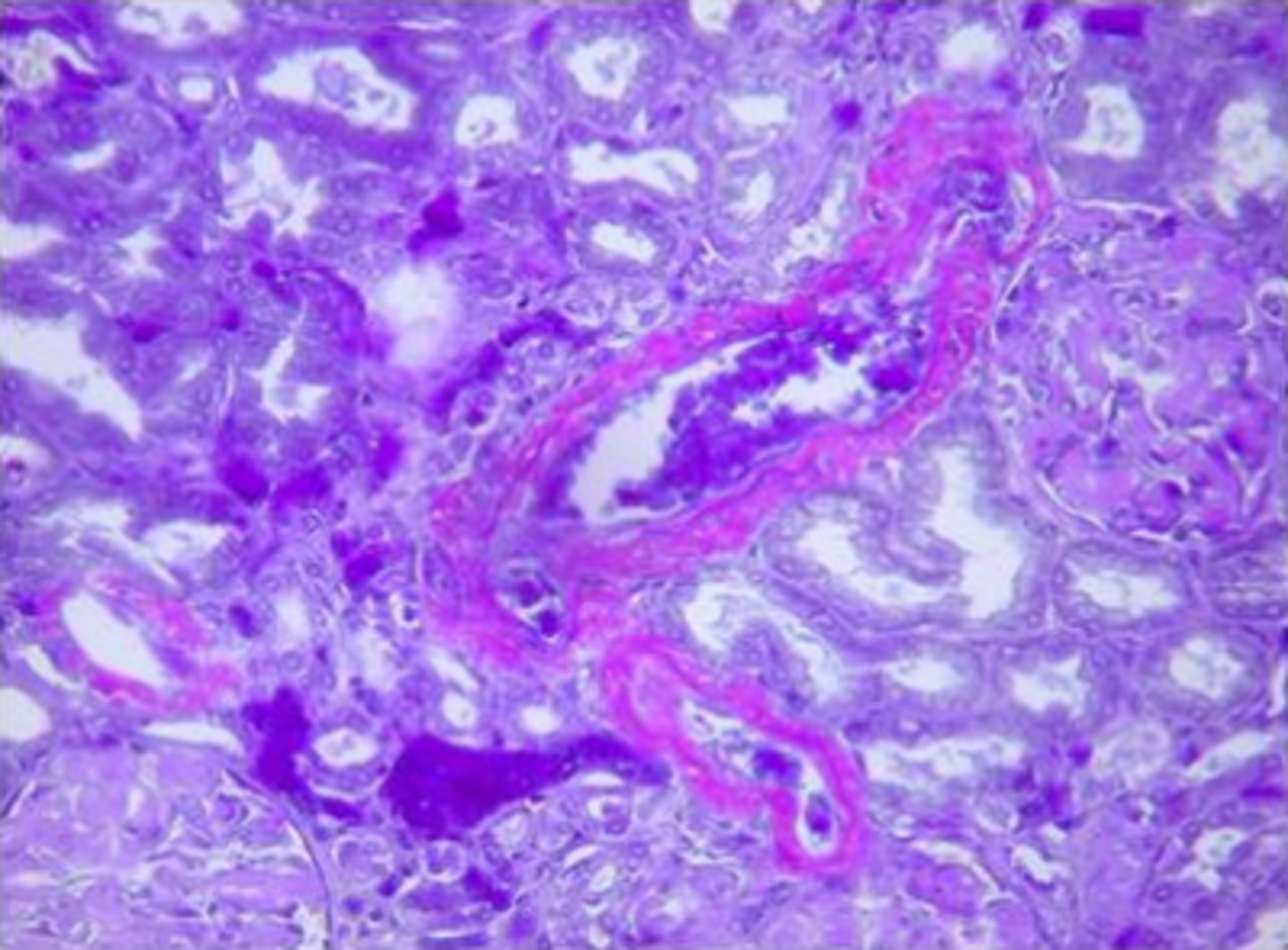
The substance stained purplish-violet in this image is most likely:
basement membrane
acidic mucosubstance
amyloid
collagen
amyloid
Feedback:
Amyloid is deposited in the blood vessel walls and stained purplish-violet by the crystal violet procedure. Some deposits can also be seen in the glomerulus of this kidney.
Human papilloma virus (HPV) is an RNA virus that causes proliferation on epidermal and mucosal surfaces.
True
False
False
Feedback:
HPV is a DNA virus that causes proliferation on epidermal and mucosal surfaces.
The neuron cell body should be colorless prior to counterstainr if the Luxol fast blue stain has been differentiated properly.
True
False
True
Feedback:
Proper Luxol fast blue differentiation will result in a colorless background and blue myelin. Therefore, a counterstain such as hematoxylin or Cresyl echt violet should be applied for neuron demonstration.
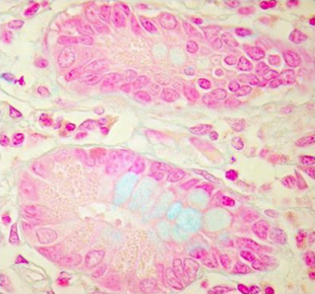
Corrective action for the problem seen in this image would involve all of the following with the exception of:
preparing a new alcian blue solution
ensuring that the solution has a pH of 2.5
washing well after counterstaining
ensuring that hydration is adequate
washing well after counterstaining
Feedback:
The alcian blue solution is most likely depleted or weakened, and should be replaced. It should have a pH of 2.5 and sections should be well-hydrated. Although washing well after nuclear fast red counterstaining is necessary, it is not the problem seen in this section.
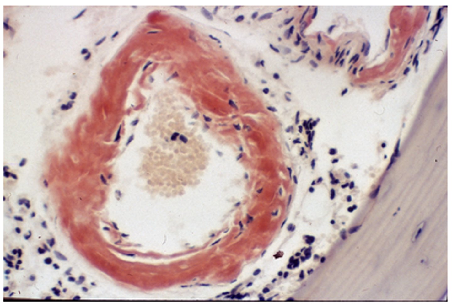
*Another technique that could be used to demonstrate the substance stained red in this image is:
mucicarmine
Sudan black B
Thioflavin T
auramine-rhodamine
Thioflavin T
Feedback:
Thioflavin T will also demonstrate amyloid, the substance stained red in this image. Mucicarmine stains acidic mucosubstances; Sudan black B stains lipids, and auramine-rhodamine stains acid-fast bacilli.
On a periodic acid-methenamine silver stain, the tubular basement membranes are stained black, but the glomerular basement membranes show only faintly gray staining. This indicates that the sections:
were overstained
were underoxidized
should have been impregnated longer
should have been stained at a lower temperature
should have been impregnated longer
Feedback:
Tubular basement membranes will blacken prior to the glomerular capillary basement membranes. For satisfactory staining,the glomerular basement membranes should be used for timing the stain.
Oligodendroglia are cells that function in the production and maintenance of myelin sheath surrounding:
axons
neuroglia
microglia
astrocytes
axons
Feedback:
Oligodendroglia produce, and appear ta maintain, the myelin sheath surrounding many axons
Fat or lipid staining is an example of this type of staining reaction.
Physical
Chemical
Physical
Feedback:
Although most stains in the histopathology laboratory are known to stain by the mechanism of a combination of both physical and chemical reactions, lipid or fat stains are known to stain solely by physical reaction.
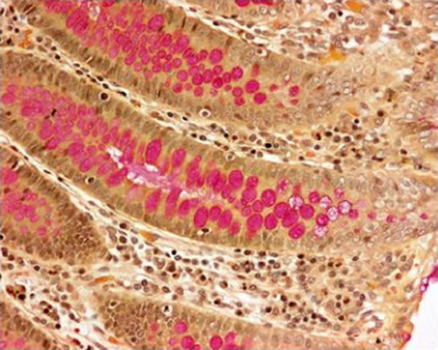
The staining technique shown in this image is:
Congo red
mucicarmne
PAS
aldehyde fuchsin
mucicarmne
Feedback:
Mucicarmine has stained the mucin in the goblet cells red and the typical counterstains of iron hematoxylin and metanil yellow have been used. Congo red would not stain the goblet cells and aldehyde fuchsin would stain them purple. Although the PAS would stain goblet cells rose, the counterstains are not commonly used with the PAS, but are used with mucicarmne.
Acidic mucosubstances can be demonstrated by the Prussian blue reaction following treatment of the sections with:
diastase
hyaluronidase
dimedone
colloidal iron
colloidal iron
Feedback:
Acidic mucosubstances attract ferric cations in a colloidal iron and solution and then may be demonstrated with the Prussian blue reaction. Diastase is an enzyme for the breakdown and subsequent demonstration of glycogen; hyaluronidase is used for the digestion and subsequent demonstration of connective tissue mucosubstances; dimedone is used in organic chemistry to determine whether a compound contains an aldehyde group.
Melanin is normally found in the:
kidney
liver
skin
stomach
skin
Feedback:
Melanin occurs normally in the retina, brain and particularly skin as yellow-brown to black granules.
The primary diagnostic use of alcian blue staining techniques is to:
detect the presence of neutral mucins in tumors
bind to basic groups present in mucosubstances
exhibit the metachromatic properties of mucosubstances
identify and classify acidic mucins
identify and classify acidic mucins
Feedback:
Alcian blue is used at different pHs and with enzyme digestion techniques to identify and classify acidic mucosubstances. Alcian blue will not stain neutral mucins, it binds to acidic groups, and it is not a metachromatic dye.
The final intensity of the color produced in the PAS reaction is dependent on the concentration of:
lipoproteins
acid mucosubstances
proteaglycans
1,2 glycol groups
1,2 glycol groups
Feedback:
In the PAS reaction, adjacent glycol groups are oxidized to aldehydes and then reacted with Schiff reagent, thus the intensity of color developed will depend an the concentration of these groups. Lipoproteins, acid mucosubstances, and pratecglycans will nat react with the PAS reaction.