ATAR Psychology - Sem 1 Unit 1 Exam (Unfinished)
1/201
Earn XP
Description and Tags
- biopsychology - lifespan psychology - developmental psychology - science inquiry
Name | Mastery | Learn | Test | Matching | Spaced |
|---|
No study sessions yet.
202 Terms
Two Types of Nervous Systems
Two Types of Nervous Systems:
Central Nervous System (CNS): Includes the brain and spinal cord.
Peripheral Nervous System (PNS): Consists of nerves outside the CNS.
The CNS consists of:
the brain
spinal cord
interneurons, which are the neurons within the brain and spinal cord
The functions of the CNS
The CNS is the control centre for the whole nervous system. The place where incoming messages are processed, and where outgoing messages are initiated.
The PNS consists of:
the nerves that connect the CNS with receptors, muscles, and glands, so nerves that carry messages in and out of the CNS.
The two main types of neurons: PNS
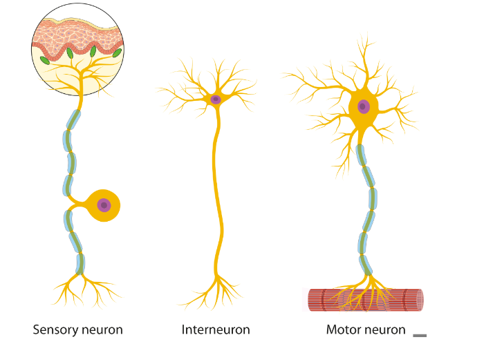
Sensory Neurons: carry messages from receptor cells into the CNS.
Motor Neurons: carry messages away from the CNS to muscles and glands.
Brain
Composed largely of brain cells that receive and transmit electrical and chemical signals all over the body.
It is the control centre - control and decision making centre of the CNS
It is the largest and most important part of your central nervous system.
Spinal Chord
Extension of brain stem
A thick bundle of nerves that runs down the middle of your spine.
It splits off into a network of nerves that run all over your body.
Channels communication between brain and peripheral nerves; coordinates reflexes.
Neuron
Neurons or nerve cells carry electrical messages all over the body
Somatic Nervous System
Carries nerve impulses (from skin, ears, eyes) to the CNS (via sensory neurons) and from the CNS to skeletal muscles and skin (via motor neurons)
Produces voluntary movement - conscious control
Sensory functions gather info from sensory receptors across the body sending info to the brain via the spinal cord
Sensory Nervous System: Sensory Input
Motor Sensory System: Motor Output
Autonomic Nervous System
Carries nerve impulses to/from the CNS to/from internal organs and glands
Regulate basic bodily functions (e.g., HR, BP, respiration, digestion, body temperature, pupil diameter, release of energy, defecation, urination, sweating)
Self regulating/involuntary functions - operates without conscious control
Concerned with maintaining a constant internal environment
Will function when we experience stress, fear or anger
2 further subdivisions
Sympathetic
Parasympathetic
SYMPATHETIC - AROUSAL
Dominates when under stress/threats (either by physiological or psychological stimuli)
Activates internal muscles to act quickly (fight or flight response)
Mobilises body for action, involves energy output - body expends energy
Responses
Increasing heart rate, dilation of pupils (allow more light in)
Digestion slowed
Release of endorphins (pain relieving hormones) to prepare for injury
Release of hormones (epinephrin) for energy
Change in electrical properties of skin (GSR)
Parasympathetic - Calming
Maintains the steady state of balanced normal functioning = homeostasis (maintenance of a constant internal environment)
Regulation of blood sugar levels, waste elimination
Releases hormones (e.g. acetylcholine) to slow heart rate
Restores body to calmness after threat
Decreasing HR, constricts pupils (less light in)
We calm the body to conserve and maintain energy
The Key Parts of a Neuron
Cell Body (Soma): Contains a nucleus that controls the activities of the neuron.
Dendrites: Extensions of the cell body that receive electrochemical messages from other neurons and transmit them toward the cell body for processing.
Axon: The long projection of a neuron or long fiber, that carries the electrochemical message away from the cell body.
Axon Terminals: The enlarged endpoints of axon branches where they effectively make contact with the dendrite of the next neuron, or effector (i.e. muscle, gland, etc) via a synapse. Axon terminals store neurotransmitters and release them into the synapse.
The myelin sheath is a fatty substance that covers the axon of some neurons, helping to speed up the transmission of nerve impulses.
The Three Types of Neurons
Sensory neurons: Transmit sensory information from receptors to the central nervous system. Myelinated
Motor neurons: Carry signals from the central nervous system to muscles and glands. Mylienated
Interneurons: Connection between sensory neurons and motor neurons, found in the CNS.

Neural Transmission
The transfer of information between neurons is called neural transmission.
Synapse
the junction that exists between neurons, so neurons don’t actually touch one another.
The Synapse Consists of
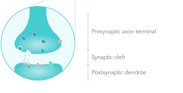
The synapse consists of a presynaptic terminal, synaptic cleft, and postsynaptic terminal.
synaptic cleft is just the fluid gap or space between the two neurons.

Synaptic Axon Terminals
The presynaptic axon terminal is that part of the preceding neuron - where the electrical impulse is coming from i.e., the sending neuron.
The postsynaptic dendrite is that part of the next neuron - where the electrical impulse is going to i.e., the receiving neuron.
Action Potential + Neruotransmitter=
‘Electrochemical’ Signal!
Neurotransmitter
A group of chemical substances released by neurons to stimulate, or not stimulate, other neurons (or muscle or gland cells).
Neurotransmitters diffuse across the synapse - thus relaying information from one neuron to the next. They're often called 'chemical messengers'.
NEURON CONTEXT
An action potential (an electrical impulse) travels along the axon of the pre-synaptic neuron, toward the axon terminals.
Within the axon terminals of the pre-synaptic neuron, the action potential triggers vesicles located in the terminals to release neurotransmitters (chemicals).
These neurotransmitters diffuse across the synaptic cleft and bind to specialised receptor sites on the dendrite of the post-synaptic neuron.
If the neurotransmitter is 'excitatory' (e.g., noradrenaline), then the post-synaptic neuron is more likely to fire off an electrical impulse.
If the neurotransmitter is 'inhibitory' (e.g., serotonin ), then the post-synaptic neuron is less likely to fire off an electrical impulse.
At the dendrites, the chemical message is converted back into an electrical impulse and the process of neural transmission occurs again.
Electrochemical Signal
an 'electrical part' moving along the axons of neurons (the action potential), and the 'chemical part' is the neurotransmitter moving across the synapse.
Main Parts of the Brain
Hindbrain
Midbrain
Forebrain
The Hindbrain
Located at the base of the brain near the back of the skull
Often referred to as the 'brain stem'.
responsible for lower-brain functions that occur without any conscious effort including:
control of basic autonomic survival functions (Medulla)
coordination of voluntary movements (Cerebellum)
Medulla Oblongata: Functions and Location
located at the base of the brain stem, in front of the cerebellum.
relays information between the spinal cord and the brain
regulates vital involuntary bodily functions by communicating with the autonomic nervous system (ANS)
Cerebellum: Functions
helps coordinate voluntary movement and balance by relaying motor information to and from the cerebral cortex
and helps to coordinate the timing and the force of the different muscle groups that act together during a voluntary movement, so that we have smooth limb and body movements.
is believed to play a role in motor learning, where motor skills are improved through practice
The Midbrain
A very small area in the middle of the brain that connects the hindbrain and the forebrain.
The midbrain plays a crucial role in processing information related to hearing, vision, movement, pain, sleep and arousal.
includes the:
Reticular Formation
Reticular Formation: Functions and Location
extends throughout the length of the brainstem, from the spinal cord to the midbrain
stimulates the brain by bombarding it with important sensory information, which keeps the cerebral cortex active and alert
nerve fiber filters into important and unimportant
also stimulate the reticular formation to send its own nerve impulses up towards the cortex, arousing the cortex to a state of alertness and activity
The Forebrain
It is the largest, most complex and highly developed region of the brain.
It contains a variety of structures that are responsible for our most complex processes, including emotions, motivations, sensations, perceptions, learning, memory and reasoning.
Includes the:
Hypothalamus
Thalamus
Hypothalamus: Functions and Location
peanut sized structure located just below the thalamus
maintains homeostasis in the body
regulates the release of hormones that help achieve particular physiological state by connecting the nervous system to the endocrine system
by the release of hormones, influences behaviours associated with basic biological needs, such as hunger and thirst
controls the brain’s internal ‘body clock’; regulate circadian rhythms, coordinate sleep-wake cycle
regulates our appetite, thirst and body temperature
Thalamus: Functions and Location
two small egg-shaped structures, positioned in the centre of the brain, on top of brain stem
it acts as a relay system for sensory messages on their way to the cerebral cortex
it conducts motor signals and relays information from brain to the cortex
coordinates shifts in consciousness such as waking up and falling asleep
regulates itself, focusing on more important inputs and stimuli, filtering irrelevant stimuli, works with the reticular formation like an on-off switch
what is cerebral cortex
the outer layer of the brain's cerebrum
Left Hemisphere Consists
controls language; spoken, reading or writing
Broca’s areas: helps in production of speech to produce understandable sentences
Wernicke’s area: understanding of language
analytical thinking, sequential or linear processing, logical reasoning, mathematical ability.
Right Hemisphere Consists
non-verbal communication: emotional or bodily gestures or expressions that contribute to our understanding of language
spatial skills: recognising patterns, shapes, faces and melodies, movement - dance or musical ability
Contralateral control of the body
how each hemisphere of the brain controls the opposing side of the body
Contralateral control: Left Hemisphere
receives sensory information from the left side of the body
the left side of the cerebral cortex see the right side of the world
controls movement in the right side of the body
Contralateral control: Right Hemisphere
receives sensory information from the right side of the body
the right side of the cerebral cortex sees the left side of the world
controls movement in the left side of the body
Corpus Callosum: Functions and Location
two hemispheres connect by a thick bundle of nerve fibres (white matter connects hemispheres)
ensures both sides of the brain can communicate and send signals to each other
Formally: the corpus callosum physically connects the hemispheres, allowing information registered in one hemisphere to be transferred to the opposite hemisphere for processing.
a combination of sensory, motor and cognitive information is constantly being transferred between hemispheres via this neural highway.
The Four Lobes
Frontal
Parietal
Temporal
Occipital
Frontal Lobe: Pre-Frontal Cortex
higher level functions - our advanced cognitive and executive functions (planning, decision making, problem solving, motivation, attention, etc.)
contributes to our personality, intelligence, and social skills.
predicts possible consequences of actions using memory to control responses.
work with the amygdala and hippocampus to regulate emotions.
Frontal Lobe: Primary Motor Cortex
controls voluntary movement of skeletal muscles
left PMC controls the right side of the body.
right PMC controls the left side of the body.
Frontal Lobe: Broca’s Area (Left Frontal Lobe)
controls the production of articulate (clear and fluent) speech.
how? by controlling facial muscles in the jaw, cheeks, lips, tongue, and even diaphragm, that produce the words.
also associated with hand and arm gestures that accompany speech.
has neural connections to wernicke’s Area.
Parietal Lobe: Somatosensory Cortex
a strip of neurons located at the front of the parietal lobe (next to the primary motor cortex).
it registers and processes sensations detected by the body’s sensory receptors in skin, skeletal muscles, and joints.
touch, temperature, pressure, pain and other somatic (body) sensations relating to muscle and joint movement and the body’s position in space are registered.
enables us to determine where objects are located in space and where our body is positioned in space
Temporal Lobe: Primary Auditory Cortex
registers and processes auditory information received by both ears and integrates it with information from other senses.
right temporal lobe is specialised to process non-verbal sounds.
left temporal lobe is specialised to process verbal sounds that are associated with language.
link to hippocampus, contribute to memory.
Temporal Lobe: Wernicke’s Area (Left Temporal Lobe)
brain area just above the left ear
primarily responsible for understanding language (language comprehension).
receives information from ears and eyes so interpretation of both speech and written words.
has neural connections to Broca’s Area and Primary Auditory Cortex
Occipital Lobe: Primary Visual Cortex
registers and processes visual information transmitted from the retinas of both eyes via the optic nerve.
contains a variety of neurons specialised to respond to specific features of visual information i.e. colour, shape and motion.
sends visual information to other brain lobes for further processing for understanding
visual information arrives at the primary visual cortex in many ‘bits’. assembles the ‘bits’ of information into a whole image or pattern that can be given meaning.
Phineas Gage
Railroad construction foreman in the 19th century.
Suffered a traumatic brain injury when an iron rod pierced his skull.
Damage to his left frontal lobe resulted in significant changes in personality and behaviour.
The case provided evidence for the localisation of brain functions.
Roger Sperry (1959-1968)
Roger Sperry:
Conducted research on split-brain patients in the late 1950s and 1960s.
Demonstrated functional specialisation of the cerebral hemispheres.
Showed that each hemisphere is specialised for certain functions.
His work advanced our understanding of brain organisation and lateralisation.
Walter Freeman (1936-1945)
Walter Freeman, assisted by James Watts, pioneered the frontal lobotomy procedure in the USA during the 1930s to address severe mental health conditions and overcrowding in psychiatric institutions.
Freeman believed that mental illness stemmed from excessive self-awareness and overactive emotions, attributing these symptoms to the thalamus as the center of human emotion.
The procedure aimed to sever neural connections between the thalamus and the pre-frontal cortex to eliminate excessive emotions and stabilise personality.
Electroencephalogram (EEG)
measures the electrical activity of the brain (brain waves) using small metal discs (electrodes) that are attached of the scalp.
the frequency ( measured in hertz) and amplitude (measured in microvolts) of various types of brain waves differ.
records changes in the amplitude and frequency of brainwaves.
detect abnormal electrical activity within the brain, such as that produced during an epileptic fit.
Low and quick conduct
Computerised Tomography (CT)
are x-rays of the brain that produce an image of the brain structure.
a rotating x-ray beam moves 360 degrees around the patient whilst taking multiple x-ray images
so, produces a single static image (still picture) that. is 2D, but many of these are pieced together to produce a 3D reconstruction.
can produce 2D or 3D images of the inside of the body - known as computerised axial tomography scan (CAT scan)
Magnetic Resonance (MRI)
technique involving magnetic fields.
use strong magnetic fields and radio waves to produce a detailed image of the brain structure.
MRI produces image of organs, tissue, or bones - the body’s interior anatomy.
An MRI of the brain and spinal cord can reveal evidence of brain injury: stroke, blood vessel damage, and spinal cord injury.
produces a single static image that is 2D
Functional Magnetic Resonance Imaging (fMRI)
technique involving magnetic fields
operates in the same principle as MRI but is a specialised form of MRI
used to examine the brain’s functional anatomy, meaning the part of the brain that handles critical functions.
so measuring blood oxygen levels to indirectly measure neural activity.
used to show specific brain areas when completely physical or intellectual tasks
helps doctor assess the effects of a stroke or other conditions affecting the brain or spinal cord.
The Ethical Guidelines
Protection from Harm - Physical and Psychological
Informed Consent
Withdrawal Rights
Deception
Confidentiality
Privacy
Voluntary Participation
Debriefing
Protection from harm - Physical and Psychological
Researchers responsibility to protect participants physical and psychological welfare
If participants does encounter distress researcher stop the experiment and provide participant access to counselling
The experimenter ensure they act professionally and with integrity at all time
Informed Consent
Researcher must obtain written informed consent from each participant
Under the age of 18 or legally unable to give consent, parent must complete the consent form
Consent forms must inform the participants about their rights (i.e withdrawal rights)
Participants (Parents/Guardians) must be informed about the true nature and purpose of the experiment
Withdrawal Rights
Participants has the right to withdraw from experiment at any time without any negative consequence
Also right to withdrawal their results
Withdrawal rights must be explained to each participants before beginning the research
Deception
Misleads or withholds information from the participants
Only permissible in some cases where giving participants information might influence their behaviour affecting the accuracy of the results
When used there must be no foreseeable harm to participants must be thoroughly debriefed at the end
Confidentiality
How
Researchers ensure information collected during the research is protected and remains private
Participants results cannot be made available to anyone outside the study unless participants consent has been obtained
Participants personal information is not identified in the results
Privacy
What
Collecting personal information that is relevant to the research and accessed by those who have permission
Cannot disclose personal information unless informed consent has been obtained
Voluntary Participation
A participant must be willing to take part or not in an experiment
Must not experience any pressure or coercion to participant
Debriefing
Must be debriefed
Must correct any mistaken attitudes or beliefs and explain any deception
Provide access to information, results and conclusions, and provide access to support through counselling
The Three R’s of Animal Ethics
reduction: comparable levels of information from the use of fewer animals
refinement: methods that alleviate or minimise potential pain and distress
replacement: methods that permit the given purpose of an activity or project to be achieved without the use of animals or with the use of non-sentient animals (those that lack a nervous system. Simulations/models also.
Identify the aim of the research
The aim is the purpose of what you are going to investigate. It is a statement that describes the purpose or reasons for why we are conducting an experiment.
Develop a research question based on the aim
A research question should express the exact question you are trying to answer with your research, including the population of interest.
Hypothesis
A testable statement that gives the relationship between both the independent and dependent variables.
You should be able to recognise the independent and dependent variables within a hypothesis.
Directional and Non-Directional (Quantitative)
Inquiry Question (Qualitative)
Hypothesis: Directional
Gives a prediction of which way results will go. Makes a prediction about the expected results of a study (that's the direction part).
For example: "It is hypothesised that students who meditate will have higher semester one exam results than those students who do not meditate".
Hypothesis: Non-Directional
States that there will be a difference between the results.
For example: "It is hypothesised that there will be a difference in the memory score for those participants that drank caffeine and for those participants who did not drink caffeine."
Inquiry Question
Qualitative inquiry questions are usually vague and look at exploring relationships.
For example: "What is the relationship between cigarette intake and mental well being?"
Types of Variables
Independent
Dependent
Controlled
Extraneous - participant, environment, researcher
Independent Variable
The independent variable is the factor that is being changed or manipulated by the researcher. We see the effect of this variable.
Dependent Variable
The dependent variable is the variable that is dependent on the independent variable and is the variable that is being measured (as a result or outcome).
Controlled Variable
Variables that are kept the same (constant) across the experiment.
Extraneous Variable
Unwanted factors that may impact/affect the dependent variable.
participant (emotions and personality)
environmental conditions (what is going on around the experiment - temperature, humidity)
researcher effects (researcher influences the experiment and participants behaviour).
Confounding Variable
variables that impact the dependent variable and also have a casual or correlational relationship with the independent variable.
can alter the relationship between independent and dependent variable and can complicate results making them difficult to interpret
possibility for someone extraneous variables that are not controlled to become confounding variable
happens if researchers do not control participants
Physical Development
advancements and refinements of motor skills (one's ability to control their bodies and voluntary bodily movements)
Includes:
•Gross motor skills: Require whole body movement and involve large muscles of the body
•e.g. walking, running, throwing, lifting, learning to balance etc
•Fine motor skills: Use small muscles in hand or wrist for precise movement
•e.g. grasping, holding, pinching, putting on shoes, cleaning teeth, picking up a pencil etc
Cognitive Development
the development of the ability to think and reason, learn, and process information.
Involves language, imagination, problem solving, and memory.
Social and Emotional Development
Social Development: one’s ability to create and sustain meaningful relationships with others.
Emotional Development: one’s ability to express, recognise and manage one's emotions, as well as respond appropriately to other’s emotions.
Neuroplasticity
Ability of neurons and neural connections growing and recognising
So, changing in structure and function due to response to stimuli experience, and injury.
Includes
Developmental Plasticity
Adaptive Plasticity
Developmental Plasticity
Most rapid changes occur in infancy and Adolescence - Brain Grows
Turns neural connections in response to environmental stimuli.
Adaptive Plasticity
Continues through the lifespan
Neural connections in the brain are altered/recognised in response to learning new information or in response to injury to compensate, for lost functions and to take advantage of remaining functions
Stages of Plasticity During Infancy
Proliferation
Migration
Circuit Formation
Synaptic Pruning
Myelination
Proliferation
is the growth and division of cells, including neurons, that leads to the increase in total cell number.
while most neurons are already formed when the infant is born, some neurons are still created during infancy
Migration
while an infant is born with around 100 billion neurons, there are still neurons being generated after birth from deep inside the brain.
newly generated neurons move throughout the brain until reaching their final positions; this positioning allows for connections between neurons to be made.
neurons migrate by following chemical trails laid down by other neurons or by moving along scaffolding fibres in the brain.
research has shown that the migration of neurons ends around the age of five months.
Circuit formation
after neurons migrated, they are able to form neural circuits whereby neurons send electrochemical messages between each other. These connection can be within clusters of neurons, as well as over larger distances within the brain.
during infancy; neural infancy develop rapidly, especially in primary sensory cortex and primary visual cortex
Synaptic pruning
as infants are born with more neurons than required, neurons that do not form active neural connections with other neurons die.
synaptic pruning increases the efficiency of the nervous system by allowing remaining neural connections to strengthen and grow in complexity.
Myelination
a fatty substance called myelin starts growing over the axons of neurons
myelination contributes to the dramatic brain growth typical in infants
myelination begins in the spinal cords, then in the hind brain, forebrain, and finally, in the peripheral nervous system.
Brain Plasticity Adolescence: Cerebellum
cerebellum continues to grow, approximately twelve years of age in females, and fifteen in males
increase in volume, synaptic pruning affects behaviour and emotion, the cerebellum taking up 10% brain volume, 50% of the total neurons in the brain
cerebellum linked to decision making, reward learning, motivation, emotional control, and processing mood
understandable teengers display impulsive decision making and have difficulties regulating emotions
Brain Plasticity Adolescence: Amygdala
amygdala grows in adolescence partly due to pubertal changes
adults, pre frontal cortex regulates amygdala, but adolescence pre-frontal cortex developing, instead of pre-frontal cortex leading actions based o rational and logical thinking.
volatile amygdala guides many of the automatic actions.
sensitive to emotional stimuli, leads to teenagers to misinterpret emotions and consequently get into accidents, behave inappropriately without thinking, such as fear, aggressive behaviour towards others.
Brain Plasticity Adolescence: Corpus Callosum
corpus callosum increases during adolescence through myelination, rather than growing uniformly the structure, various regions grow at different rates. suggests hormonal surges may account for these growth rates.
neural networks within the corpus callosum strengthen a stronger connection between two hemispheress, and behavioural and emotional regulation improves.
Brain Plasticity Adolescence: Frontal lobe
The control of voluntary behaviour is not a characteristics of teenagers as theses lobes are the last regions to mature.
the frontal lobe is not fully myelinated until the age of 30 meaning teenagers have less white matter compared to adults
myelinated neurons improve connectivity between parts of the brain, and with the frontal lobes not yet fully connected, the reduced ability to integrate information from brain region affects cognition and emotional process.
Brain Plasticity Adolescence: Pre-frontal cortex
pre-frontal cortex continues to undergo myelination during adolescence leads to increase white matter
synaptic pruning continue, reducing the amount of grey matter and allowing for increasingly complex and efficient connection to be created in the brain
synaptic pruning starts back of the brain and continues forward to the pre-frontal cortex being the last to develop. responsible for problem solving and the ability to predict the consequences of behaviours by referring to past experiences.
this makes it easier to understand why teenagers assess potential risks and ending in risky and dangerous behaviours.
process of schema formation
assimilation
accommodation
equilibrium
disequilibrium
assimilation
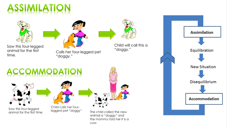
assimilation is fitting new experiences or new information into our current schemas/understanding the of the world.
OUR SCHEMA REMAINS THE SAME
eg. three year old Shaun had simple schema for a ball that incorporated everything that was round.
assimilated grapes and olives into this schema, called them ‘ball’.
Schema remains the same, just expanded to fit new additions
Equilibrium
When our existing schemas can explain what we perceive around us, we are in a state of equilibrium.
Disequilibrium
when we meet a new situation that we cannot explain, or fit into a pre-existing schema, it creates a disequilibrium .
Accomodation
accomodation is adjusting our current schema/understanding in order to understand new experiences.
accomodation is when new information changes or replaces existing knowledge.
may create new schema’s
as we interact with our world, we construct and modify our schema.
Process of Schema Formation

Piaget: Cognitive Development Stage
Sensorimotor: Object permanence
Pre-operational: Egocentrism, animism, symbolic thinking, centration, seriation
Concrete operational: Conservation
Formal operational: abstract thinking
Sensorimotor (Birth-2 years)
babies develop understanding of world through sensory and motor interactions
child seems to live in the presence; little understanding that things continue to exist if not within sight.
Object Permanence: the concept gained by infants that an object continues to exist even when it cannot be seen.