Alteration in Perfusion: Cardiac Rhythms
 Call Kai
 Learn
Learn Practice Test
 Spaced Repetition
 Match
Match Flashcards
 Knowt Play
1/37

There's no tags or description
Looks like no tags are added yet.
Name | Mastery | Learn | Test | Matching | Spaced | Call with Kai | Chat |
|---|
No analytics yet
Send a link to your students to track their progress
38 Terms
Alteration in Perfusion: Cardiac Rhythms
AV node broken- fall back is the ventricles
SA node is broken fallback is the SA
AV and SA node broken= V tech (the ventricles takes over)
Side note: sodium and potassium need to be equal
If the current starts at the Av node→ A- fib
PR =
Atrial Depolarization (Contraction)
QRS =
Depolarization of Ventricles
T Wave =
Repolarization (Resting) of Ventricles
QT =
Time it takes to complete Ventricular Depolarization & Repolarization
Dysrhythmia
Abnormal, disordered rhythm of the heart
Interruption of automaticity
Dysrhythmia why is it important
Decreases CO and O2 or high 02 or Co2
Can cause clot formation (blood can’t perfuse through,, heart can die)
Can be fatal
Pacemaker Rules
The pacemaker site with the fastest rate will generally control the heart.
Pacemakers Escape Mechanism
The normal pacemaker slows down or fails, and a lower pacing site assumes pace making responsibility.
Pacemaker Rules Inherent Rates
Sinus Node: 60-100 beats/min
AV Node: 40-60 beats/min
Ventricles: 20-40 beats/min Insufficient to sustain cardiac output
Lead Placement for Telemetry Monitoring
Right arm (RA): Near the right shoulder
Left arm (LA): Near the left shoulder
Right leg (RL): Below the lowest rib on the right side of the abdomen
Left leg (LL): Below the lowest rib on the left side of the abdomen
Chest (C,V[voltage]): In the fourth intercostal space, to the right of the sternum
![<ul><li><p><span style="background-color: transparent; font-family: "Times New Roman", serif;">Right arm (RA): Near the right shoulder </span></p></li><li><p><span style="background-color: transparent; font-family: "Times New Roman", serif;">Left arm (LA): Near the left shoulder </span></p></li><li><p><span style="background-color: transparent; font-family: "Times New Roman", serif;">Right leg (RL): Below the lowest rib on the right side of the abdomen </span></p></li><li><p><span style="background-color: transparent; font-family: "Times New Roman", serif;">Left leg (LL): Below the lowest rib on the left side of the abdomen </span></p></li><li><p><span style="background-color: transparent; font-family: "Times New Roman", serif;">Chest (C,V[voltage]): In the fourth intercostal space, to the right of the sternum</span></p></li></ul><p></p>](https://assets.knowt.com/user-attachments/dfb43486-508f-44b2-a43a-45b1d816b75b.png)
Get the Best Picture for Teles and EKG
Clip excessive hair on the chest wall.
Rub skin with dry gauze.
May need to use alcohol for oily skin
Keep away from bony prominences
The positive electrode is the place where the picture is taken
Bipolar Leads are known as
limb leads: this leads are consist of a positive electrode and a negative electrode
Bipolar/Limb leads
Lead I measures the current traveling between the right(RA) and left arm(LA). The right arm is negative(-) and left arm is positive(+)
Lead II current between RA and LL (left leg). RA is negative(-) and LL is positive(+). This gives the most upright P waves.
How long it takes for the heart to contract
Lead III-current between LA(-) and LL(+)
VR, VL, VF
aVR picture from heart looking toward R arm
aVL picture of heart looking toward L arm
aVF picture of heart looking toward L foot
P waves are
upright (positive deflection) and rounded and should look the same
PR interval
beginning of the P wave's upslope to the beginning of the QRS wave.
QRS interval:
Q-first negative deflection after P, R-first upright deflection after the P, S-first negative deflection after the R
T wave
normal is broad and rounded. If there is a QRS then a T wave must be after it.
Time…The 6 second strip
voltage - how hard the heart is contracting
6 sec strips→ gives you heart rate when beats are multiplied by 10
Each small box is 0.04 sec
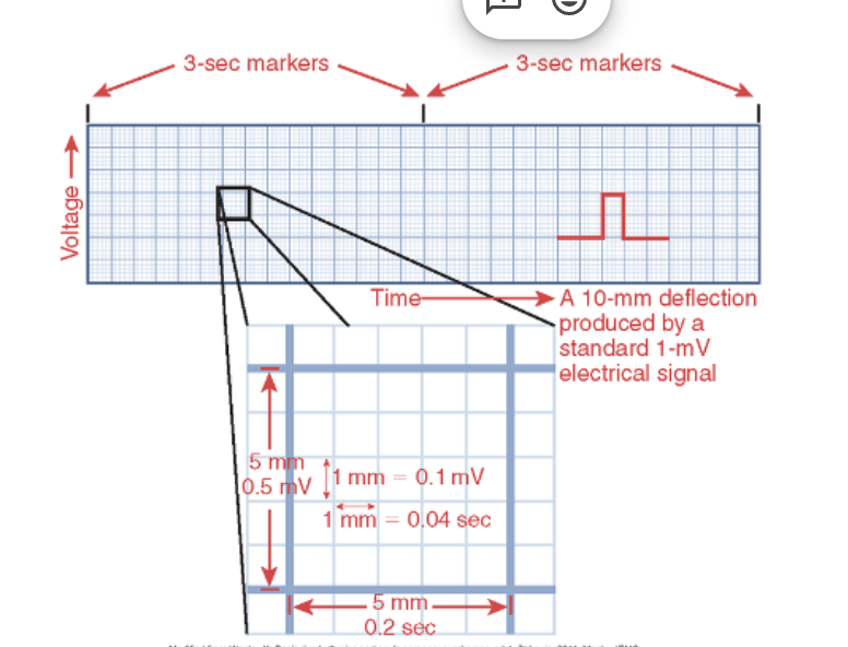
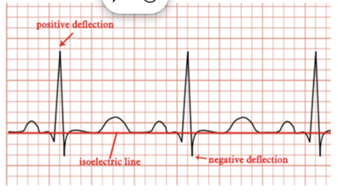
Isoelectric Line
even electricity, important because it helps identify the positive and negative deflections
Pwave (+)
Q wave (-)
S wave (-)
T wave (+)
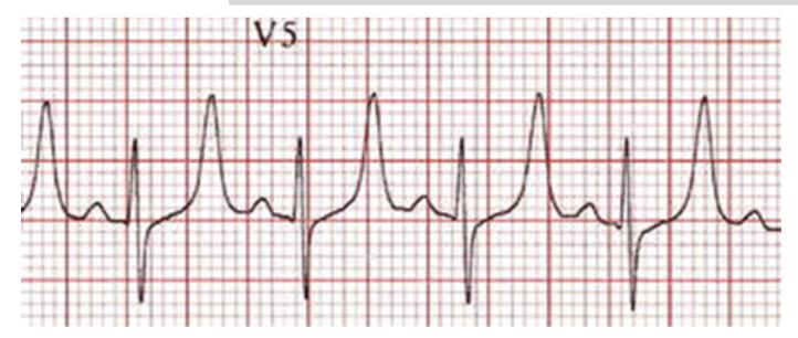
P-R interval measurement
0.12–0.20 seconds (3 to 5 small blocks). It is measured from the beginning of the P wave's upslope to the beginning of the QRS wave.
A longer PR interval could indicate a heart block.

QRS width measurement
is less than 0.12 seconds
Begins at Q and ends at the end of the S wave
Q-T interval measurement
is 0.34–0.43 seconds. A prolonged QTc is defined as QTc > 0.43 sec.
RR interval measurement
0.6–1.2 seconds.
Abnormal…U wave
Small, same direction as T wave. Usually in slow rhythms. May be low K+

Abnormal…T wave
Peaked T waves can be result of hyperkalemia
Too much k+ → high her he K+ then higher the waves
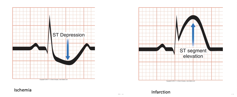
Abnormal…ST wave
ST depression (not enough O2- ischemia)
ST elevation (cell death- infraction)
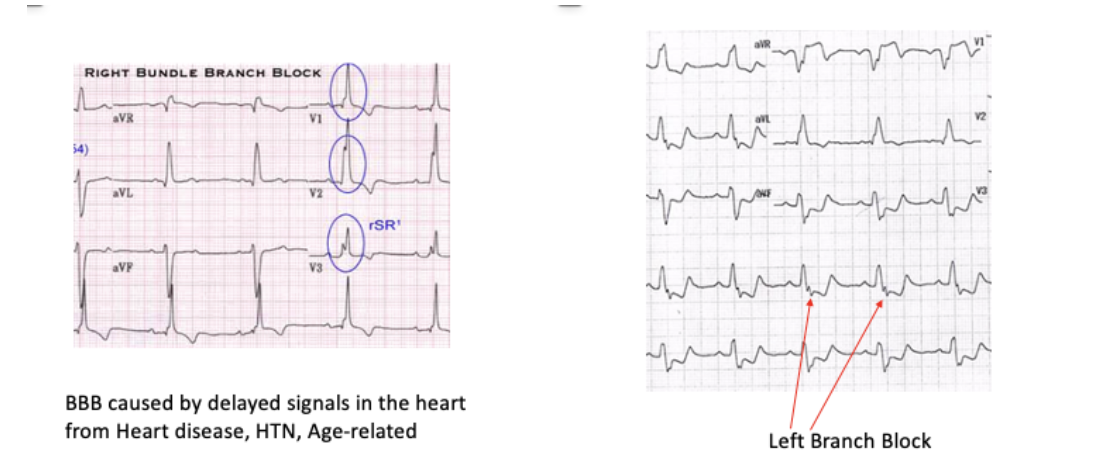
Abnormal…Bundle Branch Block
Right Bundle Branch Block→ the right ventricle effected
Left Bundle Branch Block→ the ventricle is effected

Interference
Not true rhythms because they don’t have a pattern→ means patient movement or loose electrodes
ECG Leads

ECG
Graphic representation of the electrical forces produced within the heart
aVR: upside down is normal
Look at the heart from 12 different views (12 leads ?) → no, but shows 12 views of the heart
Defibrillator and Temporary Pacing

Pacing…Single and Dual Chamber

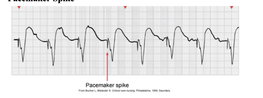
Pacemaker Spike
Pacemaker Malfunction
Pacemaker fires, but no capture occurs.
ECG has a spike not followed by a complex.
Lead damage, battery failure, dislodgement of the electrode, fibrosis at the electrode tip
Failure to Capture Pacer maker malfunctions:
Electrical charge to myocardium is insufficient to produce atrial or ventricular contraction
Which information will the nurse include when teaching a patient who is scheduled to have a permanent pacemaker inserted for treatment of symptomatic bradycardia?
A. The pacemaker prevents or minimizes ventricular irritability.
B. The pacemaker paces the atria at rates up to 500 impulses/minute.
C. The pacemaker discharges if ventricular fibrillation and cardiac arrest occur.
D. The pacemaker stimulates a heart beat if the patient’s heart rate drops too low.