CLIN PATH I: EXAM #2 (PULM - Restrictive Lung Dz)
1/92
There's no tags or description
Looks like no tags are added yet.
Name | Mastery | Learn | Test | Matching | Spaced | Call with Kai |
|---|
No analytics yet
Send a link to your students to track their progress
93 Terms
FEV1 in restrictive lung disease
FEV1 and FVC are decreased (because it is restricting how the lungs work)
Most common feature of restrictive lung disease
infiltration of the lung by inflammatory cells/fluid, causing scarring, fibrosis, and capillary obliteration
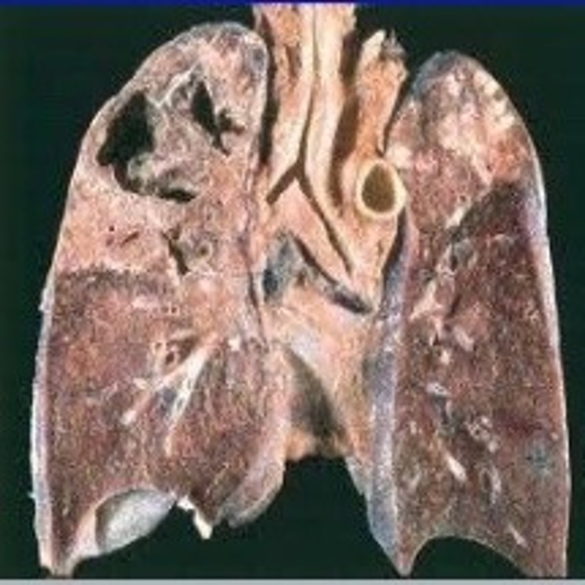
Diffuse lung fibrosis causes:
increased lung elastic recoil (increased FEV1)
decreased compliance and lung volumes (decreased FVC)
V/Q mismatch (impaired gas exchange)
lung parenchyma
space bounded by basement membranes of endothelium and epitheliu
What does the lung parenchyma contain
fibroblasts (mesenchymal cells), collagen/elastin/proteoglycans (cellular matrix molecules), leukocytes
When does idiopathic pulmonary fibrosis often present?
5th decade
Clinical presentation of idiopathic pulmonary fibrosis
chronic inflammation of alveolar walls --> fibrosis/destruction of lung architecture
**impairs perfusion and gas exchange
**progressive dyspnea and cough, as well as digital cyanosis, clubbing, and pulmonary HTN
Major causes of idiopathic pulmonary fibrosis
environmental exposure and smoking
Epidemiology of idiopathic pulmonary fibrosis
uncommon
mostly male
unremitting progression
Which diseases are indistinguishable from IPF in later stages?
Scerloderma, sarcoidosis, hypersensitivity pneumonitis
Median survival of IPF
3 years from dx
Pathophysiology of IPF
1. vascular injury
2. epithelial injury
3: leukocyte influx
vascular injury causes an increase in permeability, so more plasma proteins will get into the extravascular space, causing thrombosis
epithelial injury causes loss of barrier integrity and the release of inflammatory mediators
leukocyte influx into the activate endothelium due to infection
**causes un-uniform remodeling and fibrosis
Stages of IPF
1. Injury of epithelial wall
2. Fibroblasts come in
3. Abnormal repair response (collagen over-production to repair the holes), and fibroblasts get inside alveoli
4. Thickening of alveolar walls (repeated injury)
5.Fibrosis due to chronic inflammation
6. Irreversible loss of basement membrane, disrupted gas exchange, etc. because of the inflammatory response
Clinical manifestations of IPF
progressive dyspnea
dry persistent hacking cough (chronic irritation)
inspiratory crackles (fibrotic airways)
digital cyanosis, clubbing, decreased cap refill
IPF can lead to:
R heart failure and peripheral edema
Imaging for IPF
reduced lung volumes
increased reticular opacities (periphery)
loss of structure definition
honeycombing
Pulmonary function test for IPF
reductions in TLC, FEV1, and FVC, but maintaining a preserved FEV1/FVC ration
What is a significant contributor to exercise-induced desaturation?
diffusion impairment
Arterial pCO2 is typically ________ with restrictive disease
low (due to increased ventilation from hypoxia and increased irritant stimuli)
Grave sign of restrictive lung disease
hypercapnia (inability to maintain adequate alveolar ventilation)
Epidemiology of TB
increase in those infected with HIV, those who travel and emigrate from areas w/ increased TB, homeless people, IVDA
increased bacterial resistance to medications
What causes TB?
infection w/ Mycobacterium tuberculosis (rod shaped acid-fast bacilli)
How does TB spread
droplets (airborne)
Transmission of TB is influenced by:
1. # of bacilli in droplets
2. virulence of bacilli
3. degree of ventilation (outdoors vs. inside w/ bad AC)
4. occasions for aerosolization
The majority of the TB bacilli are trapped in:
upper parts of the airways (where mucus-secreting goblet cells are)
**cilia on the surface of these cells beat the mucus and its entrapped particles upward for removal
Bacilli in droplets that bypass the mucociliary system and reach the alveoli are quickly:
surrounded and engulfed by alveolar macrophages
Phagocytosis by macrophages (innate immunity) results in either:
latent TB
OR
primary progressive TB (active)
What are recruited during TB exposure?
T cells to begin cell-mediated immunity
Patient is ___________ to TB once exposed
sensitized
How do they make a PPD test?
Antigens from killed TB
What do you look for on a CXR w/ TB?
Ghon complex
What is the next defensive step for TB for people with intact immune systems?
formation of granulomas (to limit the replication and spread of the bacteria)
What will the eventual destruction of macrophages in TB cause?
necrotic center
By 2 or 3 weeks, the necrotic environment resembles:
soft cheese (caseous necrosis)
What are Ghon complexes
Lesions undergo fibrosis and calcification, successfully controlling the infection so that the bacilli are contained in the dormant, healed lesions, called Ghon complexes
Ghon complex are more commonly in:
apex of lung
What happens to TB lesions in people with bad immune systems?
will progress into primary progressive TB (lesions will undergo liquefaction, so fibrous walls lose structural integrity)
In immunocompromised pts, the semiliquid necrotic material can then drain into:
bronchus or nearby vessels (causing cavitation)
**sign of active TB
What will cause extrapulmonary tuberculosis
if TB spreads to blood vessels
What will cause the formation of caseous granulomas
bacilli drain into lymphatic system and collect in the tracheobronchial lymph nodes of the lung
Pott's disease
when TB spreads to the bones/joints
Where can TB cause extrapulmonary infection?
bloodstream, lymphatics, pleural, bones/joints, meninges
Factors impacting TB disease progression
HIV, DM, sepsis, renal failure, smoking, malnutrition, chemotherapy, organ transplant, corticosteroid use, old age
How to treat TB:
RIPES
Rifampin
INH
Pyramidine
Ethambuterol
Streptomycin
Clinical manifestations of TB - latent
virtually asymptomatic
Clinical manifestations of TB - active
low grade fever, cough, night sweats, fatigue, weight loss, purulent sputum
Clinical manifestations of pulmonary HTN (cor pulmonale)
dyspnea, chest pain, syncope, edema, fatigue, cyanosis
Pulmonary HTN is often seen with:
underlying disease (IPF, COPD, sleep apnea)
Epidemiology of pulmonary HTN
rare
high mortality rate if left untreated
more common in women ages 21-40
Most common cause of death with pulmonary HTN
decompensated RHF
What is pulmonary HTN defined as
>25mmHg at rest or >30mmHg with exercise
**pulmonary artery pressure
Pulmonary HTN is an issue with __________
afterload (right sided)
What happens when there is vascular injury with Pulmonary HTN
decrease NO
decrease prostacyclin
increase thromboxane
increase endothelin 1
Hallmark of PAH
plexiform lesions (vascular formations originating from remodeled pulmonary arteries)
Process of Pulmonary HTN
pulmonary vascular fibrosis and thrombosis
remodeling/decreased compliance (stiff)
increase RV afterload
increase pulmonary vascular resistance and pulmonary artery pressure (not enough blood to LA)
tachycardia to compensate for decreased CO (heart does not fill as well - decrease preload)
Best drug for pulmonary HTN
Revatio
What will cause hypoxia with pulmonary HTN
heart cannot pump hard enough as the lung pressure increases (RV and RA hypertrophy)
Impact of pulmonary HTN on the kidneys
CO is decreased, so there is less kidney perfusion (will activate RAAS and cause Na/water retention)
Cor pulmonale
Right ventricular enlargement secondary to a lung disorder that causes pulmonary artery hypertension.
**usually chronic (but can be acute/reversible)
Which valve issue can cause right sided HF
MS
Acute Cor Pulmonale is associated with?
massive pulmonary embolism
injury due to mechanical ventilation (ARDS)
Chronic Cor Pulmonale is associated with?
extensive loss of lung tissue (surgery/trauma)
unresolved pulmonary emboli
pulmonary HTN
pulmonary veno-occlusive disorders
pulmonary interstitial fibrosis
Clinical Manifestations of cor pulmonale
dyspnea on exertion
fatigue
chest pain
parasternal lift
syncope w/ exertion
pitting peripheral edema
passive hepatic congestion (anorexia or RUQ discomfort)
pulmonary emoblism
material enters the venous system and eventually gets stuck in a small vessel and forms a plug (lumen and perfusion obstruction)
Normally, the pulmonary circulation can:
remove venous emboli (but if there is a large one or many small ones it cannot)
Tx of pulmonary embolism
Anticoagulant because this is a clotting problem
**If you cannot take anticoagulants due to GI bleed - do an IVC filter (put an umbrella of mesh in the artery and makes a net for the clot)
More than 95% of PE arise from thrombi in:
deep veins of lower extremities
Most prevalent risk factor of PE in hospitalized patients:
venous stasis (prophylactic therapy)
The two most common causes of increased activation of the coagulation system:
malignancy and tissue damage
Venous thrombi are composed of:
fibrin, erythrocytes, some leukocytes, & platelets
PE w/o pre-existing cardiopulmonary disease
pulmonary artery pressure increases in proportion to occlusion (causes RV strain and EKG changes to T wave)
PE w/ pre-existing cardiopulmonary disease
pulmonary artery pressure does not correlate w/ extent of embolism
**increase in fatalities (too unstable for angiography)
V/Q in Pulmonary Embolism
reduced pulmonary perfusion distal to occlusion site increases lung segments w/ high V/Q ratio
With complete occlusion - V/Q ratio reaches:
infinity (represents alveolar dead space)
What will PE do to ventilation
causes hyperventilation
Most common finding with acute PE
low pCO2 (due to tachypnea)
**can still have a O2 sat over 90
Thromboembolus
venous thrombi that migrates from lower extremities (most common)
Air emboli
from cardiac or neurosurgery (central venous catheters - occur on right side of heart)
Amniotic fluid emboli
during pregnancy
Fat emboli
from a long bone fracture or liposuction
Septic emboli
from endocarditis (causes splinter hemorrhages) or thrombophlebitis
Tumor emboli
renal cell carcinoma w/ invasion
Risk factors of PE - Virchow's Triad
1. venous stasis
2. injury to vascular wall
3. hypercoagulability
what causes venous stasis
bed rest, immobility, arrhythmias (blood sits in LA appendage), air travel, obesity
what causes hypercoagulability
OCs, birth control, polycythemia, cigarette smoking
what causes vessel wall damage
trauma, penetrating wounds, fx of long bones, surgical procedures, manipulations during labor, burns
PE is associated with sudden onset of:
dyspnea, pleuritic chest pain, hemoptysis
Homan's sign
DVT (can cause PE - swollen, red, tender, warm)
What can be heard w/ PE
inspiratory crackles and friction rub
What occurs in severe PE
cardiopulmonary arrest
Many perfusion defects are corrected within ________
9-14 days
In some pts, the emboli can become organized, which means they are:
incorporated into the pulmonary arterial wall (epithelialized fibrous mass)
Chronic pulmonary thromboembolism
when emboli are incorporated into the pulmonary arterial wall