Biochemistry Exam 1
 Call Kai
 Learn
Learn Practice Test
 Spaced Repetition
 Match
Match Flashcards
 Knowt Play
1/83

Earn XP
Description and Tags
Chapters 1-4
Name | Mastery | Learn | Test | Matching | Spaced | Call with Kai | Chat |
|---|
No analytics yet
Send a link to your students to track their progress
84 Terms
Major Classes of Biomolecules
Proteins
Lipids
Carbohydrates
Nucleic Acids
Difference Between Nucleoside and Nucleotide
Nucleoside = Base + Sugar
Nucleotide = Base + Sugar + Phosphate
Replication
DNA → DNA
Transcription
DNA → RNA (via RNA Polymerase)
Translation
RNA → Protein (occurs at Ribosomes)
Helicase
“Unwind”
Unwinds the DNA double helix
Topoisomerase
“Relieve Tension”
Relieves tension/supercoiling caused by unwinding
Primase
“RNA Primer”
Synthesizes short RNA primers
DNA Polymerase
“Build DNA”
Builds new DNA strands
Ligase
“Seal Gaps”
Seals gaps in the DNA backbone
Plant vs. Animal Cells
Plant Cells = Plasma Membrane + Cellulose Cell Wall (same for Bacteria!!!) + Chloroplasts + Vacuole
Animal Cells = Plasma Membrane + Vacuole (no Cell Wall or Chloroplasts)
Rough ER
Has attached Ribosomes; proteins synthesized/processed and folded
Smooth ER
No Ribosomes; processes exogenous chemicals (e.g., drugs)
Golgi
Sorts proteins; carbs added to proteins
Transport Vesicles
Carry proteins from Rough ER to Golgi
Lysosome
Digestive enzymes; digests endocytosed material
Endosome
Forms during endocytosis
Secretory Granule
Destined for exocytosis/plasma membrane fusion
Protein Exit Pathway
Synthesized in Rough ER → Golgi → Secretory Vesicle → Exits via Plasma Membrane
Open Reading Frames
Harder to identify in eukaryotes due to splicing
Telomerase
Uses RNA template to extend telomeres with repeated sequences; high activity can support extended cellular lifespan and continue division
Hydrogen Bond
Hydrogen covalently bonded to Nitrogen, Oxygen, or Fluorine (N, O, or F)
Hydrophobic Effect
Nonpolar groups cluster in water; release ordered water and increases water entropy.
*Membrane formation and MUCH of protein folding are powered by this effect*
Amine

Amide

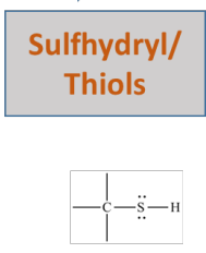
Sulfhydryl / Thiol
Ester

Pyrrolidine

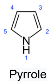
Pyrrole
Imidazole

Indole

Isopropyl
e.g., Valine

Isobutyl
e.g., Leucine

Sec-Butyl
e.g., Isoleucine

Arrhenius Acid vs. Base
Acid: Donates H+
Base: Donates OH-
Bronsted-Lowry Acid vs. Base
Acid: Proton donor
Base: Proton acceptor
Lewis Acid vs. Base
Acid: Electron-pair acceptor
Base: Electron-pair donor
HCO3- Conjugate Forms
Conjugate Base: CO32-
Conjugate Acid: H2CO3
Keq
Equilibrium Constant

Ka
Acid Dissociation Constant
*Can also be determined via Ka = Keq [H2O]
![<p>Acid Dissociation Constant<br><br>*Can also be determined via K<sub>a</sub> = K<sub>eq</sub> [H<sub>2</sub>O]</p>](https://assets.knowt.com/user-attachments/8553d5f6-44a2-426d-93bb-83f52264d603.png)
High vs. Low Ka
The higher the concentration of strong acid (i.e., [H3O+]), the higher the Ka.
The higher the concentration of weak acid (i.e., [HA]), the lower the Ka.
pKa
The lower the pKa value, the stronger the acid.
*To find Ka from pKa, use: Ka = 10 -pKa

Kw

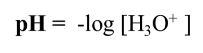
pH from [H3O+]
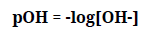
pOH from [OH-]
[H3O+] [OH-]
1.0 × 10⁻¹⁴
pH + pOH
14
Henderson-Hasselbalch Equation
Also written as log [A-] / log [HA]
*If pH = pKa, then [Base] = [Acid]
![<p>Also written as log [A<sup>-</sup>] / log [HA]</p><p></p><p>*If pH = pK<sub>a</sub>, then [Base] = [Acid]</p>](https://assets.knowt.com/user-attachments/86678dc0-46e7-4f13-9e03-73ba56478d9f.png)
Zwitterionic Form
Of an amino acid, refers to dipolar charges while in neutral pH; the NH2 is instead a positively charged NH3+, and the COOH is instead a negatively charged COO-.
Hydrophobic Amino Acids
“FLAPWIG MV”
Glycine
Alanine
Valine
Leucine
Isoleucine
Methionine
Proline
Phenylalanine
Tryptophan
FLAPWIG MV
Hydrophobic Amino Acids
Polar Amino Acids
“SYNC QT”
Serine
Threonine
Tyrosine
Cysteine
Asparagine
Glutamine
SYNC QT
Polar Amino Acids
Positively Charged / Basic Amino Acids
*Additionally, these are Hydrophilic
Lysine
Arginine
Histidine
Negatively Charged / Acidic Amino Acids
*Additionally, these are Hydrophilic
Aspartate (a.k.a., Aspartic Acid)
Glutamate (a.k.a., Glutamic Acid)
Isoelectric Point (pI)
The center of where an amino acid is neutral; represents the pH value where the concentration of each acid is equal to its conjugate base.
*For acidic amino acids, ONLY USE THE ACID pKa VALUES; for basic amino acids, ONLY USE THE BASE pKa VALUES!

Typical pKa Values (Table)

Essential Amino Acids
“MILK WITH VF”
These are amino acids that are not generated by the body and thus must be consumed from external sources.
Histidine
Isoleucine
Leucine
Lysine
Methionine
Phenylalanine
Threonine
Tryptophan
Valine
Peptide Bond
An amide bond between two amino acids resulting in the loss of H2O; in linear peptides, # peptide bonds = n - 1, where “n” is the number of amino acids.
*As such, a dipeptide = 2 amino acids + 1 peptide bond
Residue
An amino acid incorporated into a protein
Backbone
The repeating main-chain atoms; R groups are variable side chains.
Cystine
The unit resulting from two linked cysteines in a disulfide bond
Peptide Planarity
Peptide bonds have partial double-bond character and thus are essentially planar; thus, rotation around the peptide bond itself is restricted.
Also, most peptide bonds are trans.
φ (Phi) Rotation
Rotation about the Nitrogen and α-Carbon bond
ψ (Psi) Rotation
Rotation about the α-Carbon and Carbonyl bond
Torsion Angle
The rotation about the Phi and Psi bonds, also called Dihedral Angle; determines the path of the polypeptide chain.
Ramachandran Diagram
Plot depicting Phi vs. Psi values; the darker the coloration, the more sterically favorable the conformation is.
Primary Structure
Amino acid sequence; peptide bonds
Secondary Structure
Three-dimensional structure resulting from Hydrogen bonds between peptide NH and CO groups of nearby amino acids.
Examples: α helix, β sheet, and turns
α Helix Features
R groups project outward
CO of residue “i” binds to NH of residue “i+4”
1+3 and 1+7 CAN exist, but i+4 is most common
β Strand
Fully extended polypeptide segment
*Side chains are alternatively above and below the strand.
β Sheet
Neighboring β strands linked by Hydrogen bonds
*Sheets may be Parallel, Antiparallel, or Mixed; they can be flat or be twisted.
Turns and Loops
Lie on the surfaces of proteins
*Because turns move easily, they poorly image in X-Ray Crystallography
Coiled Coil
Superfamily of structural proteins, including some cytoskeleton proteins, some muscle proteins, and α-Keratin
*α-Keratin has two right-handed α helices intertwined to form a left-handed superhelix.
Collagen
Structural protein that serves as a component of skin, bone, tendons, cartilage, and teeth; consists of three intertwined helical polypeptide chains that form a superhelical cable
*Glycine MUST appear at every third residue due to its ability to sterically fit; Gly-Pro-Pro is common.
Collagen Stabilization
Collagen helices are stabilized by the steric repulsion from the Pyrrolidine rings of Proline, NOT by Hydrogen bonds; this crowding is why Glycine is necessary.
Tertiary Structure
The spatial arrangement of amino acids that are far apart in the primary structure and to the pattern of disulfide bond formation; results from R group interactions.
Globular Proteins
Such as Myoglobin, form complicated three-dimensional structures and are very compact.
The interior of globular proteins consists mainly of hydrophobic amino acids; the exterior is mainly charged and polar amino acids.
Motif
Also called “super-secondary structures,” these are recurring combinations of secondary structures that are found in many proteins.
Domain
Two or more similar or identical compact functional structures within a protein
Quarternary Structure
Proteins that are composed of multiple polypeptide chains called subunits; can be as simple as two identical polypeptide chains or as complex as dozens of different chains.
Urea
Disrupts noncovalent interactions
β-mercaptoethanol
Disrupts disulfide bonds
Optical Activity in Amino Acids
All but one amino acid are chiral and thus optically active; Glycine alone is achiral and optically inactive since it has two identical branches (two Hydrogens).