TB6: protein interactions
 Call Kai
 Learn
Learn Practice Test
 Spaced Repetition
 Match
Match Flashcards
 Knowt Play
1/215

There's no tags or description
Looks like no tags are added yet.
Name | Mastery | Learn | Test | Matching | Spaced | Call with Kai | Chat |
|---|
No analytics yet
Send a link to your students to track their progress
216 Terms
Why study protein itneractions?
Interactions between molecules defines life - proteins are key players of this
The specific, reversible intermolecular interactions are key to understanding living organisms
what are the types of different protein interactions?
Protein protein interactions
protein ligand interactions
protein nucleic acid interactions
What are protein-protein interactions involved in?
Regulation and signalling
Localisation
Assembly of multicomponent cellular machines
Eg. complexes such as ATP-synthase, chaperonins and the proteasome would not function without PPIs between different subunits of them
Generative cooperativity, allowing tuning of responses
Providing mechanical support to cells
Eg. keratin, collagen and microtubules all rely on PPIs
The immune system
what are protein-ligand interacitions involved in>
Enzyme interactioins
This can be with substrates, or with co-factors
Receptors and signalling molecules interacting to allow signal transduction
Allosteric modulators interacting with protein targets
Membrane lipids
Drugs
what are protein-nucleic acid interactions involved in?
Processes such as transcription, translation, replication, gene regulation and processing of transcripts all rely on PNIs
These are essential to biology!!
what are the properties of most protein interactions?
In biological systems, protein interactions are specific (and reversible most of the time)
why is reversibility of protein interactions important?
This reversibility allows responses to be made under different conditions, and also allows protein turnover
how is specificity achieved in protein interactions?
Specificity is achieved through the unique spatial array of chemical groups formed as a globular protein folds
This is achieved through the rich diversity of exposed chemical groups on a protein surface
Such as positive vs negative charges, polar vs apolar patches, H-bons donors vs acceptors
The range of surface amino acids are less important for folding stability as they ddo not fold into the hydrophobic core - this allows them to be much more varied to form different interactions
This variation on the surface produces a pattern of patches that convey 2+3D complementarity with other proteins - this allows different interactions
These interactions between proteins are dominated by weak noncovalent interactions - these convey specificity and reversibility of intermolecular binding
2 classes of PPIs?
reversible PPIs and obligate oligomers
describe reversible PPIs
Reversible PPIs have reversibility and can be studied to determine affinity and regulation of these interactions
These are often involved in signalling pathways and regulatory processes
describe obligate oligomers?
Obligate oligomers have low reversibility. Once the single monomers are produced they are assembled to form an oligomeric complex. They only really dissociate when the complex is turned over
The PPI here provides function
describe the physical properties of PPI surfaces
Wide range of affinities
Mostly high specificity
Shape and sizes of the PPI interfaces are large and variable
They are often flat and undulating in morphology though - this differs to active sites
For those that are buried, the amount of polar and non-polar residues is similar to non interface surfaces
This is to prevent aggregation
Interactions are largely driven through specific interactions conferred by the amino acids present:
Salt bridges
H bonds
Arrangements of small hydrophobic patches
As well as the types of amino acids present, shape complementarity is also important
Often enriched in aromatic amino acids
describe the physical properties of buried PPI surfaces?
Amount of non-polar / polar surface area buried is similar to non-interface protein surfaces
for something that isnt always oligomerised, needs to remain soluble to prevent aggregation
largely driven through specific itneractions
salt bridges
hydrogen bonds
arrangments of small hydrophobic patches (rather than a sole large one)
shape complementarity is important for these interactions
enriched in aromatic amino acids F, Y, W
generally smaller in area, less hydrophobic, and more charge, than those between subunits in obligate oligomeric proteins
How can we determine the buried surface area to determine the surface areas of PPIs interaction surfaces?
Rolling a probe sphere along the surface of the protein allows determination of the surface area
this can be done for both individual proteins and the complex to get: Buried surface area = Ax + Ay - Axy → so it is the sum of the proteins by themself subtracted from the entire protein complex area
Can see a wide range of buried surface
What different regions to PPI interfaces have?
a core
a rim
describe the core of a PPI interaction surface?
the core: centre of the interaction
water is completely excluded
water wont freely exchange
describe the rim of a PPI interaction surface?
accessible to water and water is exchangeable
how can water molecules contribute to the hydrophobic effect at PPI surfaces?
Water molecules can contribute to the hydrophobic effect
release of water from hydrophobic residues can enhance affinity
pulling waters off of charged, hydrated amino acids may be needed to form salt bridges
what affects can water have at PPI interfaces?
improve complementarity by filling gaps between the proteins
contribute to interfacial H bonds
eg. by bridging between proteins
screens unfavourable electrostatic interactions
what are hotspots in PPI surfaces?
positions in PPI interfaces that are important for complex stability
these residues have more contribution to the interface
how can we determine PPI hotspots?
can be determined with alanine scans
change each of the residues to alanine and measure the affinity
∂∂G is the change in ∂G
those greater than 1.5 in this case is considered the cut off for an important residue
what can substitutions at hot spots do?
reduce affinity
what residues are typically hotspots? what interactions may they participate in?
arg, tyr, trp
hydrophobic, aromatic and polar interactions
what interactions with pi electrons are common at interfaces?
pi electrons can interact with cations
pi-pi stacking of aromatic rings
why use kd rather than ka
this puts affinity into physically intuitive units
this is an indication at the point of which there is half saturation
how can we get interaction dynamics from ka/kd?
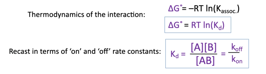
when we assume proteins interact as rigid bodies, what are we assuming? what is the equation?
we assume little or no change in structure, other than side chain rearrangements
this is a simple bimolecular association

describe the conformational changes that can occur at large interface PPIs?
loop/secondary structure rearrangements
disorder-order transitions of disordered regions
domain rearrangements in multi-domain protein
what are the different types of conformational change models for different PPIs?
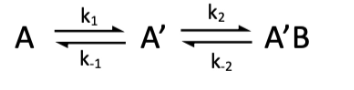
Induced fit (conformational change)
Conformational selection
describe conformational selection of protein binding
conformational selection - sometimes, the protein adopts a different conformation - this can only bind the other protein
and so the initial binding is dependent on the formation of the binding conformation
describe induced fit of protein binding
induced fit - the protein binds the other protein in a conformation that is not the most stable, forming an encountered complex. whilst bound, it rearranges, forming the most stable confirmation
equation for induced fit protein binding?

equation for conformational selection protein binding?
What aspects need to be considered when quantifying binding interactions?
Circumstances of interactions occurring
subcellular localisation
binary or within a larger complex
PTMs necessary?
Information wanted from the assay
complex formation
binding affinity - kd
is the affinity sufficiently lower than a wt - can use this in mutagenesis
binding kinetics - kon and koff
binding thermodynamics - H and S
can do SPR, ITC to determine different components of the equations
mass, stoichiometry
may need size sensitive approaches
where is the interface? what does the complex look like?
Possible sample conditions
in vivo
eg. may want to know the stage of the cell cycle
may need to do in cultuted cells
crude mixture or cell extracts
crude extracts are easier to work with, but ability to quantify the data is limited
purified component or reconstitution
What does a yeast 2 hybrid assay exploit?
exploits the 2 domain architecture of the yeast transcriptional activator Gal4p
Domains have distinct functions
can exploit this, splitting the protein in half and attaching the DNA binding domain and activation domain to different proteins
what are the 2 domains in the gal4p activator used in a two hybrid assay?
DNA binding domain binds UAS
the activation domain then recruits rnap for gene transcription
how does the yeast 2 hybrid work?
The activation domain will only be in the right position if X and Y interact
can use a reporter gene
eg. lacz- blue/white screening
eg. fluorescence expression
advantages of yeast 2 hybrid?
Low cost
easy to use
disadvantages of yeast 2 hybrid?
transcription activation is sensitive
there is a high false positive rate
proteins must be nuclear localised
yeast may not be relevant cellular context
limited to binary PPIs
Y3H exists but needs more effort
Requires a well defined hypothesis - need to know the identity of both
describe the use of in-vivo chemical cross linking to determine PPIs?
incorporate photo-activatable chemical group into bait protein (in cells)
this is the protein you already have knowledge of
Introduce UV light
this activates the cross linker, inducing non-specific covalent cross linking
this cross links interacting proteins
(traps complex)
This is done in a crude mixture. can then isolate the different cross-linked complexes
isolate/purify cross-linked complexes
detect cross-linked complexes
In gel Digest
this maintains the cross links
identify cross-linked complexes
detect via mass spec and then a databbase
describe the use of FRET to determine PPIs?
X is attached to CFP, Y is attached to YFP
if do not interact - low fret efficiency
if they do - high fret
get energy transfer
FRET via photoluminescence
This tests for bimolecular interactions
describe the use of BRET to determine PPIs?
bioluminescence resonance energy transfer
X is attached to luciferase
in the presence of luciferin and oxygen, this produces light via an oxidation reaction, and produces light
Y is labeled with YFP, should get an energy transfer
describe the use of split protein sensors to determine PPIs?
X and Y are known
we cut another protein with a reporter activity in half
fragments on their own lack activity
if the proteins interact, this brings the C and N termini together, allowing the reporter to show activity
Examples:
GFP + GFP variants
those with measurable enzymatic activity
How can protein interactions be detected in crude mixtures?
use of a pull down
describe the use of pull downs to determine PPIs
matrix anchored linker - this has a crude mixture poured over it
bait protein binds the ligand
anything binding the protein, which binds the ligand, will not come off the column (it is retained)
pros of pull downs for PPI identification?
Discover new binders
test known binders
detect large complexes, not only binary PPIs
this is due to the use of the crude mixture
cons of pull downs for PPI identification?
Detect large complexes - may be indirect
Relatively low sensitivity, not suitable for weak binders
this is because the washes through the column can induce dissociation
not in vivo
how can we get quantitative binding data? include example
Mostly measured on purified proteins in reconstituted complexes
Anything that can be detected that changes in proportion to complex formation can be used to measure kd
eg. in haeomoglobin, oxygen binding can be detected via uv-vis spectrophotometry
extinction coefficient is sensitive to whether oxygen is bound or not
the two forms oxygen bound or unbound are very different - this produces different intensities, allowing the ozygen bound to be determined
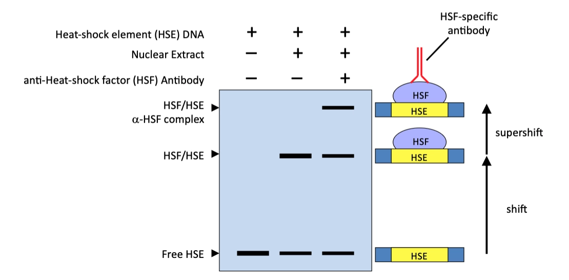
what is the EMSA used to detect?
protein DNA interactions
main principle of EMSA?
DNA bound to protein is larger than free DNA, and so moves much more slowly in the gel
set up of EMSA?
use an agarose gel
we are detecting the nucleic acids, not the protein!!
proteins binding the dna slow it down, producing a shift
this can be done as a function of protein cocnentration
the bands are quantified to determine free dna vs complexed
what are EMSA gels stained with?
Stain DNA before the gel with covalent dyes/radioisotopes. Or after the gel with a base intercalating fluorophore (eg. ethidium bromide or sybr-GOLD)
what does varying protein concentration allow for in EMSA?
we measure the change in intensity of bands for free DNA and protein bound DNA
we use this to get Kd
describe the use of competition experiments in EMSA. what do they reveal?
use target DNA, protein and an unlabelled competitor DNA
start with a labelled DNA-protein complex and add in an unlabelled competitor DNA
If there is a transition after the addition of the competitor, this indicates specificity
we vary competitor concentration, rather than protein concentration
describe the use of a super shift EMSA
Free DNA, add some protein (eg. crude mix of different TFs)
we then add abs against different tfs
this is a larger protein complex , this further perturbs movement through the gel, producing another shift
How can we analyse EMSAs?
Gel image intesities can be quantified
Total band intensities are proportional to concentration
DNA free and or DNA complex
Binding parameters can be quantified
Langmuir isotherm equation for single site ligand binding?

what do we plot when measuring single site ligand binding?
plot ñ on y vs free ligand concentration
this produces a rectangular hyperbola or a direct plot
or ñ on your vs log free ligand concentration
this produces a sigmoidal plot, where it is much easier to extract kd
main assumption for single site ligand binding plots?
assume ligand free = ligand total - this assumes ligand is in excess
what do we plot when measuring multiple site, identical and independent ligand binding?
plot ñ/Free ligand on y and ñ on x. this is a scatchard plot. this gives a straight line with gradient of -1/kd
could also do a log scale for this. a positive deflection indicates positive cooperativity
scatchard eq for measuring multiple site, identical and independent ligand binding?

what do we need to consider when quantifying binding data?
Does the signal change proportionally with complex formation?
Can we detect the signal sensitively (high signal-to noise ratio) and in the concentration range we need for detection
Do I need to label something to detect it and what are the risks of introducing artefacts?
Unlabelled (‘native’) always preferred but may not be possible.
could ;labelling artefacts prevent complex formation?
How much sample is required for the technique? Can we makenenough of it in the right form?
eg. may need specific PTMs
eg. is it folded?
Is it stable, and is it soluble at the required concentrations?
list of methods used to quantify binding data?
ITC
SPR
Thermal shift assays
fluorescence polarisation
microscope thermophoresis
NMR
stopped flow
describe thermal sift assays
when you bind something, the protein becomes stabilised
this is because there are more contacts present
can do cdmelt, DSC, DSF - diffferentways to measure melting tempertature
best done with small ligands
for PPIs, both have a specific melt point, and so can become difficult
can be done with ID blinding partners
Describe the fluorescence polarisation experiment?
Consider a cuvette containing fluorophores (eg. fluorophore labelled proteins)
Light absorbance and emitted fluorescence depends on the direction of the transition dipole moment.
so if the molecule rotates, the dipole moment also rotates
If polarized light is used to excite, this will only excites a subset of fluorophores (photoselection)
this is photoselection
those with a transition dipole aligned with the polarised light will be excited more than those not fully aligned
We use this and the emission polarisation is detected
before detection, it is passed through polarisers again
can use grids parallel or perpendicular to the inital polariser
we detect both of these
Why do we observe a change in anisotropy in FP experiments?
Use a small ligand with a fluorophores tag, this is very fast tumbling
so in the time for excitation and detection, molecules tumble through all orientations and so have low polarisation
large molecules are low tumbling
in the time for excitation and detection, the molecule has tumbled slowly, and so the orientation hasnt changed as much
this will have a high anisotropy
so a ligand binding will have a high anisotropy in comparison to the free ligand
so when the ligand binds the large protein, it will have high anisotropy - high A
this change in anisotropy is detected!!
What is the polarisation anisotropy?
The polarisation anisotropy, A is defined as the degree of depolarisation divided by the total fluorescence output
(intensity of the parallel polarised light - the intensity of the perpendicular polarised light) / the sum of the two
this is dimensionless, and between 0-0.5
equation for polarisation anisotropy?

what is the plot for anisotropy of binding? what are the different points corresponding to?
We measure anisotropy as a function of protein concentration
At no protein concentration, the ligand has low anisotropy
At fully bound, it has a high anisotropy
Smallest A → Largest A = ∂A
∂Amax = difference between fully unbound and fully bound

Equations for fitting of anisotropy data?
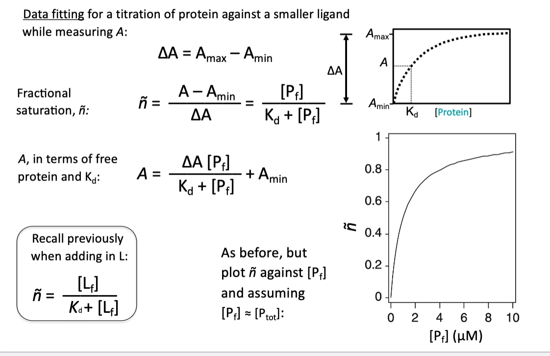
how to we get A in terms of free protein and Kd from the Langmuir isoform equation?
ñ can be scaled from the langmuir isoform to get A in terms of free protein and Kd
A = ∂A[Pfree]/Kd+[Pf] +Amin
but the values of these scaling values is not importnat, we only care that ∂A is large and that A and Amin are detectable
the Amin is because the ligand on its own will have some anisotropy
why does microscale thermophoresis work?
the impact of heat on the protein, which is dependent on the size, which is dependent on the complex and the interaction with hydration cell
describe the process of microscale thermophoresis (MST)
molecules are flown into capillaries
these often have different concentrations
IR laer introduces a temperature gradients
molecules will diffuse
we detect fluorescence in a specific region
fluorescence on y as a function of time
laser on - fluorophore diffuses away from the point of heat
turn laser off - diffuse back into the space
very simple and sensitive!!
The extent of movement depends on molecular hydration shell, charge and size
these change on binding of the ligand/protein partner
detection via covalently attached fluorophores, fused GFP or intrinsic tryptophan fluorescence
how do we normalise MST data?
Normalise using Fmin and Fmax - looking at the proportional change in relation to complex formed
critical to measure a wide range, as this allows us to define the plateau regions

what are some key considerations in experiments to quantify binding data?
many assume ligand free and ligand total are true
this only holds if there is a lot of excess ligand!!!
mutagenesis is useful
but remember there will be 2 interfaces!!
gold standard is using multiple orthogonal approaches
eg. a tm measurement and spr
need a big change in anisotropy - need to consider which components you label
What are the two classes of techniques when measuring protein-ligand interactions by NMR?
protein detected approaches
ligand detected approaches
what do protein detected NMR approaches measure?
measuring free or bound protein
what do ligand detected NMR approaches measure?
measure free ligand signal
some physical properties have been imparted to the free ligand
How can 1H 15N HSQCs be used to measure protein-ligand interactions?
Can look at chemical shift change
these are sensitive to changes in environment and conformation and therefore upon binding of a ligand there will be a shift
we can calculate the chemical shift perturbations for each residue
how do we find the CSPs via HSQCs when studying protein detected NMR for interactions?
We use an N15 labelled protein at a fixed concentration, and can introduce variable ligand concentrations
this can be done on the same sample via a titration
can also be done using many different samples with protein at different ligand concentrations to be recorded one at a time
then do spectral overlay of different spectrums with different concentratons
can use this to trace and watch the CSPs happening
not everything moves - only those involved in binding do
what happens in CSPs when a molecule only binds one specific site?
if it binds to one site, only a subset of resides should be impacted by binding
what do many residues undergoing CSP indicate in protein-ligand interactions?
indicates a conformational change or non specific binding
how do we get the CSP from HSQC?
we can determine the CSP via Euclidian geometry - we have to take into account the difference in chemical shifts for nitrogen
we do this by introducing a scaling factor of 5
what values of CSP are significant`? what does this tell us?
for the ∂delta value, anything more than 0.1 is significant
if there is a significant CSP for a residue, we asume that the ligand is binding direcly to the residue or very close by
how is chemical shift data derived from HSQCs used to determine Kd?
Using the chemical shift data, we know that the environment is changing because of the ligand binding
We use the same formulas to fit this data as before - we can use the single site binding equation to do this
Can do a multiple spectra overlay
the chemical shift information for each residue can be quantified
we do this for all to determine whether these are involved in the same binding event or not
all residues have CSPs - these need to be normalised becuase of the details of the interaction
if the kd is the same - its the same binding event
describe how CSPs look for weak interactions?
If there is a weak interaction (high koff), residues will start at the free position and move along the line to the fully bound position
we can track the movement
describe how CSPs look for strong interactions?
however if koff is very low, there is a slow exchange regime
within the timescale, the ligand doesn't come off
so we observe 2 populations - ligand free and ligand bound population
for his tight binding, the slow exchange produces an intensity drop of the free form, and an increase of the bound form - the intensities are directly proportional to the population
the slow exchange can be difficult to interpret - we dont see an incremental movement, and so assignment of the fully free and bound is difficult
are fast or slow exchange interactions easier to analyse via CSPs?
Fast exchange, weak binding events are easier to study - this is becuase peaks move in a line over the titration, making it easy to assign peaks
how can NMR HSQCs elucidate multiple different binding interactions?
When we plot the different perturbations, we can see multiple different binding events by fitting different trend lines to different residues. this can be used to validate the presence of different binding sites and determine their kd
how can 1H, 13C methyl CSPs be used for large complexes?
methyl groups only produce peaks, other proton sites are deuterated - this reduces the number of signals. and therefore reduces the number of amino acids being detected
how can structure activity relationships be determined using NMR for drug design?
protein is screened with lots of small molecule fragments
can detect some interaction and confirm specificity
this can then be independently optimised to increase its affinity
whilst bound we can screen another pool of fragments and find things that bind elsewhere, but nearby
this is again optimised to increase affinity
we then produce a chemical linker to link these together
this can them both bind, greatly increasing affinity
What are the advantages of protein detected NMR approaches?
Exploits exquisite sensitivity of chemical shifts to report on interactions
Quantitative information on affinity
2D HSQCs can be recorded quickly (minutes)
depends on the protein concentration
High resolution information on location of binding site
this is because there is one signal for every amino acid
Very good (best) at detecting weak interactions
Pooled small molecule screening possible (but requires deconvolution)
if one pool you see aa CSP, dont know which molecule binds -need to deconvolute to determine what is binding
What are the disadvantages of protein detected NMR approaches?
Requires sample that is labelled, either 15N or 13C, which means usually sample must be expressed and purified from in E. coli
e.coli may not be the best expression. system for the protein you are studying
Typical protein concentrations are 10-100’s μM so cannot easily quantify low Kd
if there is a low kd, protein concentration needs to be very below the kd
and so not good for quantification of tight binding ligands
Chemical shift assignments needed for locating binding site
Requires good quality data, well-resolved HSQCs – the size limitation of NMR can be limiting (broad signals and spectral complexity)
What are the different ligand detected approaches used in NMR to detect protein interactions?
STD-NMR
WaterLOGSY
Relaxation editing
Transferred NOE
Paramagnetic relaxation enhancement
diffusion editing
what do ligand detected NMR approaches rely on?
all detect different chemical properties depending on the bound and free ligand
unbound - fast tumbling fast diffusion and slow relaxation
Process for STD-NMR?
Protein is selectively irradiated
Upon ligand binding, some magnetisation is transferred to the ligand, and this can be detected
We can use difference spectra
we record 1D spectra with no protein irradiation - this produces no magnetisation
we then record a second experiment with selective saturation of the protein - here, the magnetisation is transferred
there will be intensity differences between the peaks on the NMR, differences can be calculated to determine which resonances are perturbed
these are used to determine which molecules bind
what 2 1D spectra are used in STD-NMR?
one where the protein is irradiated
one where the protein is not irradiated
Describe the process of water-logsy
Instead of saturating the protein, we instead saturate water
Bulk water is selective irradiated with a radifrequency pulse
Some of this is transferred to ligands through space via NOEs
however the NOE transfer depends on molecule tumbling times
small molecules that have bound a large protein will have reduced tumbling and so their NOE value will be smaller and flipped
How does relaxation editing work?
Looks at which molecules behave like large molecules
when bound to a protein, will have some properties
looking for a change in linewidth
as molecules get bigger, linewidth increases and signal decreases
based on CPMG, a way to measure t2, which is related to linewidth
what are the cons of some ligand detected NMR experiments?
techniques rely on the detection of the free ligand
transfer of properties from the bound to free satte
if there is no exchange bewteen the two, can get no results
tight binders cannot detect the diferences