Special senses
1/80
There's no tags or description
Looks like no tags are added yet.
Name | Mastery | Learn | Test | Matching | Spaced |
|---|
No study sessions yet.
81 Terms
Describe the general function of the outer, middle and inner ear.
Outer: funneling of sound
Middle: Relaying tympanic vibrations to inner ear
Inner: Transduction of vibrations of neuronal signals. Balance organs
Describe the general anatomy of the outer, middle and inner ear.
Outer: the outer bit (aka auricle) and the auditory canal
Middle: the tympanic membrane and tympanic cavity (containing the 3 small bones: malleus, incus and stapes which connect eardrum to inner ear)
Inner: inside temporal bone (locked in position and protected) full of fluids.
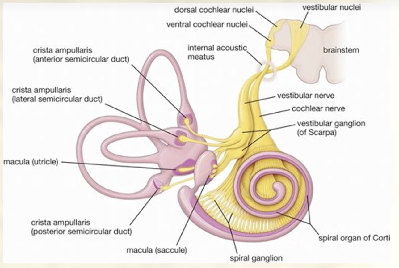
cochlea (hearing)
vestibule and semi-circular canals (balance)
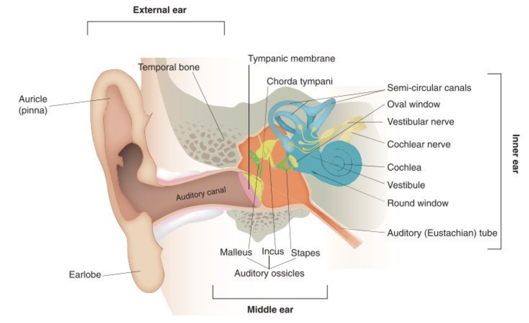
Roles of the bones in the middle ear
Malleus, incus, Stapes
transduction of vibration
amplify force of vibration
Explain the function of the tympanum (ear drum).
Focuses sound waves: Sound waves in air directed to tympanic mb (ear drum) and transduced into vibration of tympanum
Tympanum innervation
Trigeminal nerve (sensory motor)
Vagus nerve (sensory)
Describe the function of the muscles found in the missile ear.
Tensor tympani – tensioning of tympanum
Stapedius – Dampening movement of incus (when activated)
Acoustic reflex triggers tension - Reduction of vibrations/ movements during load noises = protective
Describe the function of the bones found in the missile ear.
Transduction of vibration.
amplification force of vibration
The bones are: Malleus, Incus, Stapes
Describe the function of the windows found in the missile ear.
Oval – transfer of energy to inner ear acts as second amplifier (stapes acts as actuator) inner ear fluid filled
Round – Relieves pressure waves that develop in the fluid filled cochlea. Membrane flexes ‘in and out’ to relive pressure
Briefly describe the function of the outer ear.
Funnelling of sound
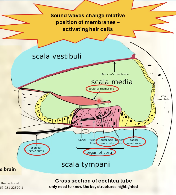
Cochlea
Cochlea: a fluid filled spiral tube
Role: transduce sound waves to nerve impulses
Key structures of cochlea: tectorial membrane, hair cells, basilar membrane, organ or Corti

Explain how the tectorial membrane, hair cells, basilar membrane, organ or Corti and are involved in transduction of sound energy to nerve impulses
Organ of Corti – supports hair cells
Hair cells – physical transduction takes place (the sound waves change the relative position of membrane-activating hair cells)
Tectorial membrane – gel-like sheet.
Hair cell tips connect to membrane
Basilar membrane – supports organ of Corti
Cochlea nerve – carries electrical signals to the brain
Cochlea
Fluid filled spiral tube that transduces sound waves to nerve impulses
Explain the role of the cochlear nerve and the spiral ganglion.
Cochlear nerve: carries electrical signals to the brain
Spiral Ganglion: contains sensory neuron cell bodies that project axon to join cochlea nerve
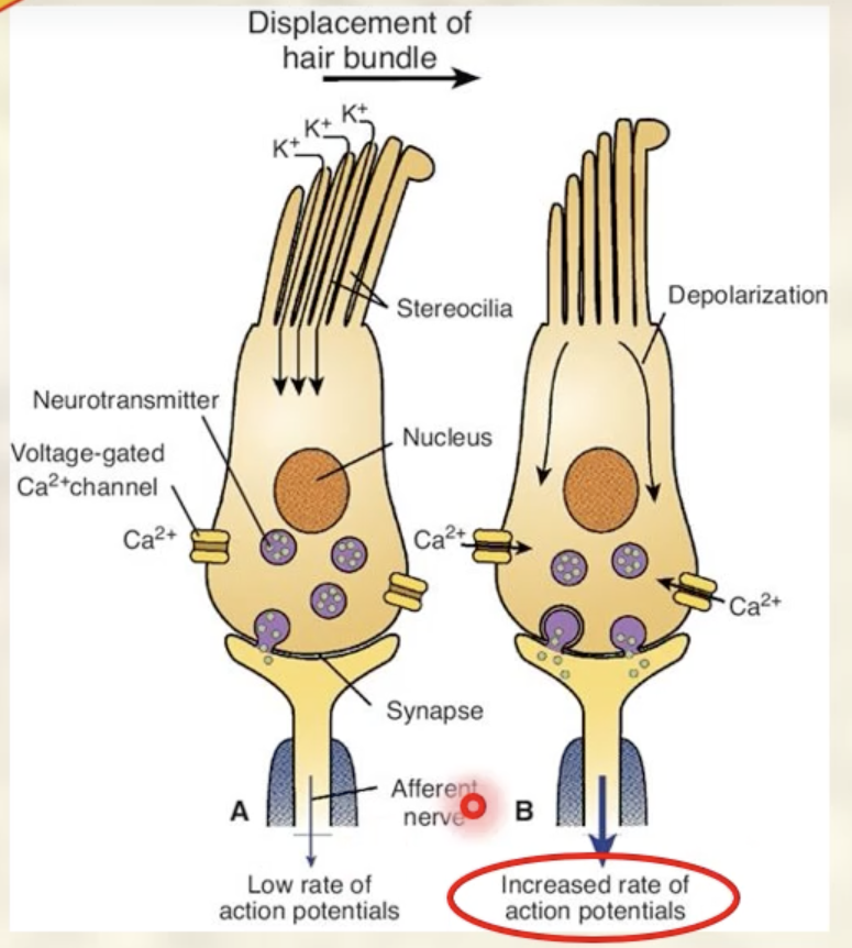
Describe how an electrical signal is generated from hearing a sound wave
Sound waves come into inner ear and displace 2 membranes (basilar membrane and tectorial membrane) which displaces the hairs
hair cells allow K+ to enter cell = depolarisation
calcium enters cell = releases neurotransmitter (glutamate) at synapse with neuron
Action potentials flow to spiral ganglion
Describe the role of the spiral ganglion
Relays the information through to the cochlear nerve which carries info to brain stem and then auditory processing centres of brain
8.Explain how hair cells of the cochlea are activated and how they can release glutamate to activate auditory nerve fibres.
Hair cells:
A. Deflection of stereocilia – K+ enters cell
B. Voltage-gated Ca++ channels open
Glutamate released at synapse with neuron
Action potentials flow to spiral ganglion
Spiral ganglion:
Contain sensory neuron cell bodies
Projects axon to join cochlea nerve
Cochlea nerve enters brain stem
Target – auditory processing centres of brain
Briefly describe the location of the vestibular apparatus.
Inner ear
Embedded in the temporal bone so that when the head moves, the vestibular apparatus move with it
Outline the general role of the vestibular apparatus.
Balance! = Sensing acceleration/ movement
Explain the function of the semicircular canals and otolith organs.
Semi-circular canals: angular acceleration
Otolith organs:
utricle: horizontal acceleration
saccule: vertical acceleration
Explain that Scarpa’s ganglion and the auditory nerve relay vestibular signals to the brain.
Scarpa’s ganglion contains neuronal cell bodies
bipolar arrangement: carries info from vestibular organs, through vestibular nerve, back to brainstem
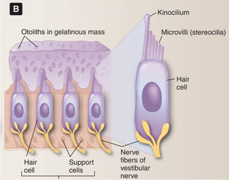
Explain how otolith organs function to sense vertical and horizontal acceleration.
Stereocilia (shortest) move away from kinocilium (tallest) – Hyperpolarisation of hair cell
Stereocilia (shortest) move towards kinocilium (tallest) – depolarisation of hair cell
Signals change in acceleration
The gel (gelatinous mas) wants to stay still but the microvilli are embedded inside it. so when you bend your head and the gel remains stationary, the hairs bend.
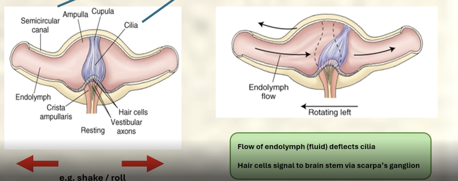
Describe how the cilia of the semicircular canals can encode angular acceleration.
Angular acceleration = vectorial measurement (direction and magnitude)
At the base of each canal = swelling called ampulla containing a cupula = gelatinous
Flow of endolymph (fluid) deflects cilia
Hair cells signal to brain stem via scarpa’s ganglion
Describe pathway of vestibular information from ear to brain
Vestibular organs
vestibular nerve
arrives in brain stem at the vestibular nuclei
Describe the four main outputs of the vestibular ganglia found in the brain stem.
1.Contribution to control of eye muscle
2.Contribution to control of lower motor neuron limb extensors
3.Contribution to cervical spinal cord - control of head movement
4.‘Balance’ information sent to cerebellum
Explain some effects of ageing on balance.
1.Reduction in hair cells – 40% by age 75
2.Deterioration of brainstem and cerebellum
3.Decline in oculomotor control – visual instability when moving head
4.‘Balance’ information sent to cerebellum
5.Can cause dizziness, disequilibrium, and an increased risk of falls
Identify and list the layers of the eye.
Fibrous: cornea and sclera (shape + structure)
Vascular: ciliary body, choroid, iris
Inner: retina (photoreceptors of neural layer and pigmented layer, macula, fovea, optic disc)
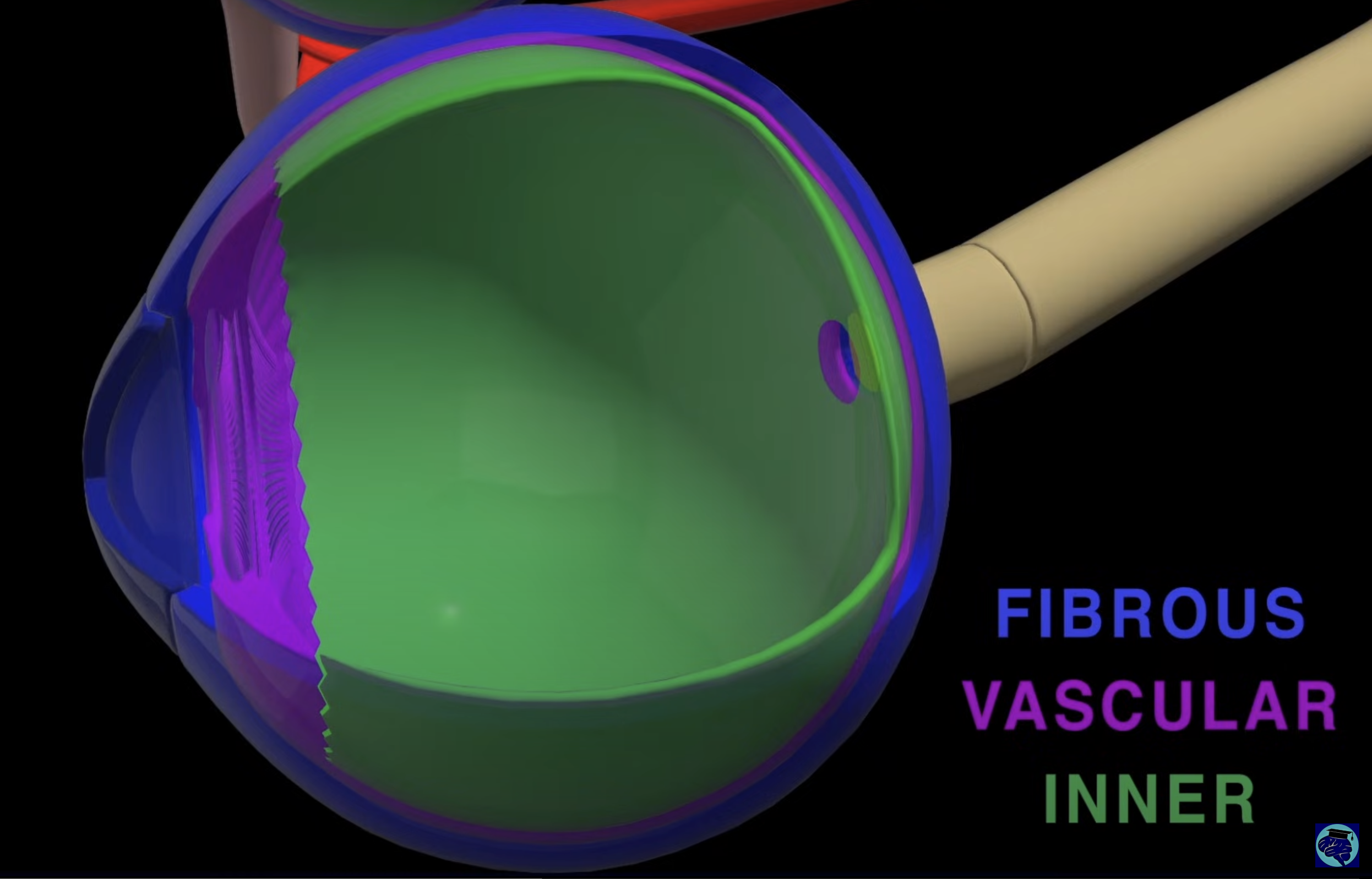
Explain the three-layer structure of the eye wall
Fibrous tunic
Sclera – heavy fibrous white of eye
Cornea – transparent / continuous with sclera
Vascular tunic
Choroid – pigmented layer
Ciliary body – ciliary muscle
Iris- colour pigment, aperture of pupil
Neural tunic
Retina – 3 layers
Optic Nerve – carries signals to brain
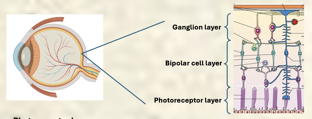
Describe and explain the three layers of the retina.
Photoreceptor layer: rods and cones
Bipolar cell layer
ganglion layer

Rods
Function: specialized for low-light
Sensitivity: extremely sensitive to light
Do not detect colour
Location: Mostly in the peripheral retina.
Role: provide night vision
Cones
Function: specialized for bright-light
Sensitivity: Less sensitive to light than rods
Allow for high acuity
Location: concentrated in centre of the retina(fovea)
Role: enable colour vision
Role of the bipolar cell layer
process signals from rods and cones
transmits signals to ganglion layer

Role of ganglion layer
converts graded potentials from retinal cells into Action Potentials
Optic nerves
visual signals from the retina to chiasm
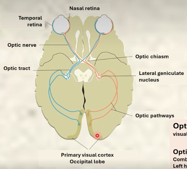
Understand the role of the optic chiasm
Combines visual signals
Left hemisphere of both eyes combine in left optic tract
Right hemisphere of both eyes combine in right optic tract
Important for binocular vision and depth perception

Describe the visual pathways of the brain.
Carry combined visual information to visual cortex.
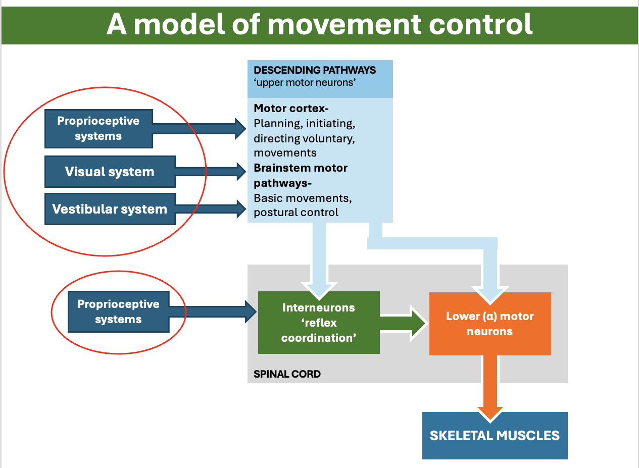
Define what proprioception is.
The body’s ability to sense its position, movement and the amount of force it is using in a space
Explain that the visual system provides proprioceptive information.
You can use your visual system for proprioception! to find out where your limbs are.
The proprioceptive system, visual system and vestibular system all play important roles in the nervous coordination
Gustation
the sense of taste/ activity of tasting
chemical sense
Structure and function of taste buds
Taste receptor
Modified epithelial cells
Replaced 10 – 14 days
Microvilli on apical surface – taste pore
Synapse with nerve cells
Support cells
Physical / metabolic support
Basal cell
Produce new taste receptors
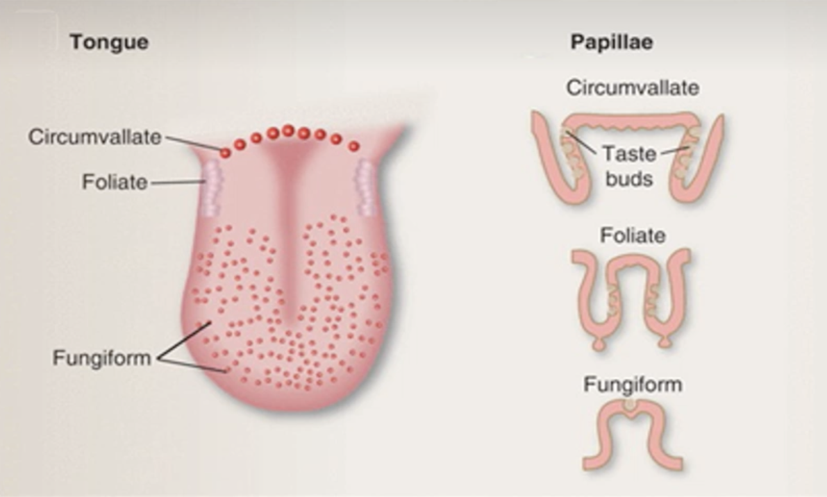
Papillae
The small natural bumps on your tongue which present taste buds:
3 types:
circumvallate (largest, dark-reddish colour)
foliate (smaller, paler)
fungiform (smallest with rich blood supply = red colour. Group of taste buds on the apex)
Different cell types found in taste buds and their functions
Taste receptors = modified epithelial cells (not nerve cells)
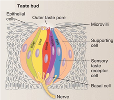
Taste buds
Components:
Taste receptors (the multicoloured cell on the image):
Support cells
Basal cells
Taste Receptors
modified epithelial cells
replaced 10-14 days
microvilli on apical surface = taste pore
synapse with nerve cell
Support cells
physical/ metabolic support
act like glial cells (support, protect, provide nutrients to neurons)
Basal cells
produce new taste receptors every 10-14 days
Describe which cell type is associated with perception of which basic taste
Type 1 cells
Support cells – glial like
possibly saltiness
Type 2 cells
Sweet
Bitter
Umami
Type 3 cells
Sour
possibly saltiness

Saltiness and tastebuds
not sure which cells trigger saltiness taste (possibly type 1 or type 3 cells)
salt = NaCl
Na+ passes through sodium channels on receptor cells so Na+ influx amplifies signals triggered by other taste molecules
Genetic variants of different taste buds
propylthiouracil tastes different to people depending on genetic variants (some taste it as bitter, others dont) - dont need to remember this!
Taste perception pathway
Cranial nerves carry info from tongue into
medulla oblongata (autonomic response like vomit reflex) and hypothalamus
amygdala and insula (emotional integration, linked to memory which makes flavour a perception thing)
Flavour
perception
Olefaction and gustation = Highly integrated to provide perception of flavour
integrated in frontal cortex
Olfaction
The activity of smelling (or the ability to smell)
one of the CHEMICAL senses where the sensed chemicals are odorants
Olfactory bulbs
base of frontal lobe
olfactory epithelium that sits high up in nasal cavity, part of brain. pokes through bone and dangles into nasal cavity.
provides direct neuronal connection to olfactory epithelium

Primary olfactory neurons
project through ethmoid bone
into olfactory epithelium
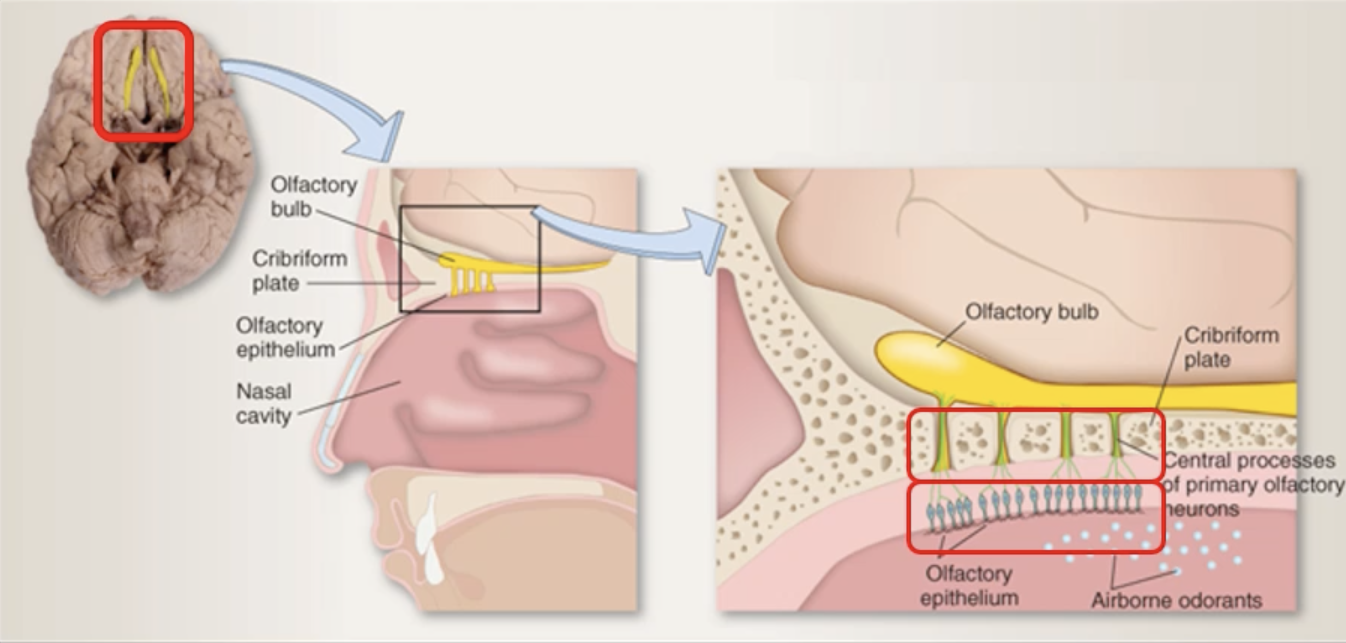
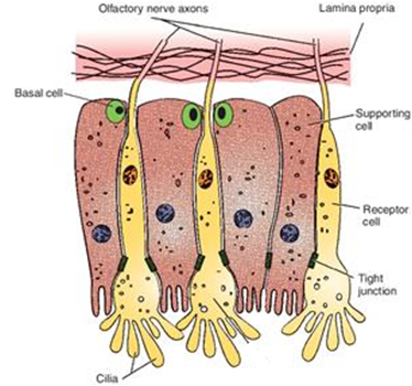
Olfactory epithelium
Receptor cells: olfactory neurones (replaced 30-60 days)
Support cells: (like glials) physical/ metabolic support, detoxify odorants
basal cells: create new support cells
Olfactory cilia: embedded in layer of mucus, odorants dissolve in mucus, then detected by cilia
odorant binding proteins: sit in mucus, detect odorants and collect them which leads to depolarisations of nerve cell
Olfactory processing
olfactory bulb: early processing/ filtering
lateral olfactory tract
olfactory cortex
together they form CNS olfactory system
sent to other areas of the brain:
hippocampus (formation of memories linked to smell)
limbic system (emotional responses, directs someone behaviour to smell)
amygdala 9associate learning between smell, emotion, situations, behaviour
signals bypasses thalamus
Explain the locations of the three different forms of lingual papillae

Explain why food tasted bland if olfaction is compromised
Gustation and olfaction highly integrated to provide perception of flavour – no smell, not much flavour
Key area for integration in frontal cortex
Understand that cranial nerves carry gustatory signals into the brain
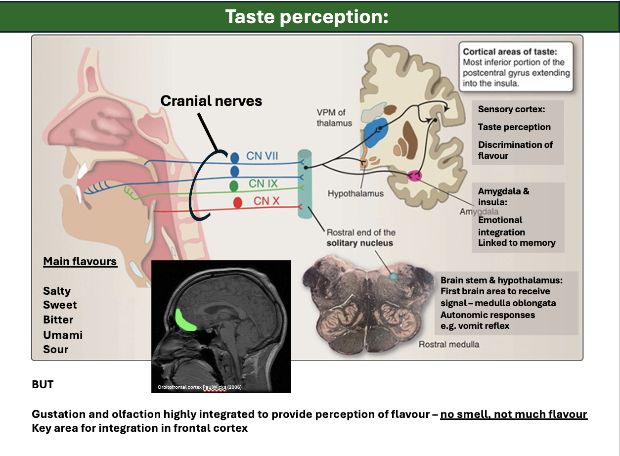
Explain the main brain areas involved in gustation and taste perception
Explain the general role and location of the olfactory bulbs
general role: provide direct neuronal connection to olfactory epithelium
location: base of the frontal lobe
Describe the olfactory epithelium and explain the role of each cell type found within it
Receptor cells – olfactory neurons (replaced – 30 to 60 days)
Support cells -
Physical / metabolic support
Detoxify odorants
Basal cells - Create new support cells
Olfactory cilia –
Embedded in mucus
Odorants dissolve into mucus
Odorant binding proteins – ‘collect’ odorant molecules
Receptors bind odorant molecules. depolarisation and AP to brain
Describe the olfactory tract and how it’s main parts function to facilitate olfactory signal processing
Explain the role of the hippocampus in olfactory processing and responses
Formation of memories linked to smell
Explain the role of the limbic system in olfactory processing and responses
Emotional responses to smell
Directing some behavioural responses to smell
Explain the role of the amygdala in olfactory processing and responses
Associative learning between smell, emotion, situations and behaviour
Explain the role of the Thalamus in olfactory processing and responses
Signals /information bypasses thalamus
Label the layers of the retina
Sensory layer (pigment epithelium layer): absorbs light detected by rods and cones
bipolar layer: transparent - light passes through most of retinal thickness
ganglion layer: transparent: light projected from lens
Note: Choroid layer - provides nutrients and removes waste

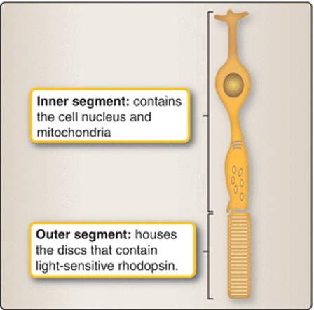
Rod cells
Light sensitive discs contain rhodopsin (~1000)
Rhodopsin responses - same for all wavelengths of light
So – CAN’T distinguish between colours
Highly light sensitive
Blunt rod-like shape
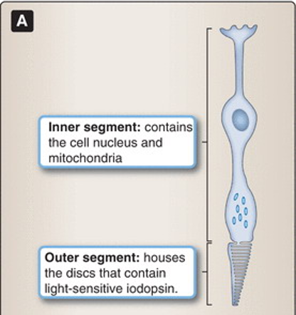
Cone cells
Light sensitive discs contain iodopsin
Iodopsin responses – encode different wavelengths of light
So – CAN distinguish between colours
Less light sensitive ~100x
Pointed tip
But shorter than rods
No disks – folds instead
Allows for rapid replacement of pigment layers
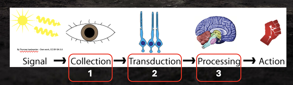
Define the three steps of sensation, collection, transduction, processing
Light entering eye is refracted
Image reversed and inverted
Neural processing corrects this
Lens changes shape – focusing light
Ciliary muscle control lens
Rounder lens – visual field closer
Flatter lens – visual field distant
Explain the dorsal and ventral visual streams
Explain the two main retinal specialisation, the fovea and the optic disk
Fovea:
Most light projected onto fovea
Retinal layers pushed aside
Direct access to photoreceptors
Mainly cones
High acuity and colour sensitive
Optic Disk:
Axons from ganglion cells
Converge on optic disk to create optic nerve which carries signal to brain
Briefly explain the role of the pupil
Controls ‘amount’ of light
Dark – pupil dilates
But this allows ‘stray light’ to degrade focus / depth of field
Describe how accommodation works
The focussing of light- by the lens changing shape
Ciliary muscle control lens
Rounder lens – visual field closer
Flatter lens – visual field distant
Explain why the retinal image is inverted because of refraction and that neural processing corrects for this
Pupil constriction
controls the amount of light that enters eye
problematic: scatterable light = not quite as accurate in dark
Retinal specialisations
optic disc
fovea
Fovea
most light is focussed to
2 outer layers of retina (ganglion and bipolar layer) are pushed aside to provide better access to photoreceptors
high acuity and colour sensitive
Lateral geniculate nucleus
relay center
there is some facsilitation to focus and concentrate on specific areas of visual field
Outputs from visual cortex
Visual info and proprioceptive info integrated in parietal cortex
dorsal visual stream (the “where/’how” pathway): occipital lobe to sensory associated areas. allows us to have spacial awareness in movements and relationships
ventral visual stream (the “why” pathway): insula and temporal lobe memory areas (what is this thing that I am looking at, what does it mean to me?), object and facial recognition
3 steps to the broad understanding of our senses
collection
transduction
processing