Lecture 3 - Cells
 Call Kai
 Learn
Learn Practice Test
 Spaced Repetition
 Match
Match Flashcards
 Knowt Play
1/44

There's no tags or description
Looks like no tags are added yet.
Name | Mastery | Learn | Test | Matching | Spaced | Call with Kai | Chat |
|---|
No analytics yet
Send a link to your students to track their progress
45 Terms
Functions of the Plasma Membrane
Mechanical barrier that separates the inside (intra-) and outside (extra-) cell fluids
Selective permeability that controls what goes in and out
Electrochemical gradient that maintains the inside of the cell being more negative than the outside
muscle and nerve cells can change their gradient
Communication that allows for cell-to-cell recognition
Cell signaling that interacts with extracellular chemical messengers and relays messages to the inside of the cell
Plasma Membrane Components
Phospholipids: two layers with hydrophobic tails on the inside and hydrophilic heads on the outside
Glycolipids: only on the outer membrane surface and contain a sugar group
Cholesterol: adds stability


What are integral proteins?
most span the entire membrane (transmembrane)
have both hydrophobic (interact with lipid tails) and hydrophilic regions (interact w water)
function as transport proteins, enzymes, or receptors
What are peripheral proteins?
loosely attached to integral proteins
function as enzymes and cell-to-cell connectors
Tight Junctions
think of as a seal so things can’t pass through
integral proteins on cells touching together that fuse to form an impenetrable junction that prevents fluids and most molecules from moving between cells

Desmosomes
think of as velcro between cells that withstands tension so cells don’t rip
formed when linker proteins (peripheral proteins) of neighboring cells interlock like the teeth of a zipper
are anchored to its cell through thickened “button-like” areas on the inside of the membrane called plaques

Gap Junctions
think of as a small tunnel between cells for small things to pass
transmembrane proteins that form tunnels that allow small molecules to pass through from cell to cell
spreads ions, simple sugars, and electrical signals to be passed quickly from one cell to the next

Passive Transport
does NOT require energy
ALL forms of diffusion are passive
simple, facilitated, osmosis
filtration is a form —> it usually occurs through capillary walls
Diffusion
movement of molecules from an area of high concentration to low concentration (down their concentration gradient)
smaller molecules = faster diffusion
bigger molecules = slower diffusion
higher temps = faster diffusion
lower temps = slower diffusion
Simple diffusion
non-polar and lipid-soluble/hydrophobic substances diffuse through the lipid bilayer cell membrane
Carrier-mediated facilitated diffusion
substances bind to protein carriers
think of a revolving door opening on one side to let the molecule in then turning and reopening on the other side to let it out

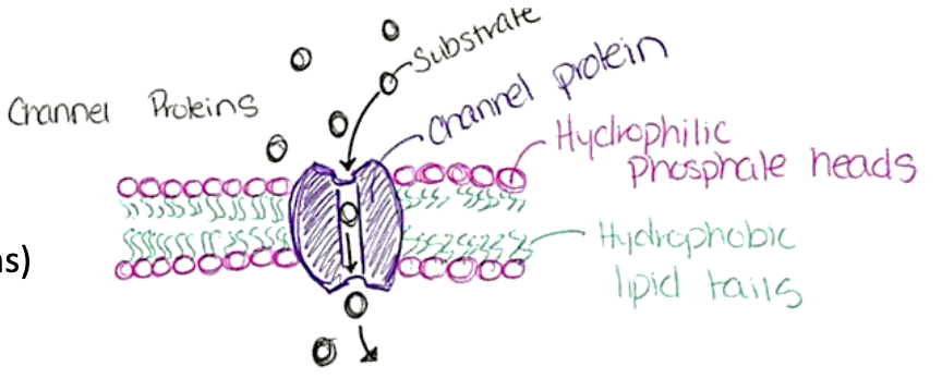
Channel-mediated facilitated diffusion
substances move through water-filled channels
leakage channels are always open
gated channels are controlled by chemical or electrical signals
Osmosis
is a form of diffusion = does not require energy
movement of water across the plasma membrane in 2 ways
via diffusion through the lipid bilayer
through small water channels called aquaporins
water moves from low concentration to high concentration based on substance concentration
Water is _____
hypotonic
What happens to cells in an isotonic solution?
they retain their same size and shape because the solute and water concentrations are the same inside and outside
What happens to cells in a hypertonic solution?
the outside solution has a higher concentration of solutes so the cell shrivels up as fluid leaves the cell to try to balance the concentrations
What happens to cells in a hypotonic solution?
the inside of the cell has a higher concentration of solutes so the cell expands as fluid enters it to try to balance the concentrations
may cause the cell to burst if it gets too full
Tonicity
the ability of a solution to change the shape/tone of cells by altering the cell’s internal water volume
Active Membrane Transport
requires energy to move solutes across a plasma membrane when solute is too large/non-polar, not lipid soluble, or is moving against its concentration gradient
uses carrier proteins but differs from carrier mediated facilitated diffusion
ATP hydrolysis changes the shape of the carrier protein to allow the solute to be pumped across
Ex: Sodium-Potassium Pump
the enzyme Na+-K+ ATPase pumps Na+ out of the cell and K+ into the cell AGAINST concentration gradients
maintains electrochemical gradients = essential for muscle and nerve tissues
Vesicular Transport
think as a carrier bubble that moves substances
takes energy!!!
4 Processes
Endocytosis: transport into the cell
1. Phagocytosis: for large things like bacteria
2. Pinocytosis: small things like extracellular fluid
3. Receptor-mediated endocytosis: only when something binds to a receptor on the plasma membrane to initiate endocytosis
Exocytosis: transport out of the cell
Transcytosis: transport into, across, then out of the cell
Vesicular Trafficking: transport from one organelle in a cell to another

Non-Membrane Organelles
ribosomes, cytoskeleton, centrioles
Mitochondria
provides ATP for the cell to use as energy via cell respiration (requires oxygen)
has a double membrane (inner membrane has folds called cristae)
contain their own DNA, RNA, ribosomes
resemble bacteria —> can do same cell division called fission
Ribosomes
performs protein synthesis
non membranous
2 globular subunits
made of protein and ribosomal RNA (rRNA)
2 Forms
free ribosomes: free floating that synthesize proteins that function is cytosol or other organelles
membrane-bound ribosomes: attached to the rough ER and synthesize proteins that are incorporated into membranes or lysosomes

Endoplasmic Reticulum
made of flattened membranous tubes that enclose fluid-filled interiors called cisterns
continuous with the outer nuclear membrane
rough and smooth parts
Rough ER
rough surface bc has ribosomes attached
synthesizes proteins that attach to plasma membrane
synthesizes phospholipids
final protein is then enclosed in a vesicle and sent to golgi apparatus
Smooth ER
continuous with rough ER
contains enzymes within its membrane that function in:
lipid metabolism: synthesizing, breaking down, and transporting fats in cells to store energy, build cell membranes, and regulate body functions
cholesterol synthesis & making lipids for liver cells (lipoproteins)
synthesis of testosterone hormones in testes
absorption, synthesis, and transport of fats (in intestinal cells)
detoxification of chemicals and drugs (in liver and kidney cells)
breakdown of glycogen to glucose (in liver cells)
storage and release of calcium (in skeletal & cardiac muscle cells)
Golgi Apparatus
stacked and flattened membranous sacs
modifies, concentrates, and packages proteins and lipids from the rough ER
1. Transport vesicles from ER fuse with the inner side of Golgi
Proteins or lipids taken inside are further modified, tagged, sorted, and packaged
Golgi is “traffic director” controlling which of three pathways final products will take as new transport vesicles pinch off the outside side of Golgi
Lysosomes
main function is to digest things like bacteria, viruses, and toxins, break down old organelles, break down and release glycogen, break down and release calcium from bones
spherical membranous bags containing digestive enzyme
What is autolysis
when a cell digests itself
often happens when a cell is injured and the lysosome ruptures
Peroxisomes
spherical membranous sacs containing powerful enzymes
main function is to neutralize toxic substances and neutralize free radicals (which are toxic, highly reactive molecules that are by products of cell metabolism)
Oxidase: uses oxygen to convert toxins into H2O2
Catalase: converts H2O2 to water
a high amount found in liver and kidneys!!
Cytoplasm
elaborate network of rods that run through cytosol
microfilaments
intermediate filaments
microtubules
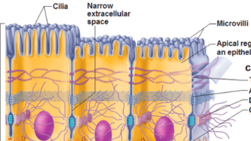
Cellular extensions of cytoplasm
Flagella: aid in the movement of the cell; sperm are the only cells w flagella
Cilia: move materials across the surface of the cell - in respiratory tract
cells in respiratory tract push mucus up the tract to the nose to dispose of it
are bigger than microvilli in length & width
Microvilli: fingerlike projections that increase surface area for better absorption - in digestive tract (small intestine) and kidneys
Nucleus
largest organelle
nuclei of all cells have the instructions for all cell types in them but they only use the one needed for their specific type of cell
“control center” - responds to signals dictating the kind and amount of proteins that need to be synthesized
most cells are uninucleate (1 nucleus)
skeletal muscle, some bone cells, and some liver cells have multiple nuclei
red blood cells have no nucleus
3 Structures
1. nuclear envelope: double-membrane barrier that encloses the nucleoplasm
2. nucleolus: contains the DNA codes for rRNA and synthesizes it
3. chromatin: tightly packed DNA
Role of DNA Protein Synthesis
DNA has the code for protein synthesis and dictates the AA sequences
Gene: segment of DNA with the code for 1 polypeptide
What is a triplet code?
3 sequential bases that code for a particular amino acid
ex: GCC, GCA, ACA, etc
What is RNA
acts as the messenger for protein synthesis because DNA cannot leave the nucleus
is a copy of the DNA code that is carried to ribosomes
RNA formed in the nucleus
single stranded, has Uracil and not Thymine, has ribose sugar
What is mRNA?
Messenger RNA
is an exact copy of the DNA we want to make (copied from template strand)
What is rRNA?
Ribosomal RNA
forms ribosomes (location of protein synthesis)
What is tRNA
Transfer RNA
carries the produced amino acids to ribosomes
Transcription - 1st Step of Protein Synthesis
DNA info coded into mRNA
RNA polymerase unzips DNA and adds complementary nucleotides to the sequence of the template strand onto the new RNA strand so that it becomes the RNA version of the coding strand
occurs in nucleus
Translation - 2nd Step of Protein Synthesis
the language of mRNA is translated to the language of proteins
pairs pf base sequence translated into amino acid sequence
each triplet code in DNA = called codons in mRNA (are both 3 base sequences)
multiple codons code for the same AA to protect against transcription errors
essentially mRNA codons are paired with tTNA anticodons to form amino acid sequences to synthesize proteins
anticodon sequence of tRNA is identical to DNA (except has U instead of T)
tRNA in Translation
Structure
has an anticodon (3 bases) on one end and the opposite ends to the corresponding amino acid
the anticodon end pairs with the mRNA codon that is complementary
ex: codon AUA bonds with anticodon UAU
Function
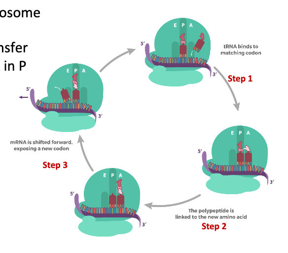
tRNA binds codon on A site of ribosome
ribosomal enzymes attach polypeptide chain from tRNA in P site to the A site where the amino acid of tRNA is attached
ribosome displaces tRNA
A site moves to P site
P site moves to E site
E site is ejected from ribosome
How to differentiate between triplet code, codon, and anti-codon charts
triplet code chart has T bases in it
if the chart has U bases in it you know it is either codons or anti-codons but cannot differentiate unless told it is mRNA or tRNA
mRNA = codons
tRNA = anti-codons