Exam #2 (Nervous system introduction, neurophysiology, and synapses)
1/111
There's no tags or description
Looks like no tags are added yet.
Name | Mastery | Learn | Test | Matching | Spaced | Call with Kai |
|---|
No analytics yet
Send a link to your students to track their progress
112 Terms
somatic sensory receptor
- Monitors things that are in
- Skeletal, joints, skin, muscles
- Monitored by receptors
somatic sensory visceral
- Monitor your internal organs
- Blood vessels, heart, lungs, bladder
- Monitored by receptors
Types of receptors:
- Chemoreceptors (oxygen levels, CO2 levels, pH levels, calcium levels)
- Baroreceptors (blood pressure, pressure in hollow organs like bladder and lugs)
- Thermal receptors (body temperature, inner and external)
PNS = Peripheral nervous system:
- Neurons that are outside of the brain and spinal cord (afferent division) going into the brain or spinal cord (afferent pathway)
- Everything outside of the brain and spinal cord
Control center (integration center) = brain and spinal cord:
- This is your CNS (central nervous system)
- Makes the decisions to change things, to maintain homeostasis within the body
- Sends information out alongside the PNS (efferent division (motor))
- This is your efferent pathway (leads to effectors)
- One goes to the somatic system and the effectors for this is the skeletal system
- Another division it goes to is the autonomic system which goes into two divisions (parasympathetic nervous system and sympathetic nervous system)
Parasympathetic nervous system and sympathetic nervous system:
- Both have smooth muscle, cardiac muscle, adipose tissue, and glands
Central nervous system:
- Brain + spinal cord
- Acts as an integration center (meaning they get much info from the neurons)
- Processes the info and sends signals out to the effectors
Afferent nervous system:
receptors to CNS
efferent nervous system
CNS to effectors
somatic nervous system
- Under efferent nervous system
- Affects skeletal muscle
Autonomic nervous system:
- Under efferent nervous system
- Smooth muscle, cardiac muscle, adipose tissues, glands
Parasympathetic nervous system:
- Under autonomic system
- Controlling the calm state of the body (rest + digest)
Sympathetic nervous system:
- Under autonomic system
- Prepares you for action (fight or flight)
- Gets your respiratory rate up
Neuroglia or glial cells:
- Support, nourish, and protect our neurons
- Help maintain the nervous system
- Outnumber the neurons 10 to 1
- Don’t generate nerve impulses
Astrocytes (CNS):
- Largest and most humorous
- Maintain blood-brain barrier (protects brain from pathogens)
- Tail is hydrophobic and heads are hydrophilic
- If it can’t fit through the cell membrane, then it has to go through the membrane protein channel (water, ions like sodium, chloride,
Oligodendrocytes (CNS):
- Wrap around axons of neurons (multiple layers with myelin)
- Provides myelination (multiple layers)
- Can’t help regenerate or repair damaged axons
Microglia cells (CNS):
- Phagocytes (engulf and destroy)
- Gets rid of cellular debris, waste products and pathogens
Ependymal cells (CNS):
- Found in ventricles of brain and spinal cord central canal
- They make the cerebrospinal fluid that circulate the ventricles and goes down the central canal
Satellite cells (PNS):
- Their job is to regulate nutrient and waste exchange
- Making sure neurons stay healthy
- Ganglia – where you find cell bodies of neurons together
Schwann cells (PNS) :
- Myelinate axons of neurons in PNS
- Help regenerate damaged axons (like arms, the trunk, the legs)
neurons
- continue to create action potential along the length of the axon
- They also have no centrioles
- They don’t undergo mitosis, so they can’t make copies of themselves
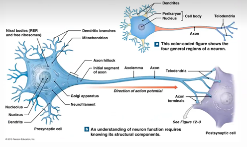
cell body
synthesizes proteins
perilkaryon
Nissl bodies
rough ER
dendrites
receive signals, transmit info to cell body
axon
action potential (AP)
axon hillock
generates action potential between the cell body and axon
initial segment of axon
generates action potential between the cell body and axon
axoplasm
cytoplasm in axon
axolemma
cell membrane of axon
axon collaterals or telodendria
segment before the synaptic terminal or axon terminal
axon terminal or synaptic terminal
attaches to the effector cell, looks like a bulb
node of Ranvier
where two Schwann cells meet
If the neuron or axon has myelin near the oligodendrocyte or Schwann
it is considered myelinated
- not all neurons are myelinated, some of unmyelinated
Benefits of myelination
- Action potentials travel faster and further
axoplasmic transport:
- Neurons have a way to transport stuff down to the collaterals
- We also need a way to transport waste products from the collaterals back to the cell body to get rid of it
- Cell body to the terminals is called anterograde – uses a protein called kinesin
- Terminals going to cell body is called retrograde – uses a protein called dynein
- Kinesin and dynein lay down like a pathway and gets walked on by a motor protein carrying a vesicle
Multipolar neurons
most common – skeletal muscle – multiple structures attached to the cell body
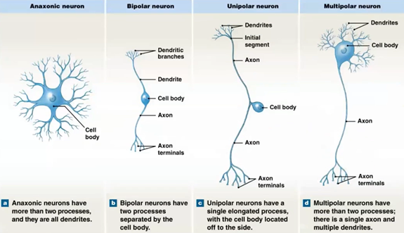
bipolar neurons
special senses – eyes, ears, nose, tongue – only two projections

unipolar neurons
PNS – sensory – going into the CNS – only has one projection going from the cell body to wherever, cell body sits off to the side of the axon

anaxonic neurons
without an axon – found in the brain – cannot produce action potential – just the cell body

motor (efferent) neurons
transmit nerve impulses from the CNS to effectors
somatic motor neurons
innervate skeletal muscles
visceral motor neurons
innervate smooth muscle, cardiac muscle, glands, and adipose tissue
interneurons (association neurons)
distribute sensory information and coordinate motor activity; information processing; memory, planning and learning; brain and spinal cord
protons
have a positive charge

electrons
have a negative charge
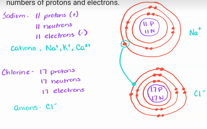
ions
have a net charge because of unequal number of protons and electrons

the movement or flow of charges make up an
electric current which is similar to the flow of water through pipes

voltage or potential difference
when we separate positive and negative electrical charges
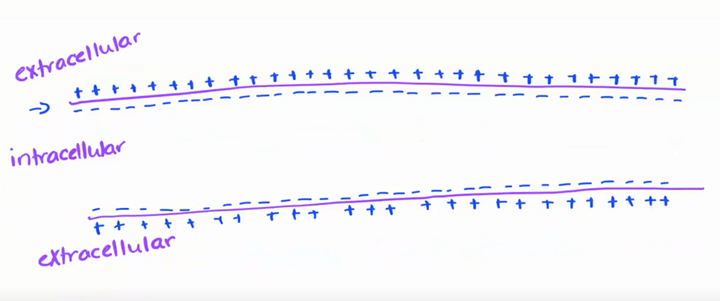
transmembrane potential
positive and negative charges are separated across cell membrane
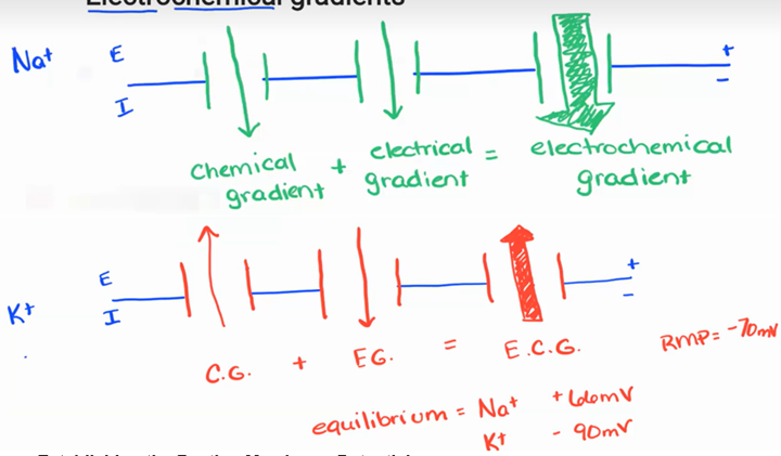
because the inside is more negative than the outside we say the transmembrane potential of a resting neuron (resting membrane potential) is –70 mV
also means the neuron is not transmitting signals

ions are distributed
unequally
extracellular cation
Na+ and Cl-
Intracellular cation
K+
proteins are what charge
negatively charged
cell membranes are
semipermeable
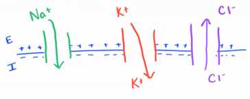
What causes the ions to flow into or out of the cell if the membrane channels are open?
chemical gradients
chemical gradients
move from high to low concentration
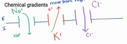
electrical gradient
opposites attract
movement based on charge
electrochemical gradient
when both chemical and electrical gradient are working at the same time
how do cells return to RMP after a change in membrane potential
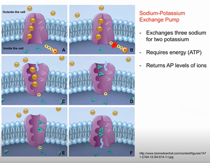
Sodium-potassium (Na+ - K+) ATPase pump
Sodium-Potassium (Na+ - K+) ATPase pump
- Move against concentration gradient
- 3 Na+ out
- 2 K+ in
- Keeps it at -70 RMP
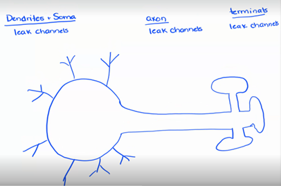
- Found alongside the entire neuron (dendrites + soma, axon, and terminals)
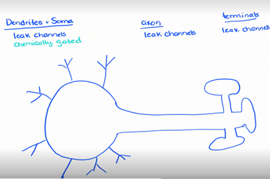
passive channels or leak channels
- Always open
- K+ leak channels
- Na+ leak channels
- Found on the dendrites, soma, axon, and terminals
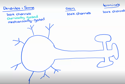
chemically regulated (gated)(ligand regulated) channels
- Open or closed in response to a specific chemical
- Found in the dendrites and soma
mechanically regulated channels
- Going to open or close in response to a membrane distortion – touch, pressure, vibration
- Found only in dendrites
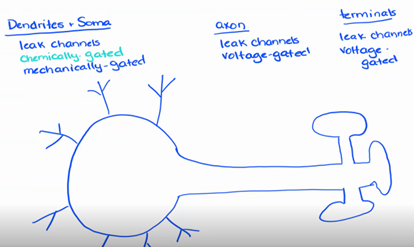
voltage regulated channels
- Open or close in response to a change in the transmembrane potential – change in voltage
- Found on the axon and terminals
When a neuron is stimulated by a signal from another neuron…
a ligand binding to a chemical channel or a shape change in a mechanically regulated channel it causes small local disturbances in the membrane potential.
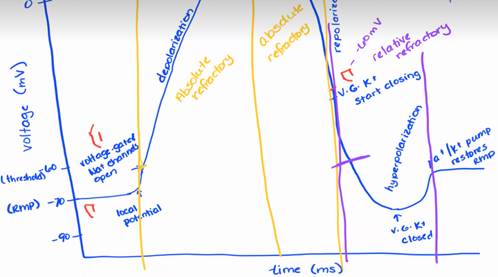
depolarization
- Overall goal: axon hillock to reach threshold (-60mV)
- Ways to reach the goal would be to open channels for a more stronger stimulus until it reaches the axon Hillock or open a channel more closer to the axon hillock
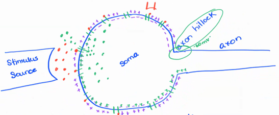
local potential - short distance
Incoming Na+ ions diffuse short distances from the initial site producing a current along the dendrite and cell body toward the axon hillock or trigger zone

graded local potential
strength varies in magnitude dependent on stimulus – open more channels or keep them open longer
decremental local potential
signal weakens the further it travels
reversible local potential
remove the stimulus, you stop the signal and allows the neuron to go back into resting membrane potential
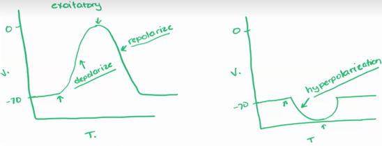
excitatory
Open Na+ channels
depolarization

Inhibitatory
Open K+ channels
Hyperpolarization
repolarization
Na+-K+ ATPase pumps return cell to resting membrane potential
hyperpolarization
to make more negative

neurons can generate
an electrical signal or action potential
voltage-gated channels
ion channels that produce action potential
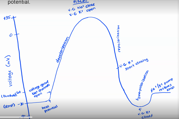
action potential
Local potential at axon hillock increases until it rises to threshold
Neuron produces an action potential; voltage-regulated Na+ channels open; more and more Na+ gates open as Na+ enters the cell; K+ gates open more slowly when threshold is reached (rapid depolarization)
When 0mV is reached/passed, Na+ gates are; voltage peaks at approx. +35mV (0mV in some, +50mV in others)
K+ gates now fully open; K+ leaves the cell repolarizing the membrane; causing shift back to negative inside and positive outside
K+ channels remain open a little longer than the Na+ channels and more K+ leaves than Na+ came in causing a 1 or 2 mV overshot or hyperpolarization
characteristics of action potential
all or none rule
no signal degradation
irreversible
all or none rule
if threshold reached, action potential will occur
no signal degradation
when action potentials are created along the length of the axon, those action potentials will remain the same strength all the way down
irreversible
once we start action potential, removing the signal will not stop the process
refractory period (impossible or difficult to make another action potential on a membrane segment)
During an action potential and a few msec after, it is difficult or impossible to stimulate to produce another action potential
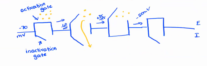
absolute refractory period
- No matter what, we cannot generate another action potential
- Threshold to 35+ mV – all Na+ channels are open
- +35mV to -50mV the inactivation gate is closed and won’t reopen

relative refractory period
Very strong stimulus may generate another action potential
Unmyelinated fibers (continuous propagation)
will always go towards terminal and never towards soma because of absolute refractory
Myelinated fibers (saltatory propagation)
- Skipping parts of the membrane for depolarization
- Can only do at the nodes of Ranvier with Schwann cells
Axon diameter and propagation speed
- Myelinated = faster
- Larger diameter axon = faster
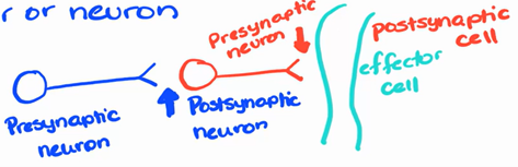
Synapse
a specialized site of contact between two neurons or a neuron and an effector (gland or muscle) that allows one-way flow of neural impulses
Neuron —> effector or neuron
Neuromuscular junction
neuron to muscle cell

Neuroglandular junction
neuron to a gland

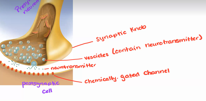
synaptic bulb
· tips of the presynaptic neuron that contain synaptic vesicles containing a neurotransmitter that will aid in signal transmission across the synaptic cleft (20-30nm in length)
*top says axon collateral and presynaptic neuron
excitatory
depolarization and sodium channels
inhibitatory
hyperpolarization and potassium or chloride channels
acetylcholine (ACh)
- found in the CNS and PNS
- can have excitatory or inhibitory effects
- binds to the heart (inhibitory effect by slowing down heart rate)
- binds to smooth muscle within the digestive system (excitatory effect by speeding up digestion)
- really depends if they bind to a sodium channel or a potassium or chloride channel
- cholinergic synapse (where this neurotransmitter is released)
norepinephrine (NE)
- found in PNS
- can be excitatory or inhibitory
- binds to heart to speed up heart rate (excitatory)
- binds to digestive system to slow down digestion (inhibitory)
- released by adrenergic synapse
dopamine
- found in the brain
- can be excitatory or inhibitory
- loss of dopamine is associated with Parkinson’s disease
serotonin
- found in CNS
low levels can lead to emotions like depression, attention span
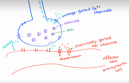
Synapse transmission - excitatory cholinergic synapse
· Action potential arrives at the synaptic knob; voltage-regulated Ca2+ channels in the synaptic membrane open
· Ca2+ enters synaptic knob and triggers exocytosis of ACh
· ACh diffuses across the cleft and binds chemically (ligand)-regulated Na+ channels; channels open allowing Na+ in = depolarize postsynaptic membrane 20ms
· influx of Na+ produces a local potential that carries to the axon hillock and if strong enough will generate an action potential
*top is the presynaptic neuron
Synapse transmission – inhibitory GABA-ergic synapse
- chemically gated Na+, K+, or Cl-
- norepi or ach (neurotransmitters), or GABA
- GABA opens chloride channels à inhibitory effect meaning hyperpolarization
Cessation (stopping) of the synaptic signal
While it is important to stimulate or inhibit the postsynaptic cell with a neurotransmitter, the stimulus must be turned off otherwise the effector will continue responding to the signal when it is inappropriate and this can be life threatening