Vitreous
1/58
There's no tags or description
Looks like no tags are added yet.
Name | Mastery | Learn | Test | Matching | Spaced | Call with Kai |
|---|
No analytics yet
Send a link to your students to track their progress
59 Terms
what is the largest structure of the eye?
- vitreous
- 80% volume of the eye
what is the size of the vitreous chamber in a newborn?
- 10.5
what is the size of the vitreous chamber in an adult?
- 16.5
what is 98% of the vitreous made up of?
- water
what are the non-aqueous component of the vitreous?
- collagen and glycosaminoglycans
- form the vitreous into visoelastic gel
what occurs to the gel content of the vitreous as you age?
- decreases
what does the existence of gel in the vitreous depend on?
- interaction between GAGs and collagen
what type of collagen is in the vitreous?
- 75% Type 2 Collagen
- 10% Type V/XI
what happens with liquifaction at age 18 and as you get older?
- liquifaction is already at 20% at 18 y/o
- progresses to 50% around the 80th decade
what difficulties arise from trying to investigate the vitreous?
1. the tissue we are trying to visualize in order to define as designed to be invisible
2. Previous techniques are combined with artifacts, so it is hard to make interpretations on the true in vivo situation
how is the primary vitreous formed during embryology?
- optic cup is occupied by the len's vesicle
- the cup grows, and the space is filled by fibrillar material secreted by embryonic retina.
- hyaloid artery penetrates and more fibrillar material from blood vessel cell wall fills the space
- the mass in the end is the primary vitreous
how is the secondary vitreous formed during embryology?
- the size of the vitreous cavity increases and hyaloid vascular system regresses developing the secondary vitreous
what is the canal that remains when the hyaloid disappears?
- Cloquet's Canal
what is cloquet's Canal?
- a tube of primary vitreous surrounded by secondary vitreous running from the retrolental space to the optic nerve
what are the suspensor fibrils that are developed from the fibrillary materal?
- zonules
what are termed the tertiary vitreous?
- zonules
what is the shape of mature vitreous body?
- spherical except at anterior end which is concave
what is the outermost part of the vitreous called?
- cortex
what is the base of the vitreous?
- three dimensional zone extending from 2mm anterior to 3mm posterior to the ora serrata; it is several mm thick
what is densely packed at the base of the vitreous?
- collagen fibrils
what is included in the outer part of the vitreous cortex?
- retina ILM and anchoring fibrils
how thick is the internal limiting membrane?
- 1-3microns thick
what is the internal limiting membrane composed of?
- mainly type 4 collagen and proteoglycans
what is considered the basal lamina of the mueller cells?
- ILM
where is the vitreous cortex firmly attached to the ILM?
- at the vitreous base
- around the optic disc (Weiss Ring)
- at the vessels
- in the area surrounding the foveola
what percentage of preretinal stands were found in non-symptomatic cases?
- 60%
- there was no biomicroscopic evidence of posterior vitreous detachment (PVD)
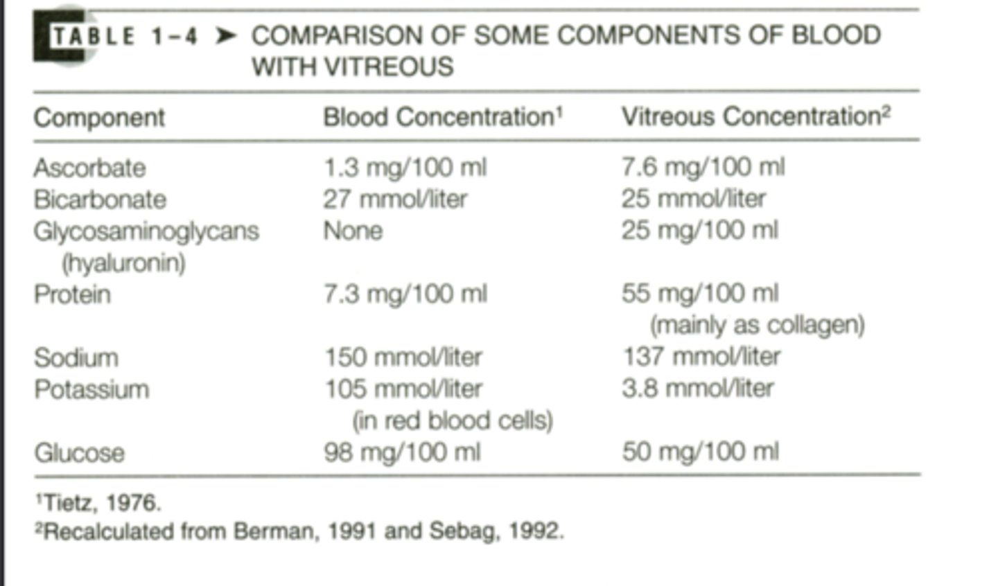
how do components of blood compare to the vitreous?
- vitreous has higher components of ascorbate, glycosaminoglycans (not in blood), protein(mainly as collagen)
- blood has higher components of bicarbonate, sodium, potassium and glucose
what does the gel structures of the vitreous act as for solutes?
- barrier against the movement of solutes
- substances move by: diffusion or bulk flow
what can be used as tracer substance to detect for diffusion?
- fluorescein
what is bulk flow?
- result of a pressure gradient from the anterior to posterior pole of the eye
- large, high molecular weight substances move due to this gradient
does bulk flow play a significant role for distribution of low molecular weight substances in the vitreous?
- no
what is the main change of the vitreous as we age?
- liquefaction of the gel structure, aka synchysis
where is synchysis most notable?
- in the center of the vitreous
what happens to gel structure during synchysis?
- gel structure is dissolved and replaced with aqueous lacunae (these melt together)
what happens to vitreous collagen molecular weight as you age?
- increases
- secondary to new covalent cross-links
what is Maillard Reaction?
- insoluable proteins produced by a covalent bond between an amino group and glucose
how does glucose in the vitreous of a diabetic patient compare to the vitreous of healthy patient?
- 2x more glucose
what is the function of the vitreous?
1. support for the retina and filling up function of the vitreous body cavity
2. Diffusion barrier between the anterior and the posterior segment of the eye
3. metabolic buffer function
4. establishment of an unhindered path of light
how does vitreous support function for the retina?
- helps prevent a large retinal detachment
- absorbs external forces to protect globe deformation
what is the incidence of retinal detachments in the US?
- 12 per 100,000
what is macula on retinal detachment vs macula off?
- macula on: macula not affected, VA is fine
- macula off: macula has detached, bad vision
is mac on or mac off retinal detachment considered ocular emergency?
- mac on because macula is not affected yet, still has chance of macula being affected, surgery that day
- mac off, the macula is already affected
what is posterior vitreous detachment?
- the central degeneration is large and causes a collapse
- the cortex sinks to the center of the vitreous body
is PVD considered a normal aging phenomenon?
- yes
what can result if there is a strong attachment between posterior cortex and the ILM?
- retinal tear
- 1st step in rhegamatogenous retina detachment
what is Weiss Ring?
- where vitreous is firmly attached to optic nerve head
is blood retinal barrier normally tight or loose?
- tight
what occurs to blood retinal barrier in diabetic macular edema?
- increased passive permeability and decreased outward active transport may lead to edema formation
what improved VA in patients who have diabetic macular edema?
- vitrectomy
do substances delivered from anterior segment easily reach the posterior part of the eye?
- no they have difficulties reaching high concentrations in the posterior part of the eye
- vitreous gel also prevents topically administered substances from reaching the retina and optic nerve
how does the removal of the lens and an anterior vitrectomy affect diffusion barrier between anterior and posterior segments of the eye?
- exchange between the anterior and posterior part of the eye is fast and easy
how does vitrectomy affect preretinal oxygen tension in diabetic patients?
- preretinal oxygen tension is improved in diabetic patients
- oxygen transport increases and retinal neovascularization and macular edema regress
what does the vitreous act as for the ciliary body and the retina?
- acts as metabolic buffer and reservoir for metabolism
- glucose and glycogen when needed for metabolism for the retina
- Vitamin C is present in high concentration and acts as a reservoir of antioxidants
can normal function of the retina be obtained after total vitrectomy?
- yes, the metabolic function of the vitreous do not seem to play a significant role
- retina substances are diluted by diffusing into the retina
what happens if the vitreous acts as a diffusion barrier to retinally produced vasoproliferation?
- they are retained in high concentrations close to the retina- depends on the state of vitreous
what could a vitrectomy cause ?
- could cause the vasoproliferation factors to move from the posterior to the anterior pole leading to neovascularization in the anterior segment
what is optical transparency of the vitreous produced by?
- low concentration of structural macromolecules
- degeneration of the vitreous with generation of opacities interferes with the path of light
what conditions interfere with transparency?
- synchysis scintillations
- asteroid degeneration
- hemorrhages
- inflammatory material
- fibrous tissue
- lack of regression of the hyaloid artery