Ocular Pathophysiology Exam 1 Review Content
 Call Kai
 Learn
Learn Practice Test
 Spaced Repetition
 Match
Match Flashcards
 Knowt Play
1/183

There's no tags or description
Looks like no tags are added yet.
Name | Mastery | Learn | Test | Matching | Spaced | Call with Kai | Chat |
|---|
No analytics yet
Send a link to your students to track their progress
184 Terms
systolic:
What is the range for a normal BP?
systolic: 120-129 AND diastolic:
What is the range for an elevated BP?
systolic: 130-139 OR diastolic: 80-89
What is the range for Stage 1 hypertension?
systolic: >140 OR diastolic: >90
What is the range for Stage 2 hypertension?
average based on two different readings on two separate occasions
Before labeling an individual with hypertension, what measurements must be taken?
tapping
What is heard during phase 1 of korotkoff sounds?
flow of blood from narrowed artery under the cuff to the wider artery distal to the cuff
What causes the tapping heard during phase 1 of korotkoff sounds?
systolic BP
What does phase 1 of korotkoff sounds, designated by tapping, represent??
phase 1, systolic
While taking the blood pressure of your patient, you first hear tapping. What phase of korotkoff sounds does this represent? What does this tell us?
murmur/swishing
What is heard during phase 2 of korotkoff sounds?
arterial wall vibration
Why is murmur/swishing heard during phase 2 of korotkoff sounds?
phase 2
While taking the blood pressure of your patient, you hear murmur/swishing. What phase of korotkoff sounds does this represent?
tapping/knocking
What is heard during phase 3 of korotkoff sounds?
phase 3
While taking the blood pressure of your patient, you hear tapping/knocking. What phase of korotkoff sounds does this represent?
muffling/swishing
What is heard during phase 4 of korotkoff sounds?
phase 4
While taking the blood pressure of your patient, you hear muffling/swishing. What phase of korotkoff sounds does this represent?
no sound
What is heard during phase 5 of korotkoff sounds?
phase 5, diastolic
While taking the blood pressure of your patient, you notice the disappearance of sound. What phase of korotkoff sounds does this represent? What does this tell us?
phase 5, diastolic - 2
While taking the blood pressure of your patient, you notice the last sound heard. What phase of korotkoff sounds does this represent? What does this tell us?
132/87
When measuring blood pressure of your 66 year old patient, you note tapping at 132 mmHg and the last sound heard at 89 mmHg. What blood pressure was measured?
132/89
When measuring blood pressure of your 66 year old patient, you note tapping at 132 mmHg and the disappearance of sound at 89 mmHg. What blood pressure was measured?
systolic: >180 OR diastolic: >120
What is defined as a hypertensive crisis?
urgency, emergency
What are the two categories of a hypertensive crisis?
systolic: >180 OR diastolic: >120, (-) TOD
What is defined as a hypertensive urgency?
systolic: >180 OR diastolic: >120, (+) TOD
What is defined as a hypertensive emergency?
acute ischemic or hemorrhagic stroke, acute myocardial infarction, acute heart failure, acute renal failure, disc edema
What are five examples of target organ damage?
disc edema
What is one example of target organ damage presenting in the eye?
do you have chest pain, dyspnea, headaches, altered mental status
A patient enter clinic with a BP of 181/111. When looking at the health of his eye, you notice papilledema and retinopathy. What other questions should you ask?
hypertensive emergency (due to presenting with TOD)
A patient enter clinic with a BP of 181/111. When looking at the health of his eye, you notice papilledema and HTN retinopathy. What is the classification of his blood pressure?
tell patient to go to the ER for an immediate BP reduction (released once 140/90 mmHg is reached)
A patient enter clinic with a BP of 181/111. When looking at the health of his eye, you notice papilledema and HTN retinopathy. You classify this case as a hypertensive emergency. What should you do?
do you have chest pain, dyspnea, headaches, altered mental status
A patient enter clinic with a BP of 180/125. When looking at the health of his eye, you notice no signs and symptoms of target organ damage. What questions should you ask the patient?
hypertensive urgency (no TOD)
A patient enter clinic with a BP of 180/125. When looking at the health of his eye, you notice no signs and symptoms of target organ damage. You ask the patient if they have any chest pain, dyspnea, headaches, or altered mental status, to which they reply no. What would you classify this case?
see PCP in 24 to 72 hours
A patient enter clinic with a BP of 180/125. When looking at the health of his eye, you notice no signs and symptoms of target organ damage. You ask the patient if they have any chest pain, dyspnea, headaches, or altered mental status, to which they reply no. You classify the patient has having a hypertension urgency. What do you recommend to the patient?
auscultatory gap
sound free period during the early phases when blood pressure is taken with a manual cuff
between phase 1 and 2
When does an auscultatory gap occur?
auscultatory gap (sound free period between phase 1 and 2)
When taking blood pressure, you notice tapping. As your continue blood pressure, the sound stops. after 10 mmHg, you note murmur/swishing. What occurred during the measurement?
measure palpebral systolic pressure and inflate 30 mmHg above to start measuring BP
How can you avoid the auscultatory gap?
radial, thumb
Palpebral Systolic Pressure 1: The first step is to palpate the __________ pulse 2 cm below the ________.
30
Palpebral Systolic Pressure 2: The first step is to palpate the radial pulse 2 cm below the thumb. You then have to inflate the cuff _____ mmHg above this value.
palpable systolic BP
Palpebral Systolic Pressure 3: The first step is to palpate the radial pulse 2 cm below the thumb. You then have to inflate the cuff 30 mmHg above this value. Then deflate the cute and note when the pulse returns. The pulse returning signifies the ________ ________ _____.
decrease in AV ratio, color change of vessels, AV crossings, hemorrhages, cotton wool spots, radial exudates
What are six ocular findings of hypertensive retinopathy?
decreases
With hypertensive retinopathy, the AV ratio decreases/increases.
cooper, silver
What are the vessel color changes that occur with hypertension retinopathy?
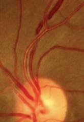
true
True or False: AV crossings can occur with ocular hypertension, leading to CRVO.
flame shaped (due to being in NFL)
What hemorrhages are visualized with hypertensive retinopathy?
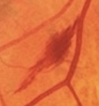
located in NFL
Why are hemorrhages visualized with hypertensive retinopathy flame shapped?

disrupted axoplasmic flow in RNFL, leading to ischemia
Why do cotton wool spots occur with hypertensive retinopathy?
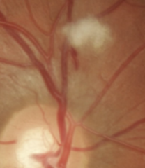
venous stasis and abnormal vascular permeability, leading to deposition of lipid and lipoprotein
Why do radial exudates occur with hypertensive retinopathy?

I
Type I/II diabetes is defined as the inability of the pancreas to produce insulin.
II
Type I/II diabetes is defined as ineffective insulin receptors.
80-130 mg/dl
For a diabetic individual, was is the goal FBS?
For a diabetic individual, was is the goal FBS 2 hours after a meal?
For a diabetic individual, was is the goal AbA1c?
fluctuating refractive error
What is the quintessential ophthalmic finding in undiagnosed diabetes?
myopia (increased glucose pulls water into the lens, leading to swelling and a myopic shift)
What shift in refractive error has been reported after an individual with diabetes has a hyperglycemia episode?
hyperopia
What shift in refractive error has been reported after an individual with diabetes has a hypoglycemia episode?
true
True or False: Even though a diabetic patient is experiencing shifts in refractive error due to changes in blood sugar levels, they will still commonly see 20/20.
hexokinase
What enzyme may not be present in diabetic patients, leading to increased water in the lens due to sorbital production?
sorbitol (aldose reductase converts glucose to sorbitol; can also lead to cataracts)
What pulls water into the lens of a diabetic?
re-refract 2-4 weeks after stabilization, refract 2-3x over 6 weeks, recommend temporary correction
You want to refract a patient who is having a myopic shift due to hypoglycemia, but you know this won't produce an accurate result. What should you do?
increase
Pathogenesis of Diabetic Retinopathy 1: When there is a(n) decrease/increase in blood sugar levels, there is a decrease in O2.
increased glucose leads to glycated hemoglobin, leading to decreased availability for O2 to bind
Pathogenesis of Diabetic Retinopathy 2: When there is an increase in blood sugar levels, there is a decrease in O2. Why does this occur?
vasodilation
Pathogenesis of Diabetic Retinopathy 3: When there is an increase in blood sugar levels, there is a decrease in O2. This occurs because hemoglobin becomes glycated, leading to decreased availability for O2 to bind. This will lead to chronic vasodilation/vasoconstriction.
thickening
Pathogenesis of Diabetic Retinopathy 4a: When there is an increase in blood sugar levels, there is a decrease in O2. This occurs because hemoglobin becomes glycated, leading to decreased availability for O2 to bind. This will lead to chronic vasodilation. This will lead to basement membrane thinning/thickening making it more difficult for O2 to travel through the BM.
pericyte
Pathogenesis of Diabetic Retinopathy 4b: When there is an increase in blood sugar levels, there is a decrease in O2. This occurs because hemoglobin becomes glycated, leading to decreased availability for O2 to bind. This will lead to chronic vasodilation. This will lead to ____________ degeneration.
endothelial
Pathogenesis of Diabetic Retinopathy 4c: When there is an increase in blood sugar levels, there is a decrease in O2. This occurs because hemoglobin becomes glycated, leading to decreased availability for O2 to bind. This will lead to chronic vasodilation. This will lead to ____________ cell proliferation.
BM thickening, pericyte loss, endothelial cell proliferation
Pathogenesis of Diabetic Retinopathy 4d: When there is an increase in blood sugar levels, there is a decrease in O2. This occurs because hemoglobin becomes glycated, leading to decreased availability for O2 to bind. This will lead to chronic vasodilation. What three changes will occur to the blood vessels?
small, large
Diabetes mellitus primarily effects small/large vessels and rarely impacts small/large vessels.
aggregation of RBCs, elevated prostaglandin, elevated thromboxane
What are three hematologic factors that occur with diabetes?
capillaries (impacts small blood vessels first)
Where are the earliest changes view with diabetes?
retinal ischemia, neovascularization
What are the two outcomes of capillary loss associated with diabetes?
microaneurysms, intraretinal hemorrhages, circinate hard exudates, edema, cotton wool spots
What are five ocular changes that occur with non-proliferative diabetic retinopathy?
false (just focal dilation, not actual hemorrhages yet)
True or False: The microaneurysms associated with NPDR is actually hemorrhages within the retina
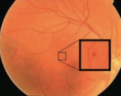
microaneurysms
What is the earliest sign of retinopathy with NPDR?

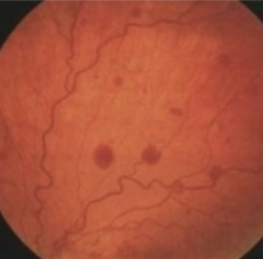
dot/blot
What type of hemorrhages are associated with NPDR?
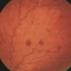
INL/OPL (due to capillaries being damaged)
Which retinal layer do dot/blot hemorrhages associated with NPDR occur?
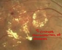
circinate rings
How do the exudates appear with NPDR?
true
True or False: CWS occur with both HTN and diabetic retinopathy.
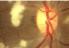
neovascularization, pre-retinal vitreous hemorrhage
What are two ocular changes that occur with proliferative diabetic retinopathy?
iris, angle, disc, elsewhere
Where can neovascularization occur with PDR?

edema (reversible, most common cause of vision loss with DR), ischemia and capillary damage, photoreceptor damage
What are three ocular changes that occur with CSME?
true (can cause decreased visual acuity)
True or False: CSME can occur at any time during diabetic retinopathy
flat, sharp borders
What is the normal physiology of the optic disc?
elevated, blurred margins
What is pathophysiology of the optic disc?
papilledema, disc edema, optic neuritis
What are differential diagnoses that you can make with the optic disc demonstrates elevated, blurred margins?
normal C/D ratio
What is the normal physiology of the optic cup?
enlarged C/D
What is pathophysiology of the optic cup?
glaucoma
What is the differential diagnoses that you can make with the optic cup if it has an enlarged C/D?
pink, good color
What is the normal physiology of the NRR?
pallor, hyperemic
What is pathophysiology of the NRR?
atrophy
What is the differential diagnoses that you can make if the NRR has pallor?
acute optic neuropathy
What is the differential diagnoses that you can make if the NRR has hyperemia?
papillitis
What is anterior optic neuritis termed?
retro-bulbar
What is posterior optic neuritis termed?
swollen
How does the optic nerve appear with anterior optic neuritis?
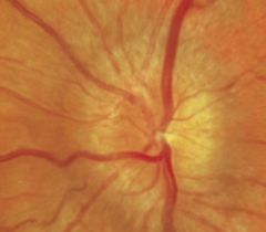
normal
How does the optic nerve appear with posterior optic neuritis?
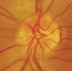
false (always unilateral, has (+) RAPD)
True or False: Optic neuritis is always bilateral, so it doesn't present with RAPD.
demyelinating disease, infectious
What are the two causes of optic neuritis?
MS (in 20% of MS patients)
What disease is optic neuritis most commonly associated with?
MRI (acute lesion of optic nerve, acute and chronic lesion in brain)
What test can patients suspected with a demyelinating disease be tested for?