Unit 3 Exam Study Guide - BIO 1121 - Anatomy & Physiology I - CSCC
1/211
There's no tags or description
Looks like no tags are added yet.
Name | Mastery | Learn | Test | Matching | Spaced |
|---|
No study sessions yet.
212 Terms
What are the three main functions of the nervous system?
- Sensory input
- Integration
- Motor output
sensory input
Information gathered by sensory receptors about internal and external changes
sensory integration
The process by which the brain integrates the information it receives from sensory input and determines what type of output or information it will send to the muscles, glands or organs.
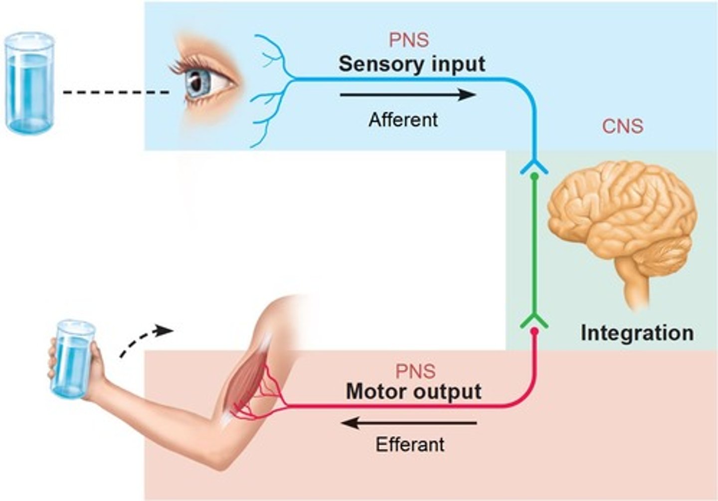
motor output
A response to integrated stimuli; the response activates glands or organs (effectors).
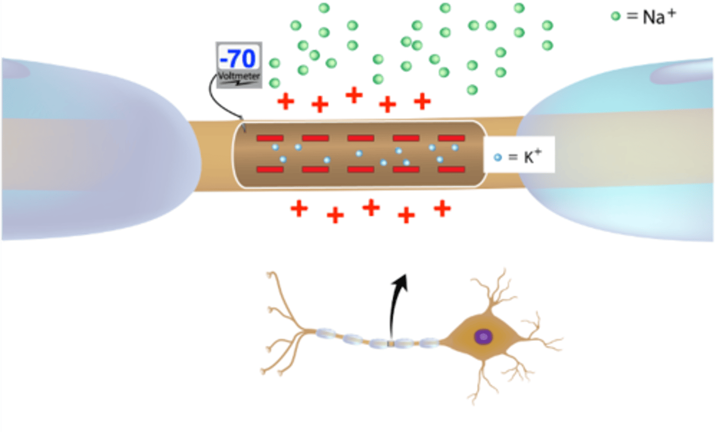
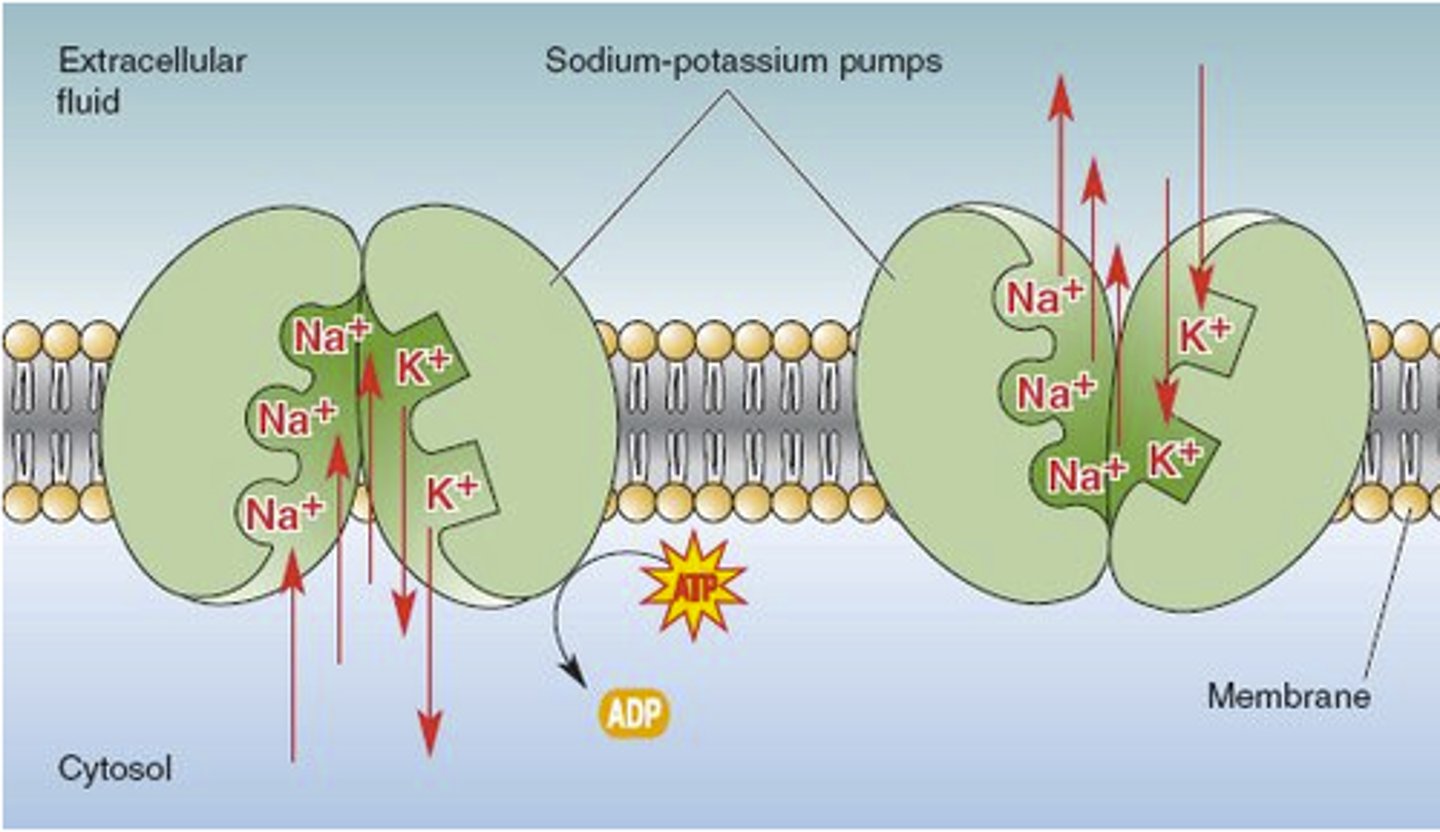
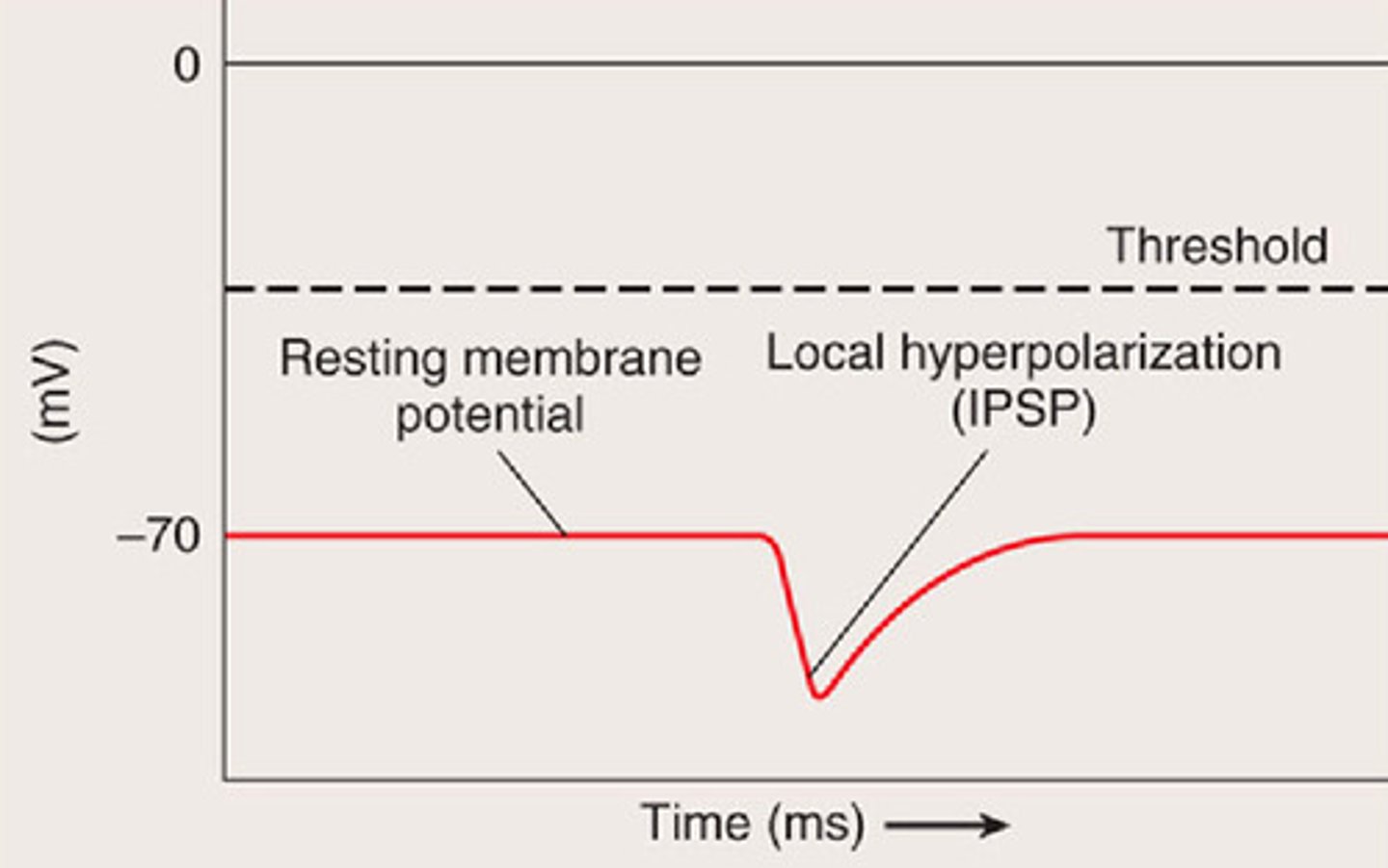
Resting Membrane Potential
The charge across the axonal membrane when the axon is at rest.
It is equal to -70mV
It is due to the relative permeability of the membrane (more permeable to K+ than to Na+) and the Na/K pumps. The result of these is that there are more positively charged ions leaving the cell than are entering the cell, causing the inside to be negative.
There is more potassium (K+) in the cell than sodium (Na+).
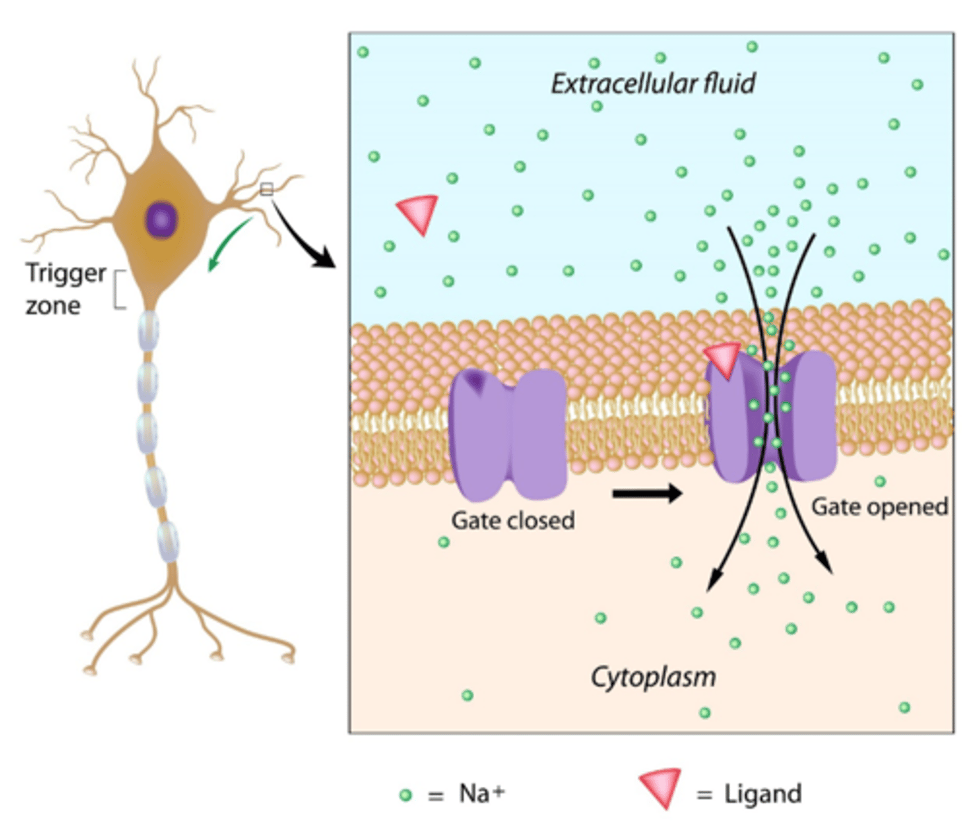
local / graded potential
Occurs when a neuron is stimulated. It is a message within one neuron; it does not travel through the nervous system.
This causes ligand-gated channels in the neuron membrane to open up.
These may be depolarizations or hyperpolarizations
If ligand-gated Na+ channels open, this graded potential will be a depolarization as Na+ diffuses into the neuron
If ligand-gated K+ channels open, this graded potential will be a hyperpolarization as K+ diffuses out of the neuron
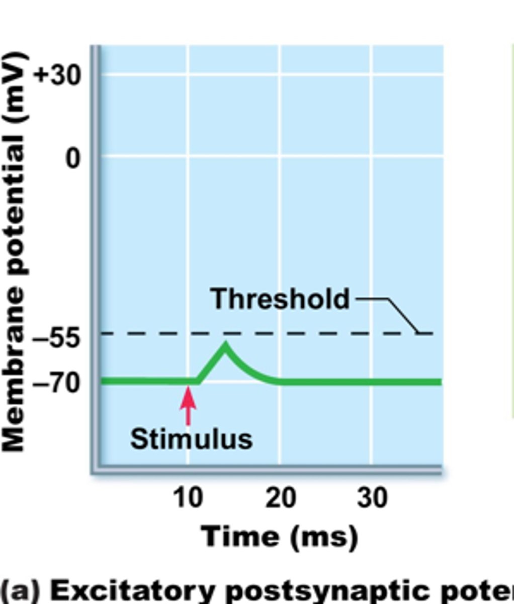
It can cause an action potential if it is a depolarization that causes the membrane voltage to get to -50 or -55 mV (this charge is called the threshold value and, at this charge, the voltage-gated Na+ channels open).
threshold
The point where enough graded potentials cause a neuron to conduct an action potential.
May occur with a depolarizing graded potential
The threshold is approximately -55 mV
Will cause VOLTAGE gated Na+ channels to open
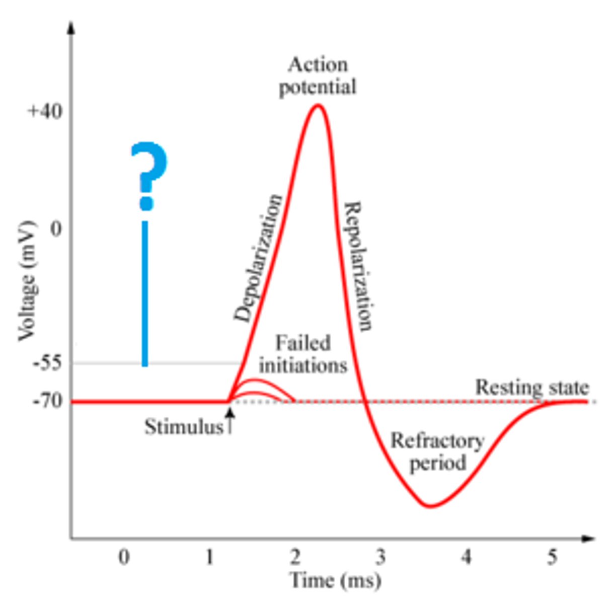
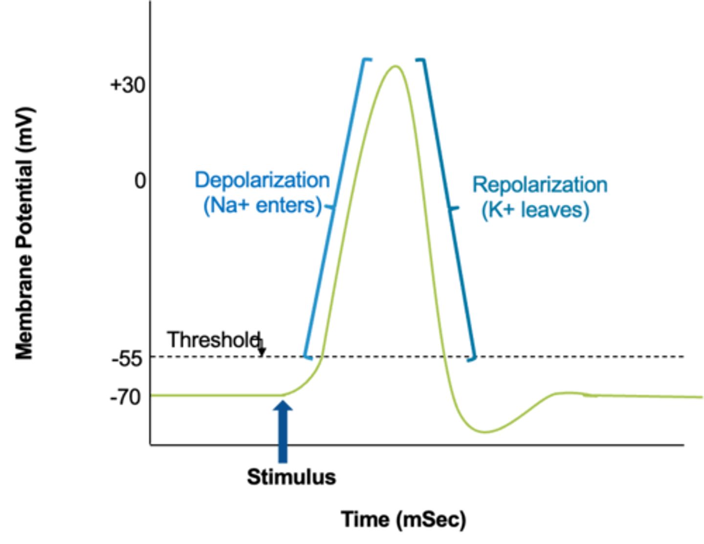
action potential
The electrical impulse that will travel the length of the axon and then be conducted on to the next cell.
Two phases of an action potential
1. depolarization
2. repolarization
depolarization
The process during the action potential when sodium is rushing into the cell causing the interior to become more positive.
- Voltage-gated Na+ channels open (threshold reached)
- Na+ diffuses into cell
- +30 mV inside cell
- Voltage Gated Na+ channels close

repolarization
- Voltage – gated K+ channels open
- K+ diffuses out of cell
- Voltage returns to normal (-70mv, RMP)
- But ions (Na+ and K+ have not returned to normal yet)
restoration phase
k+ close ion concentration is restored by Na+/K+ pump. return to normal state
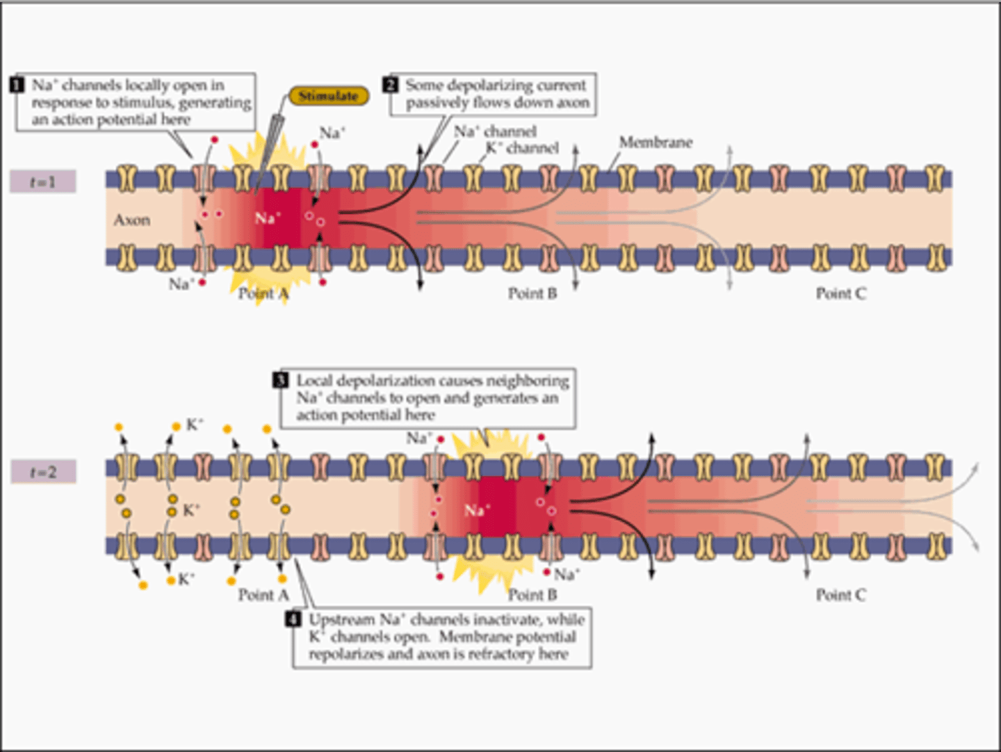
action potential propagation
The processes of depolarization and repolarization repeat, causing the action potential to "travel" down the axon, toward the axon terminal.

Steps of the Action Potential
1. Some stimulus occurs (before this, the membrane was at rest, -70mV)
2. Chemically gated Na channels open on the dendrites and cell body
3. Na+ enters the nerve cell
4. The inside is now more positive, -50 mV, which is threshold
5. Voltage gated Na+ channels at the axon hillock open up (and voltage gated K+ channels start to open).
6. Na+ enters the axon, making the charge in there more positive (+30mV). This is called the depolarization phase of the action potential.
7. Next, voltage gated K+ channels fully open
8. K+ will leave the axon, causing the voltage to go back down to -70mV. This is called the repolarization phase of the action potential.
9. The voltage is good now, but the ions (Na+ and K+) are in the wrong places.
10. So, the Na+/K+ pumps will work to fix the ion concentrations. Those pumps will pump Na+ back out of the axon and K+ back in to the axon.
11. This whole thing (steps 4-9 above, the action potential) just happened on one small section of the axon.
12. BUT once it happens, it will travel down the entire length of the axon, to the axon terminal. It can travel by local conduction (if the axon does NOT have myelin) or by saltatory conduction (if the axon has myelin)
13. When the action potential gets to the axon terminal, it causes the release of a chemical called a neurotransmitter.
14. The neurotransmitter will then bind to a receptor on the next cell (the postsynaptic cell)
15. That will cause chemically gated channels to open on the postsynaptic dendrites or axon
16. That then starts the whole process (back to step 1) on that next neuron.
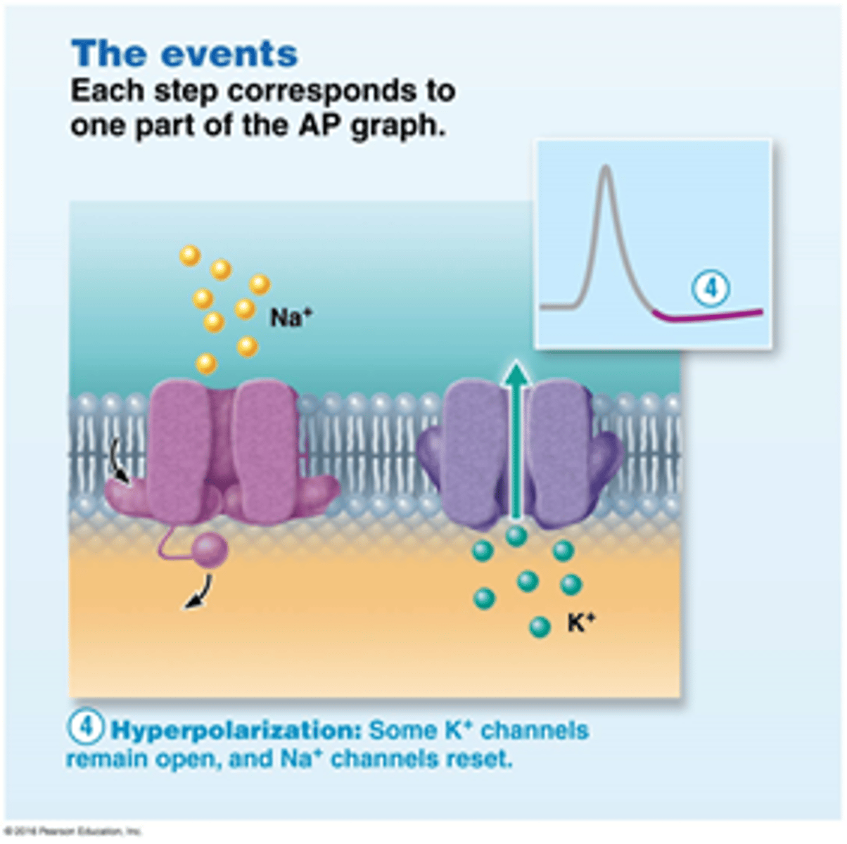
hyperpolarization
Membrane potential temporarily becomes more negative than resting membrane potential
Some K+ channels remain open, and Na+ channels reset.
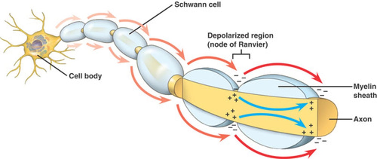
saltatory conduction
The way in which an electrical impulse travels in a myelinated axon.
The electrical impulse "jumps" from one Node of Ranvier to the next.
In contrast, in continuous conduction in an axon without myelin, the impulse must travel the entire length of the axon without "jumping" and therefore travels at a slower rate of speed.
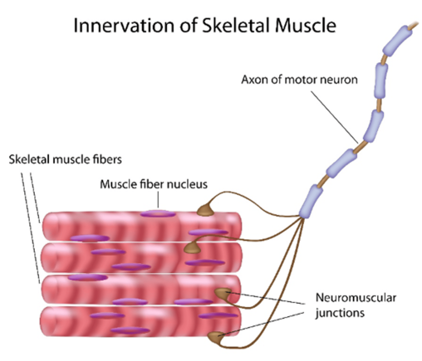
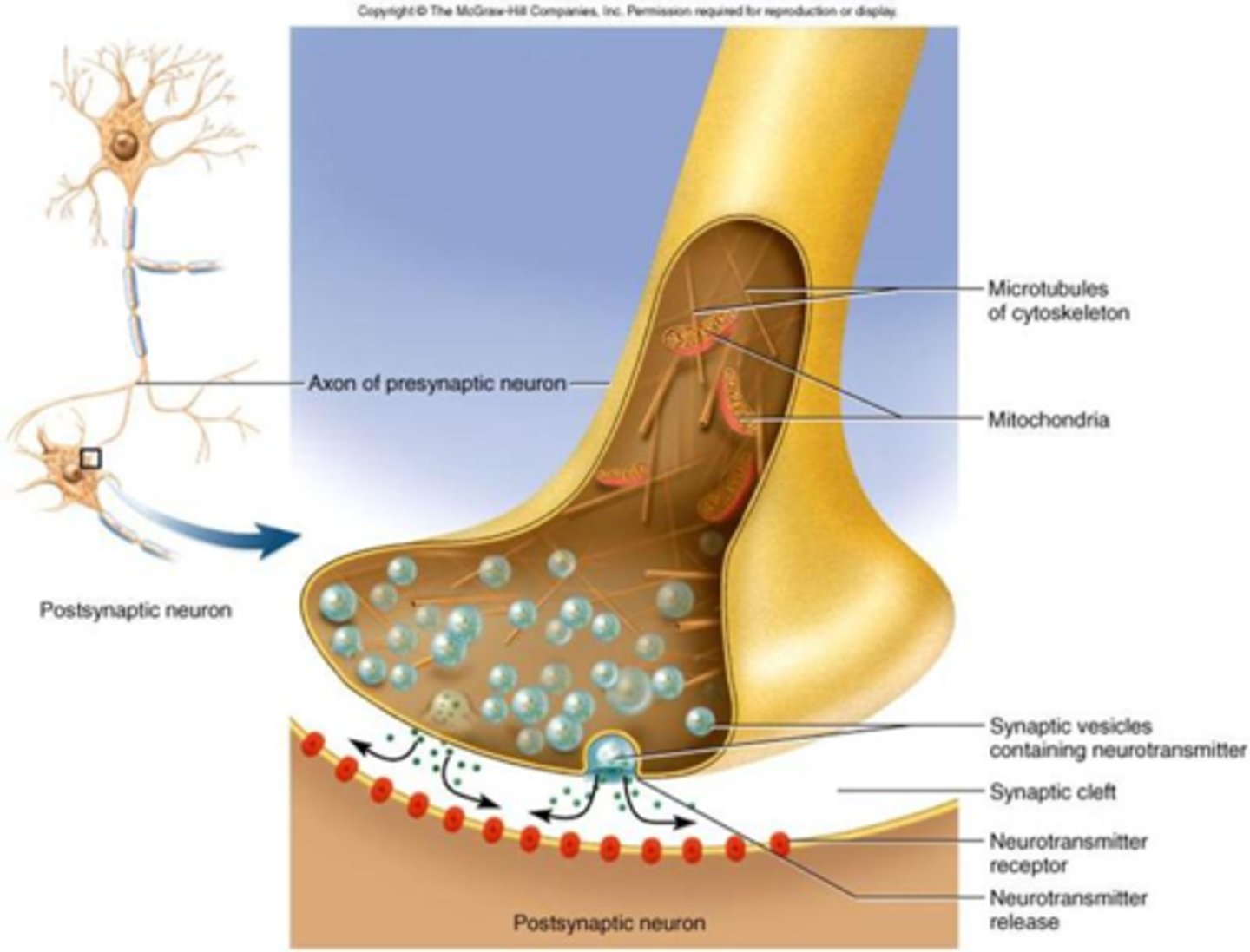
synapse
A space or gap between an axon terminal and another neuron or effector (muscle or gland).
The neurotransmitter is released into this space and then binds to a specialized receptor on the postsynaptic neuron or effector.
The neuromuscular junction is the name for the space between a nerve cell and the skeletal muscle fiber that it supplies.
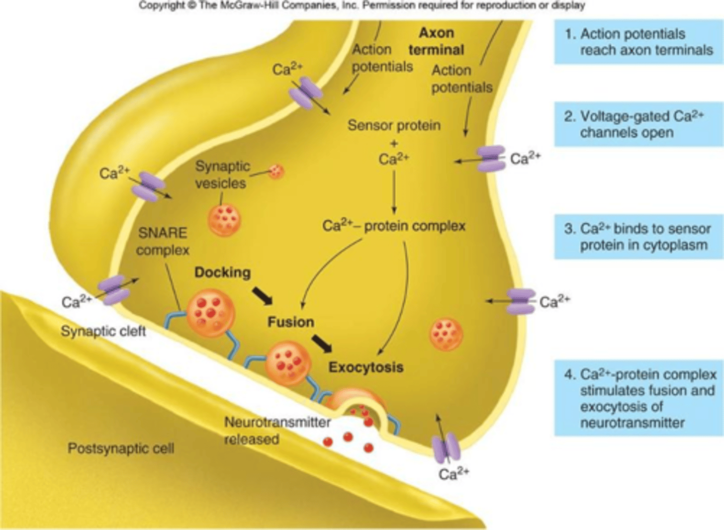
presynaptic events
1. Action Potential arrives at the axon terminal
2. Causes voltage-gated Ca+2 channels to open
3. Ca+2 enters axon
4. Causes release of neurotransmitter into the synaptic cleft
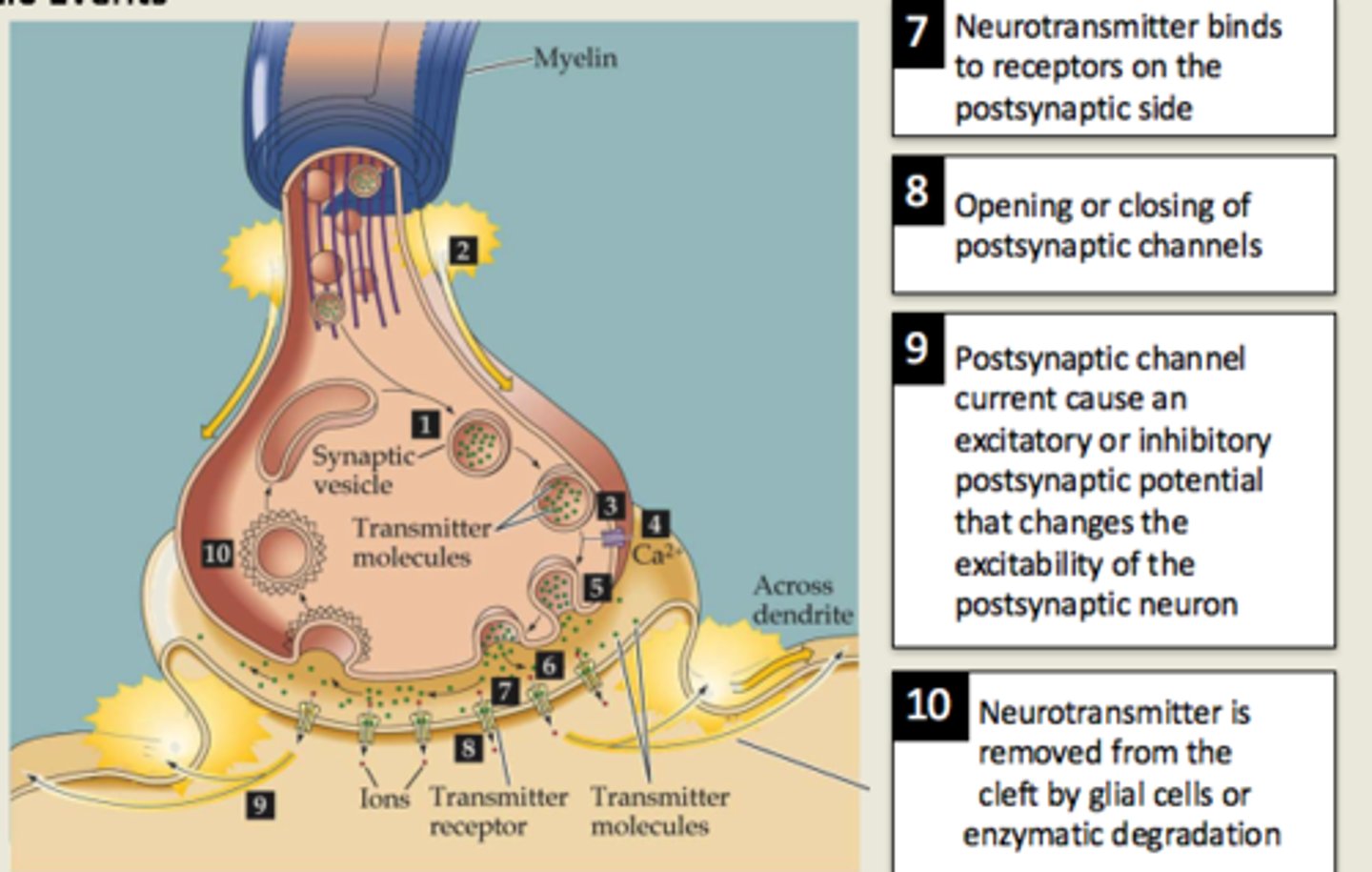
postsynaptic events
1. Neurotransmitter binds to receptors and causes ligand-gated channels to open.
2. This can cause:
- EPSP (Depolarization) – if ligand-gated Na+ channels open (and Na+ enters the postsynaptic cell)
OR
- IPSP (Hyperpolarization) – if ligand-gated K+ channels open (and K+ leaves the postsynaptic cell)
excitatory postsynaptic potential (EPSP)
A slight depolarization of a postsynaptic cell, bringing the membrane potential of that cell closer to the threshold for an action potential.
inhibitory postsynaptic potential (IPSP)
Graded hyperpolarization of a postsynaptic membrane which decreases the likelihood that the neuron will fire.
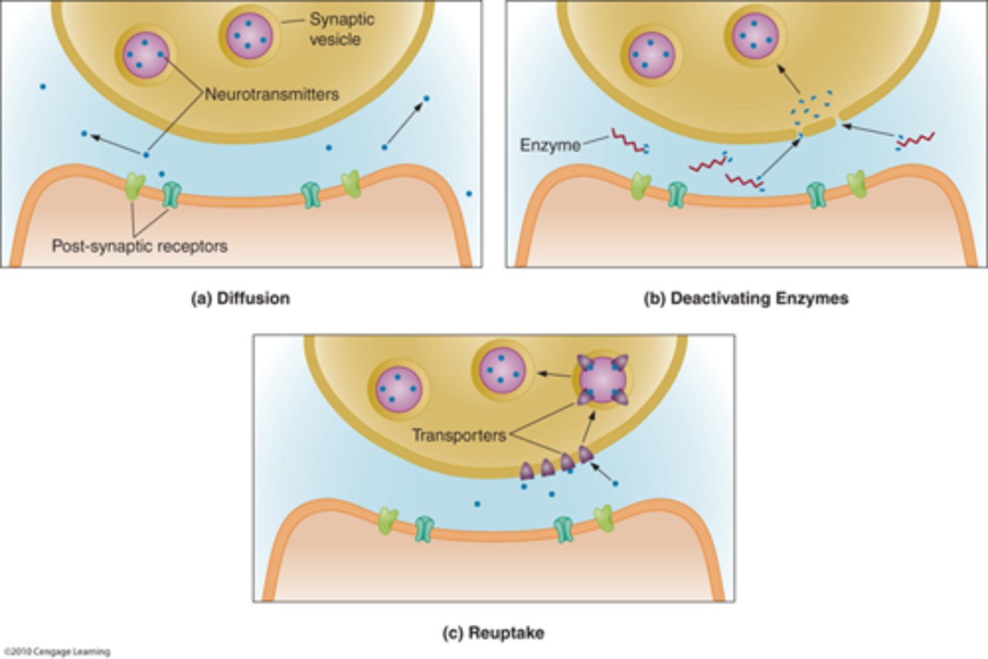
What three things can happen to a neurotransmitter after it has been released from its receptor?
Once the neurotransmitter is released into the synaptic cleft, it remains there for only a very short while.
Neurotransmitters are quickly removed from the synaptic cleft by the following mechanisms:
1. Diffusion
2. Reuptake - ex. The neurotransmitter serotonin is taken back to the presynaptic neuron by reuptake.
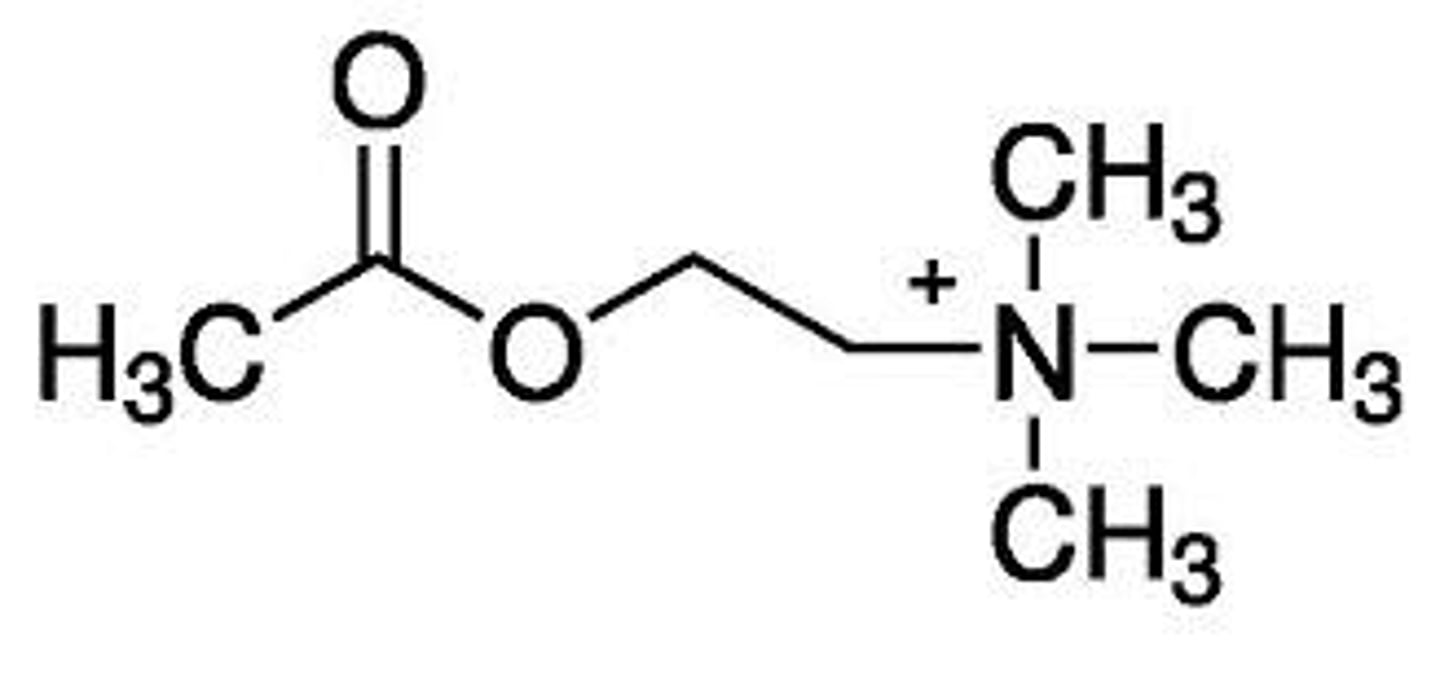
3. Degradation by synaptic enzymes – ex. The neurotransmitter acetylcholine is broken down to acetate and choline by the enzyme acetylcholinesterase.
Acetylcholine (ACh)
The main neurotransmitter of the parasympathetic nervous system and so it causes a decrease in heart rate, a decrease in blood pressure, an increase in digestive tract activity, constriction of the pupils (all the "Resting and Digesting" actions)
It can be either excitatory or inhibitory within the central nervous system (CNS).
It is the neurotransmitter at the neuromuscular junction, which causes skeletal muscle to contract.
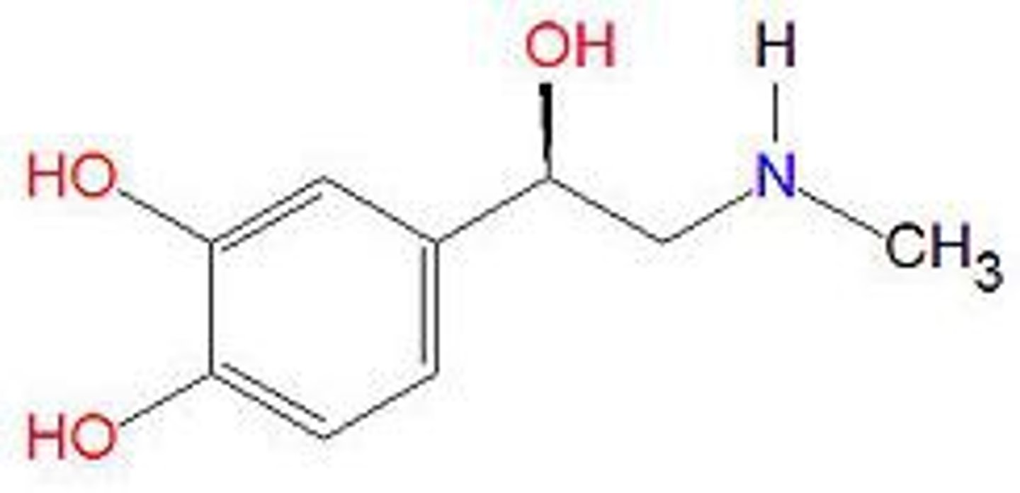
Epinephrine / Norepinephrine
The main neurotransmitter of the sympathetic nervous system. There, it causes an increase in heart rate, an increase in strength of muscle contractions, increased sweating, increase in blood pressure and decrease in digestive tract activity (all the "Fight or Flight" actions)
It can either be an excitatory or an inhibitory (due to its actions at different receptors).
Sympathetic stimulation increases secretion of this neurotransmitter.
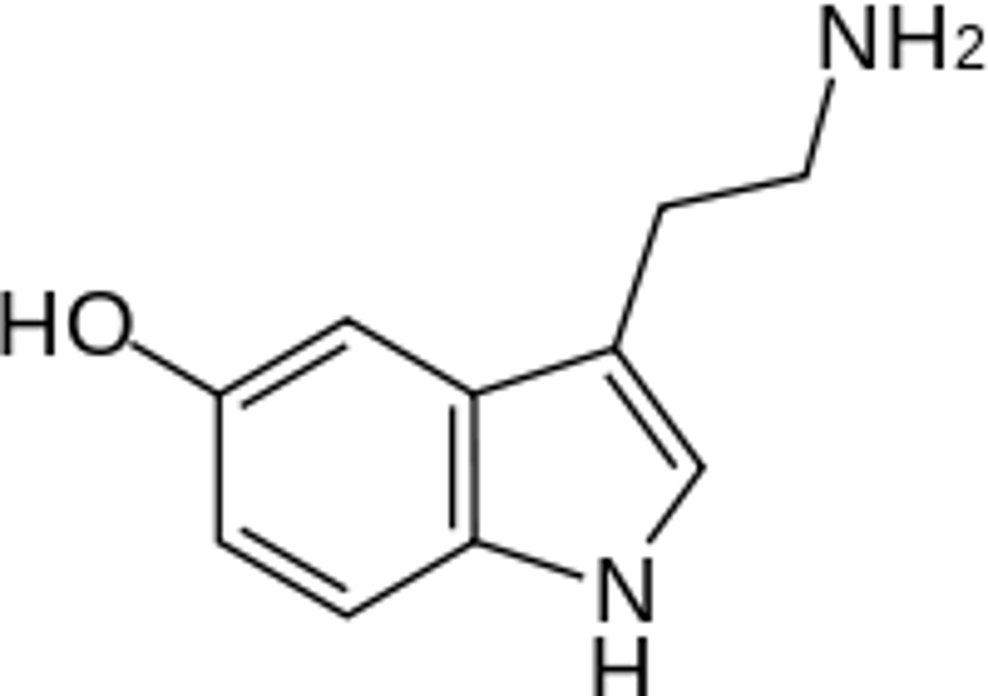
Serotonin
An inhibitory neurotransmitter in the CNS brain stem.
It helps with inducing sleep, anxiety, mood, and thermoregulation.
Levels are often too low in individuals suffering from depression.
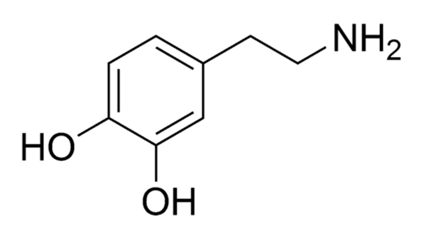
Dopamine
An excitatory neurotransmitter of the CNS basal ganglia area of the brain.
The basal ganglia help with the initiation and termination of voluntary motor movements.
Levels of this neurotransmitter are low in patients with Parkinson's Disease.
It is involved in “emotional reward.”
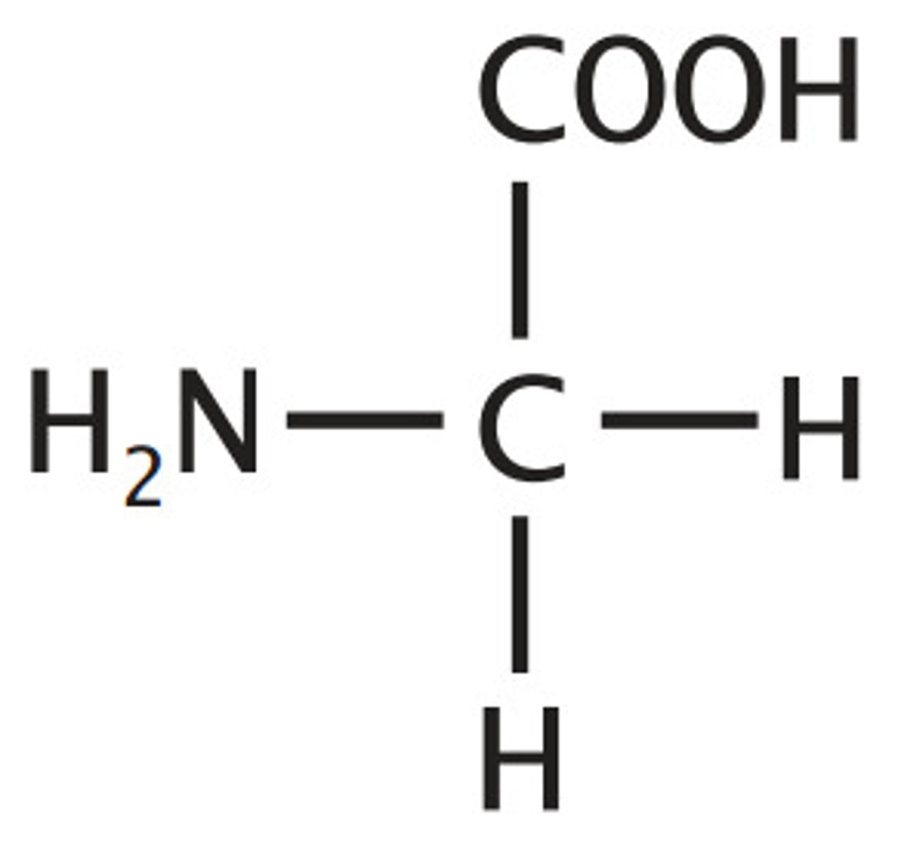
GABA (gamma-aminobutyric acid)
An inhibitory neurotransmitter in the CNS that controls localized motor, sensory and cognitive functions.
Glycine
An inhibitory amino acid neurotransmitter of CNS that works to help control localized motor function.
Endorphins
Inhibitory neurotransmitters of CNS and PNS.
They work by down-regulating pain and producing euphoria.
They are often considered the body's "natural pain killers" or the chemicals that cause the "runner's high" or "second wind."
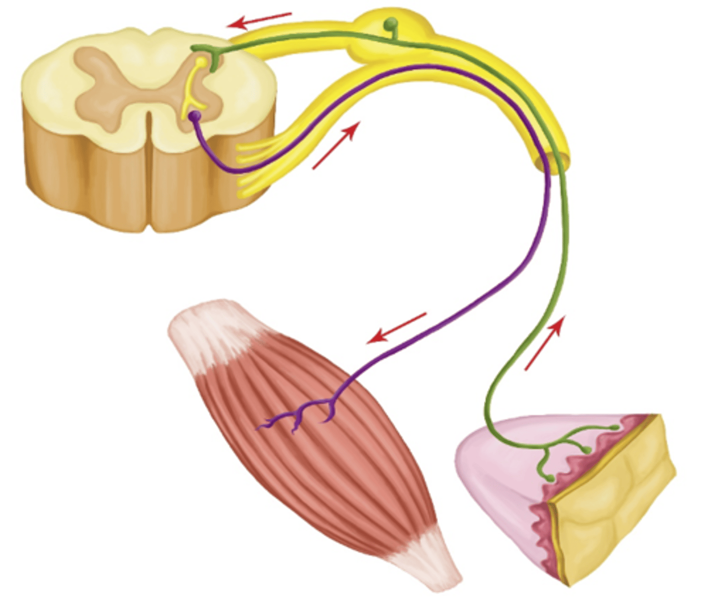
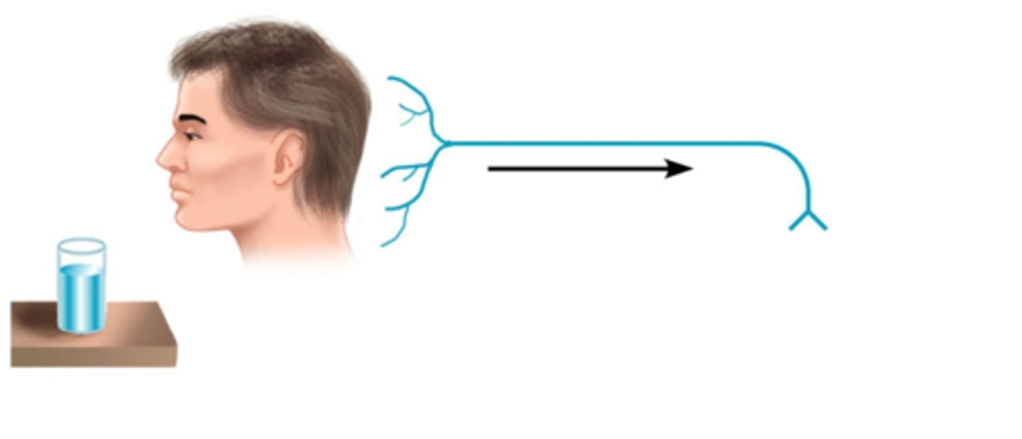
sensory neurons
Carry information TOWARD the CNS
"Afferent Arrive"
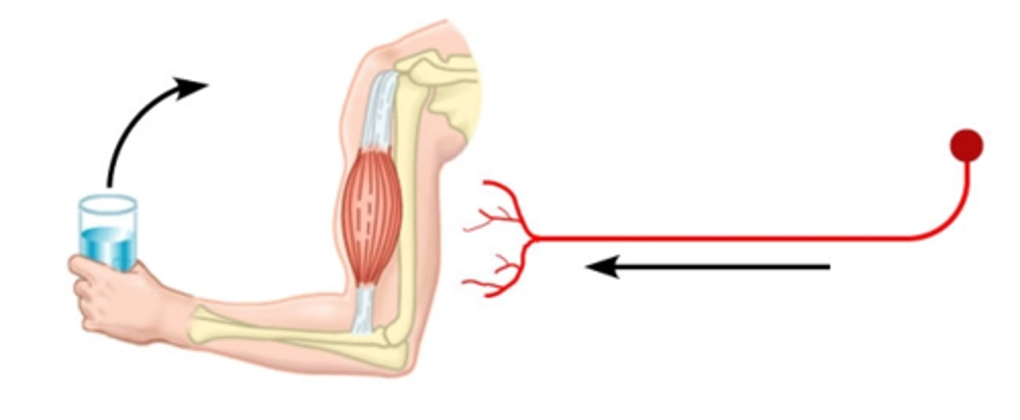
motor neurons
Take information AWAY FROM the CNS
"Efferent Exits"
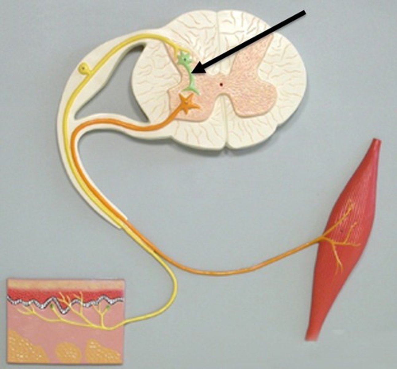
interneurons
CNS neurons that internally communicate and intervene between the sensory inputs and motor outputs
Entirely within the CNS
99% of all neurons are this type of neuron.
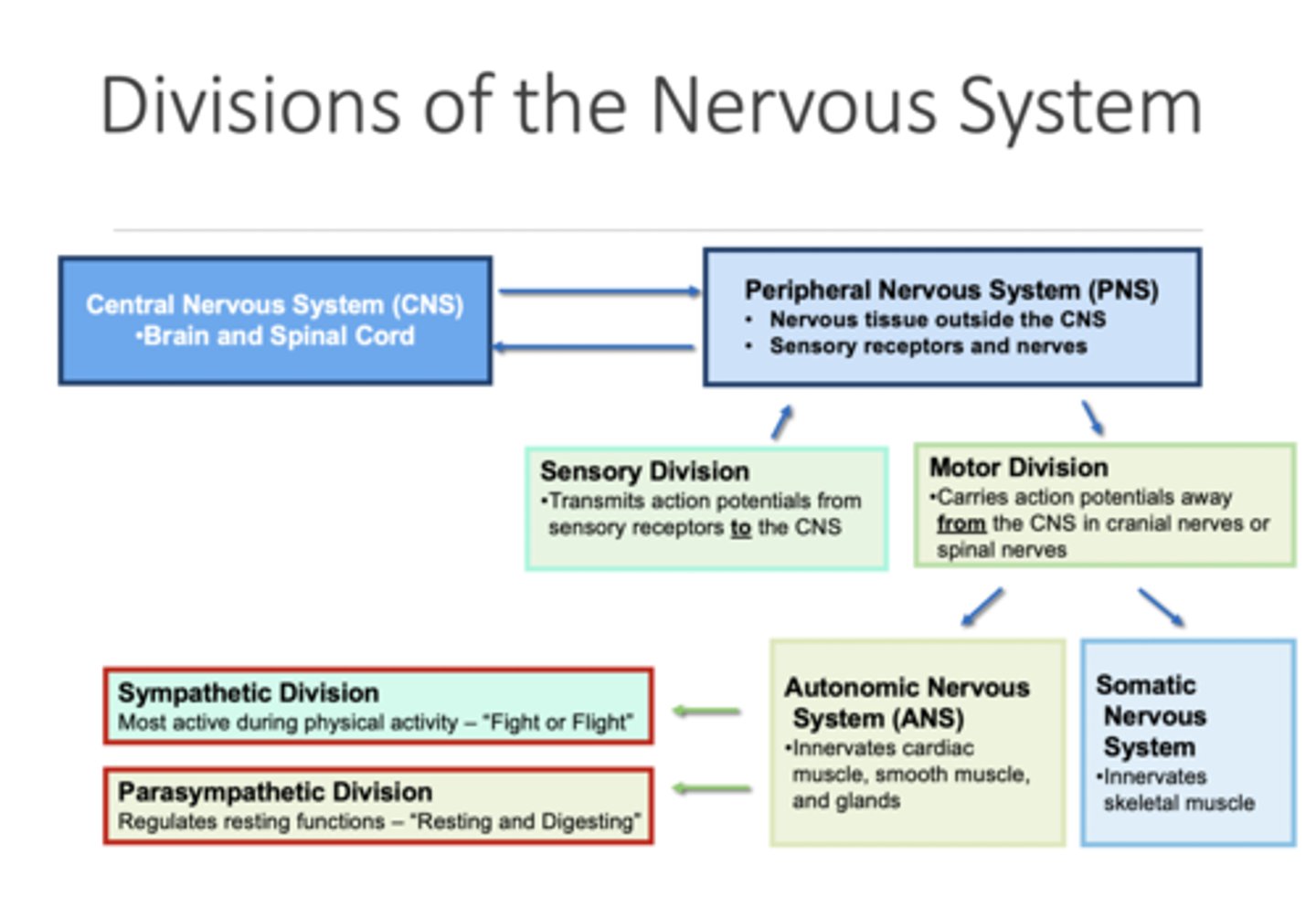
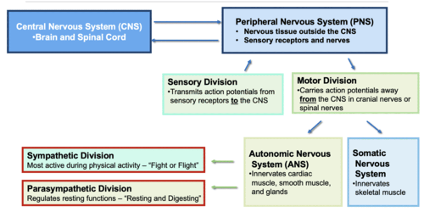
What are the two anatomical divisions of the nervous system?
1. Central Nervous System (CNS)
2. Peripheral Nervous System (PNS)
Central Nervous System (CNS)
Brain and spinal cord
Encased in bone
Processes, integrates, stores, and responds to information from the PNS.
Peripheral Nervous System (PNS)
Nervous tissue outside of the CNS
Consists of sensory receptors and nerves
Detects stimuli and transmits information to the CNS and receives information from the CNS
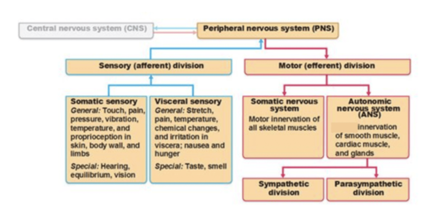
What are the two divisions of the Peripheral Nervous System (PNS)
Sensory division transmits action potentials from sensory receptors to the CNS
Motor division carries action potentials away from the CNS in cranial or spinal nerves (two subdivisions)
- Somatic nervous system innervates skeletal muscle
- Autonomic nervous system (ANS) innervates cardiac muscle, smooth muscle, and glands (three subdivisions)
◦Sympathetic division is most active during physical activity (fight or flight division)
◦Parasympathetic division regulates resting functions (rest and digest division)
sensory division of PNS
Transmits action potentials from sensory receptors to the CNS.
motor division of PNS
Carries action potentials away from the CNS in cranial or spinal nerves.
It has two subdivisions:
- Somatic nervous system (SNS) innervates skeletal muscle
- Autonomic nervous system (ANS) innervates cardiac muscle, smooth muscle, and glands (two subdivisions - sympathetic and parasympathetic)

Somatic Nervous System (SNS)
The part of the peripheral nervous system that controls voluntary movement of skeletal muscles
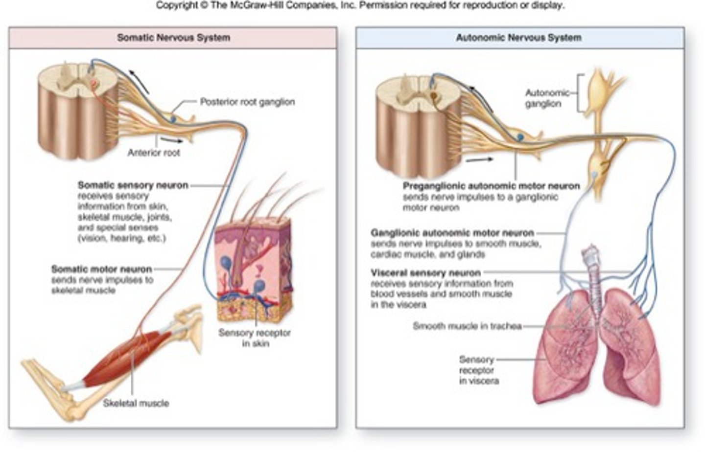
Autonomic nervous system (ANS)
The part of the peripheral nervous system that controls the glands and the muscles of the internal organs (such as the heart).
Its sympathetic division arouses; its parasympathetic division calms.
Autonomic nervous system (ANS) subdivisions
sympathetic and parasympathetic
sympathetic division of the autonomic nervous system (ANS)
This branch of the ANS is often termed the "fight or flight" nervous system since it increases fuel and energy for the body during times of stress or physical harm. Most active during physical activity.
Functions such as heart rate, respiratory rate, and metabolism all increase to make the energy necessary for the body to "protect" itself during physical emergencies
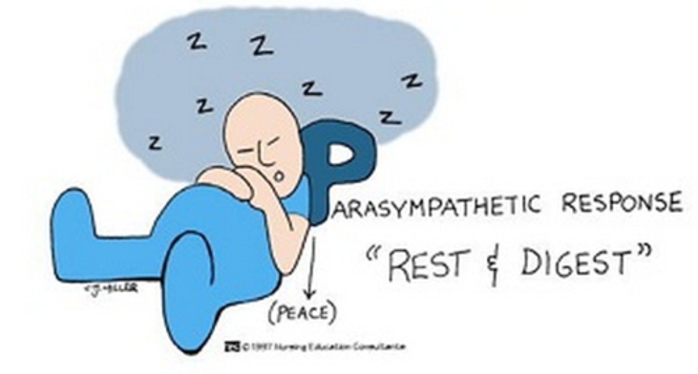
parasympathetic division of the autonomic nervous system (ANS)
This branch of the ANS is often termed the "rest and digest" nervous system since it is used during periods of rest or non-emergency periods.
It works to slow the body processes down or is the primary influence during processes such as salivation, lacrimation, urination, defecation, and digestion.
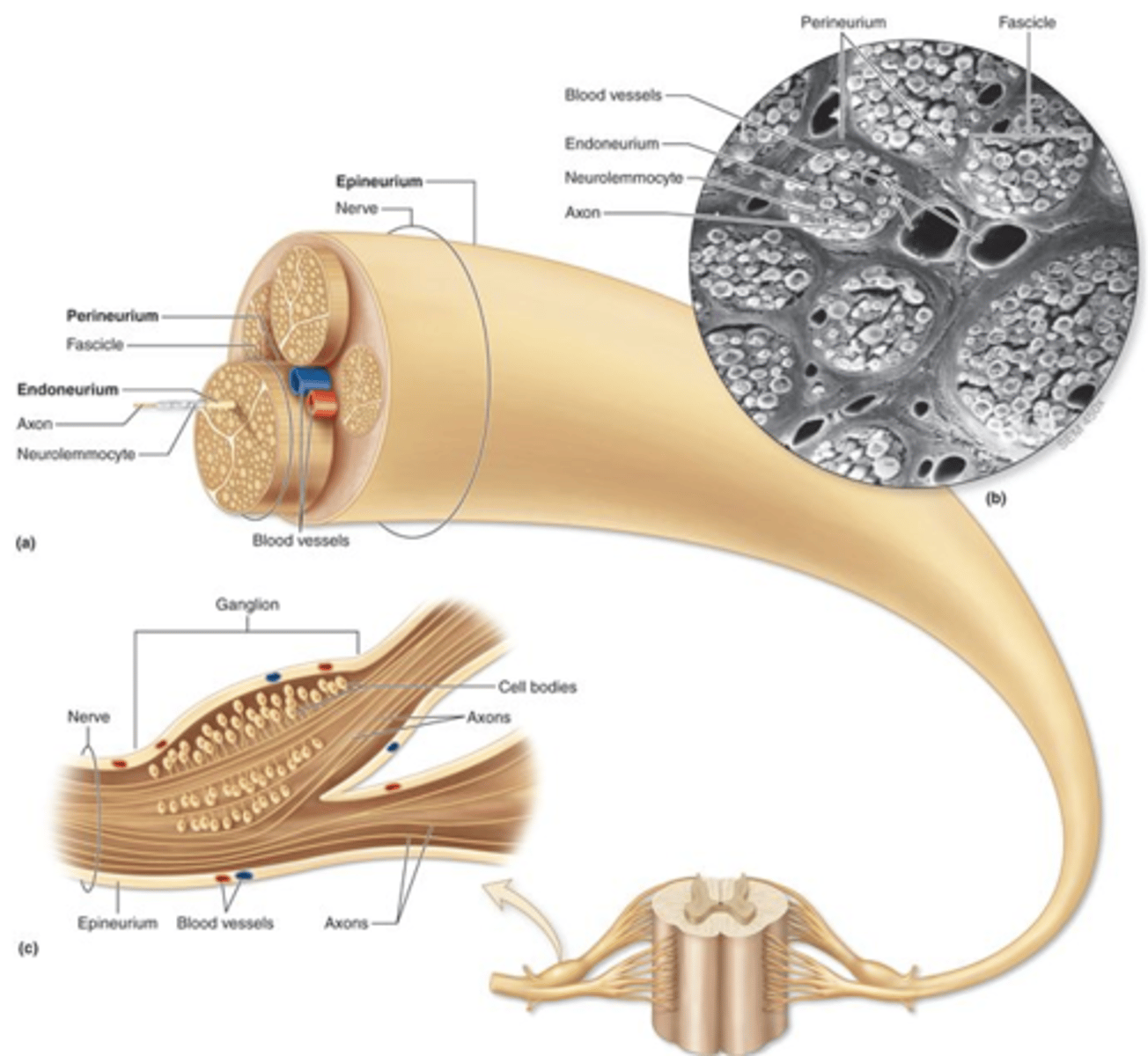
nerve
A bundle of nerve processes outside of the central nervous system.
They are made up of many axons of neurons, wrapped up in connective tissue.
They are found in the peripheral nervous system.
The cell bodies of these neurons lie within the CNS (either in the brain or the spinal cord).
There are cranial nerves (which come off of the brain) and spinal nerves (which come off of the spinal cord).
Examples include the sciatic nerve or the radial nerve or the facial nerve
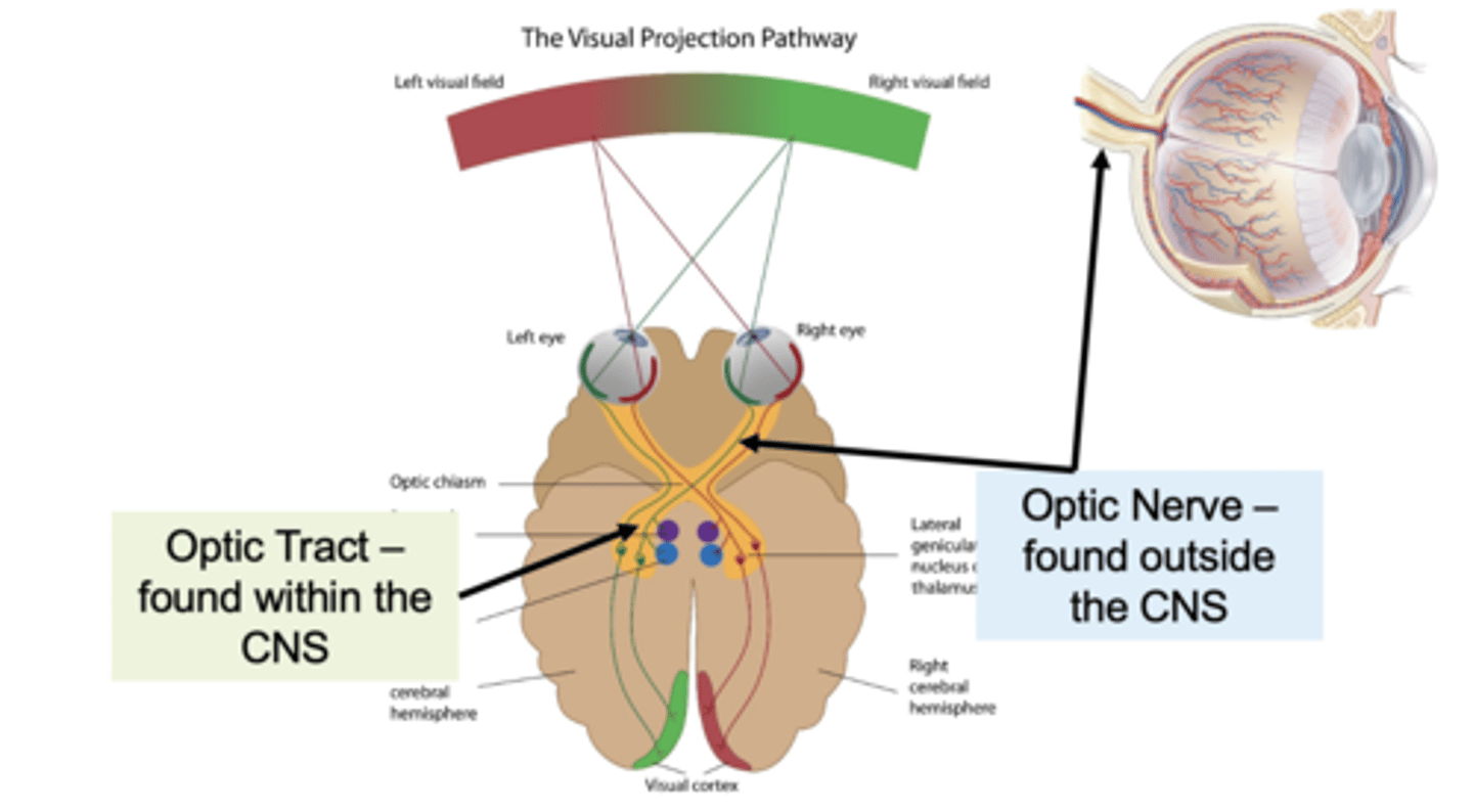
tract
undles of axons found in the central nervous system (CNS).
These axons have a similar function and a common origin and destination.
tract vs nerve
Tract: Group of axons in CNS
Nerve: group of axons in PNS
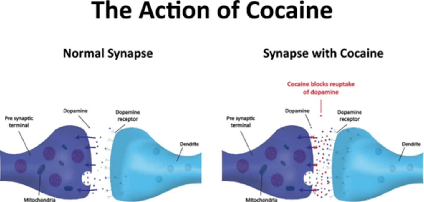
Cocaine Addiction
Cocaine blocks the reuptake of dopamine which results in increased dopamine at the synaptic cleft
Dopamine is involved in emotional reward (reward-motivated behavior)
So the increased dopamine at the synapse causes the user to want to repeat the behavior (take cocaine again)
risk factors for cocaine addiction
Genetics (family history) - anybody who has a close relative with an addiction problem has a higher risk of eventually having one themselves.
Gender - a significantly higher percentage of people addicted to a substance are male.
Having a mental illness/condition - people with depression, ADHD (attention-deficit hyperactivity disorder) and several other mental conditions/illnesses have a higher risk of eventually becoming addicted to drugs, alcohol or nicotine.
Peer pressure
Family behavior - young people who do not have a strong attachment to their parents and siblings have a higher risk of becoming addicted to something one day, compared to people with deep family attachments.
Loneliness
The nature of the substance - some substances, such as crack, heroin or cocaine can bring about addiction more rapidly than others. For some people trying a substance even once can be enough to spark an addiction.
Age when substance was first consumed - studies of alcoholism have shown that people who start consuming a drug earlier in life have a higher risk of eventually becoming addicted, than those who started later. Many experts say this also applies to nicotine and drugs.
Stress - if a person's stress levels are high there is a greater chance a substance, such as alcohol may be used in an attempt to blank out the upheaval. Some stress hormones are linked to alcoholism.
How the body metabolizes (processes) the substance - in cases of alcohol, for example, individuals who need a higher dose to achieve an effect have a higher risk of eventually becoming addicted.
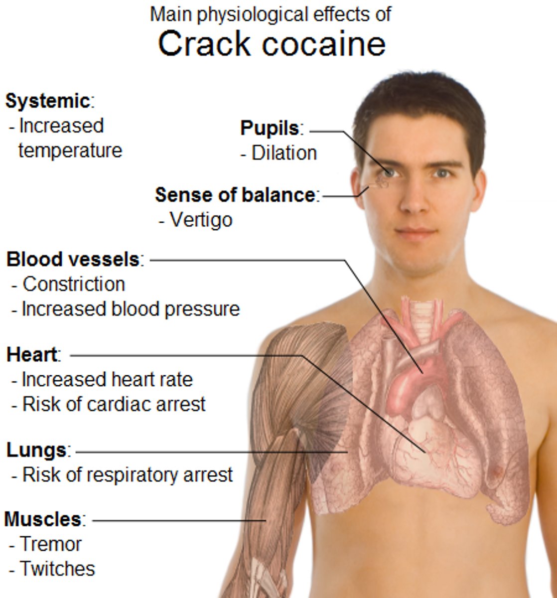
Manifestations of cocaine use
Cocaine will cause a person to have a sense of euphoria or pleasure as well as the enhanced sympathetic effects and energy.
Amphetamines mimic the sympathetic nervous system stimulation and hence affect overall energy levels.
Other sympathetic nervous system effects = high blood pressure, high heart rate
The adverse effects of cocaine include: headaches, abdominal pain and nausea, loss of the sense of smell, nosebleeds, and increased body temperature, heart rate, and blood pressure.
Cocaine users can also experience acute cardiovascular or cerebrovascular emergencies, such as a heart attack or stroke, which may cause sudden death.
Cocaine-related deaths are often a result of cardiac arrest or seizure followed by respiratory arrest.
Treatment for cocaine use
A person who is addicted to crack cocaine has several treatment options to choose from. These include:
Detoxification, which deals with the physical but not the psychological aspects of crack cocaine addiction
Short-term inpatient or residential programs, which often take place in a hospital and are usually based on the Twelve Step approach to drug addiction recovery
Long-term residential programs, usually at residential treatment centers, where the patient spends up to a year as part of a program like therapeutic communities where they deal with their drug addiction and often learn life skills to help them adjust better to a drug-free life and society after treatment
Outpatient therapy, which involves the patient going to a facility for individual or group counseling - this type of therapy is usually less expensive than inpatient forms of therapy and appropriate for people with jobs and good support networks
When you go to the dentist and he wants to drill on your tooth to fill a cavity, how does he first "numb" the area?
The medication the dentist uses is a Na+ channel blocker.
If sodium channels are blocked, no action potentials can be initiated and so there are no messages to send to the brain. Even though tissue damage is occurring and there is a painful stimulus, the brain doesn't know that as you feel "numb" in the area.
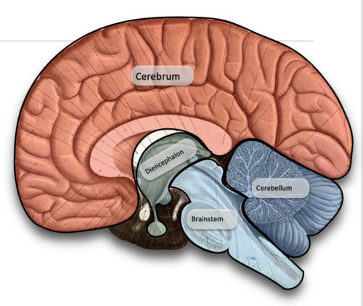
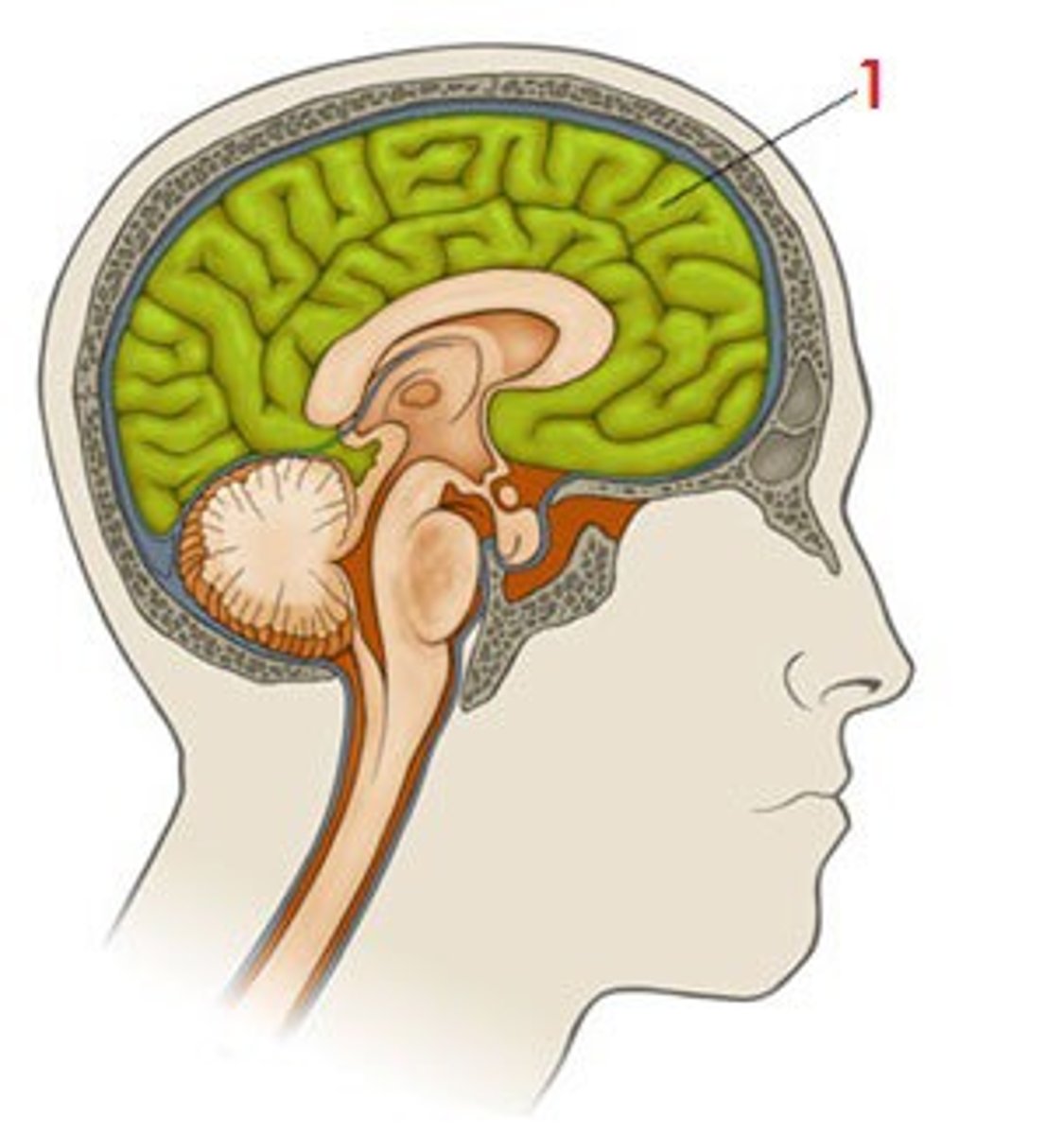
Four major subdivisions of the brain
1. Cerebrum
2. Diencephalon
3. Cerebellum
4. Brainstem
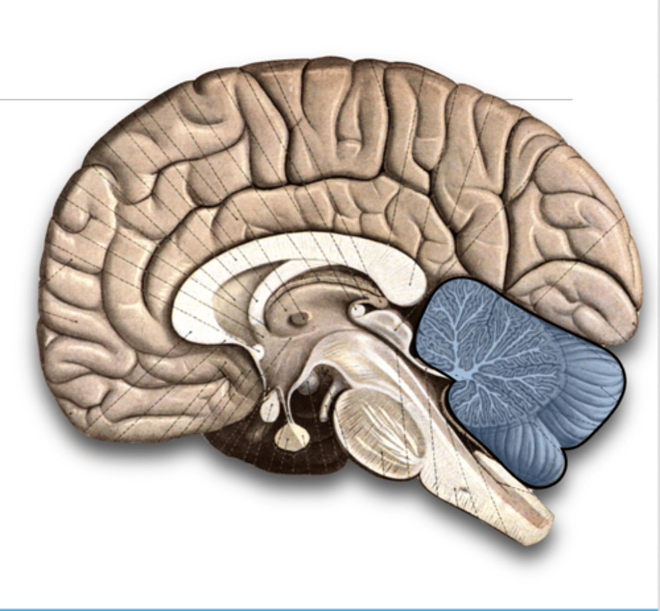
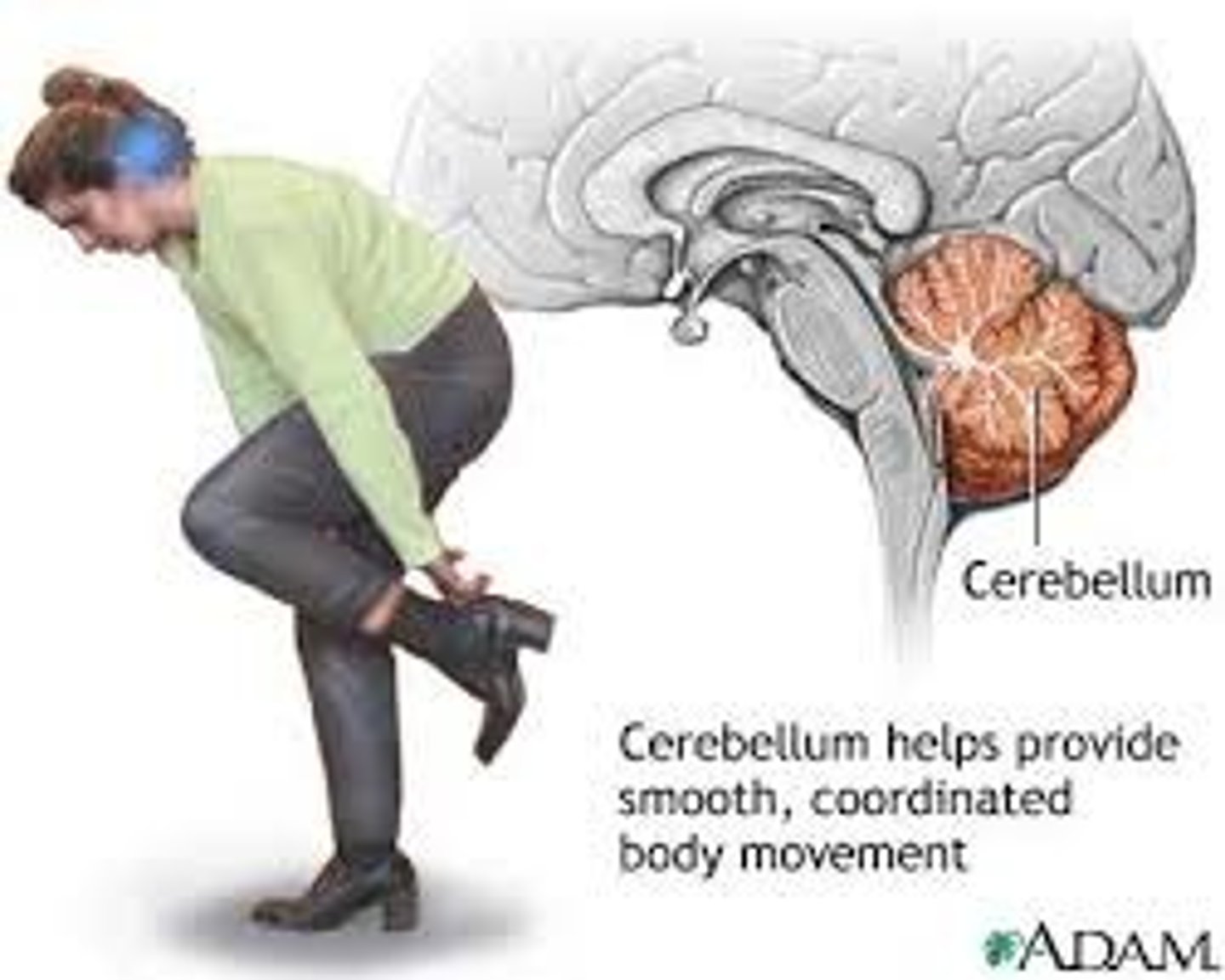
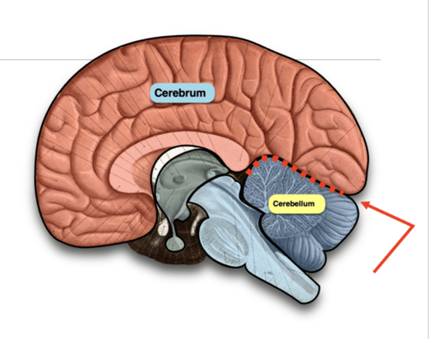
Cerebellum
Responsible for maintaining balance, posture, and motor coordination.
Its name means "little cerebrum" because it also has superficial gray matter and deep white matter.
It is separated from the occipital lobe by the transverse fissure
Cerebellum function
Fine-tunes the timing of motor function — this is necessary for:
◦Maintaining posture and balance
◦Normal walking gait
◦Coordinating movements
The cerebellum is always receiving afferent (sensory) information from muscles and joints and it uses this information to affect efferent (motor) neurons to reduce errors in movements.
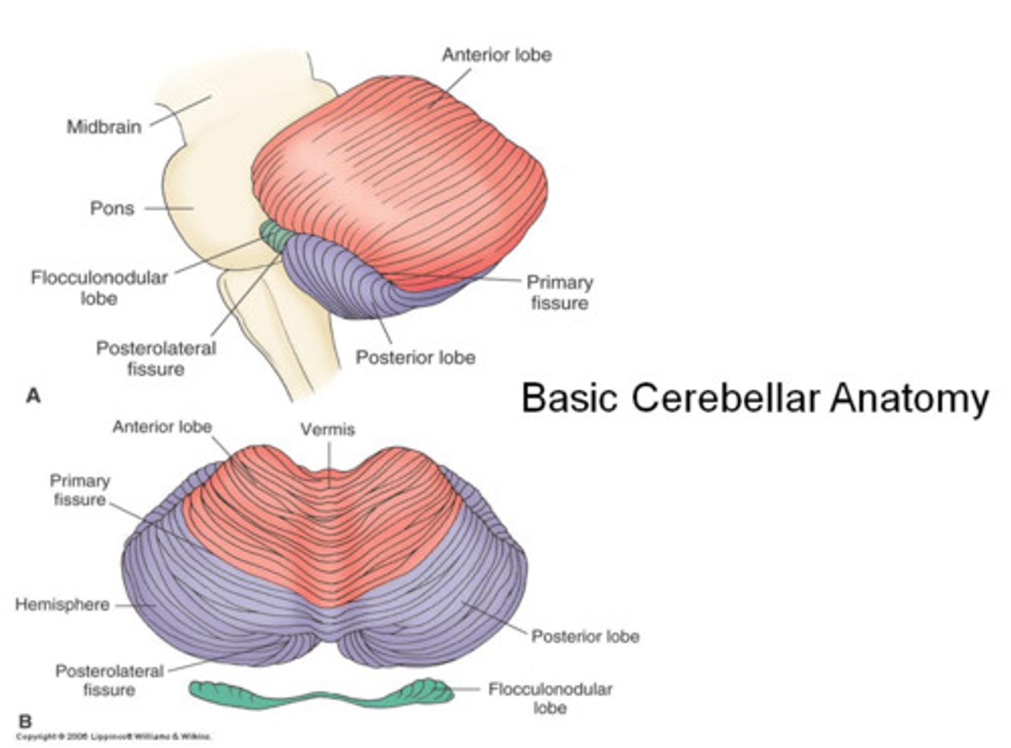
cerebellum hemispheres
It has two hemispheres (Left & Right)
Each hemisphere has three lobes:
- Anterior (A)
- Posterior (P)
- Flocculonodular (F)
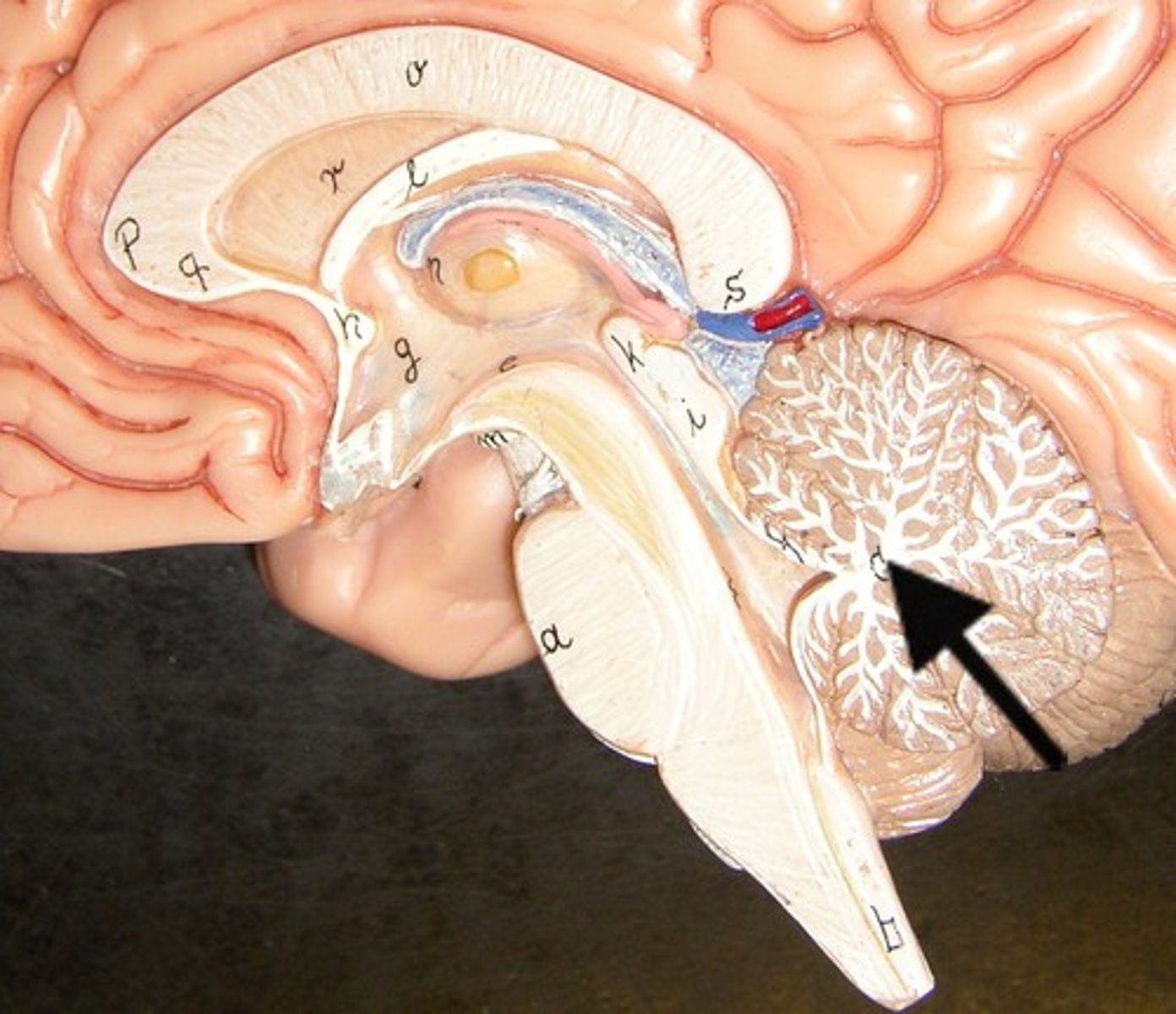
arbor vitae
Deep white matter of the cerebellum
(arbor = tree, vitae = life); due to its similar appearance to a specific shrub
Diencephalon
The area deep within the brain that contains the thalamus, hypothalamus, and epithalamus and is the link between the cerebral hemispheres and the brainstem; responsible for directing sensory information to the cortex
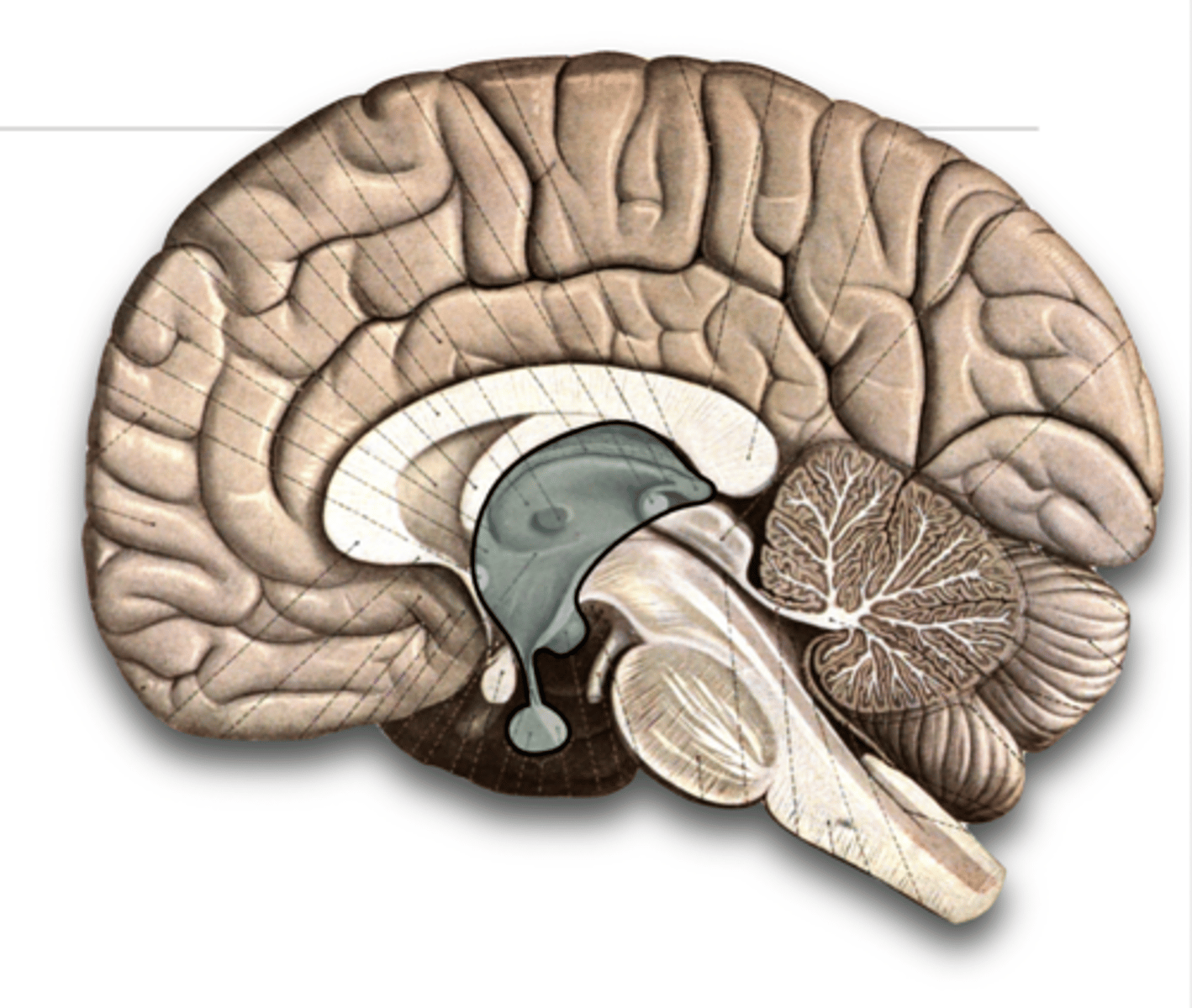
Three portions of the diencephalon
1. Thalamus - serves as a sensory relay center
2. Hypothalamus - involved in the maintenance of homeostasis
3. Epithalamus (pineal gland) - secretes the hormone melatonin, involved in sleep-wake cycles.
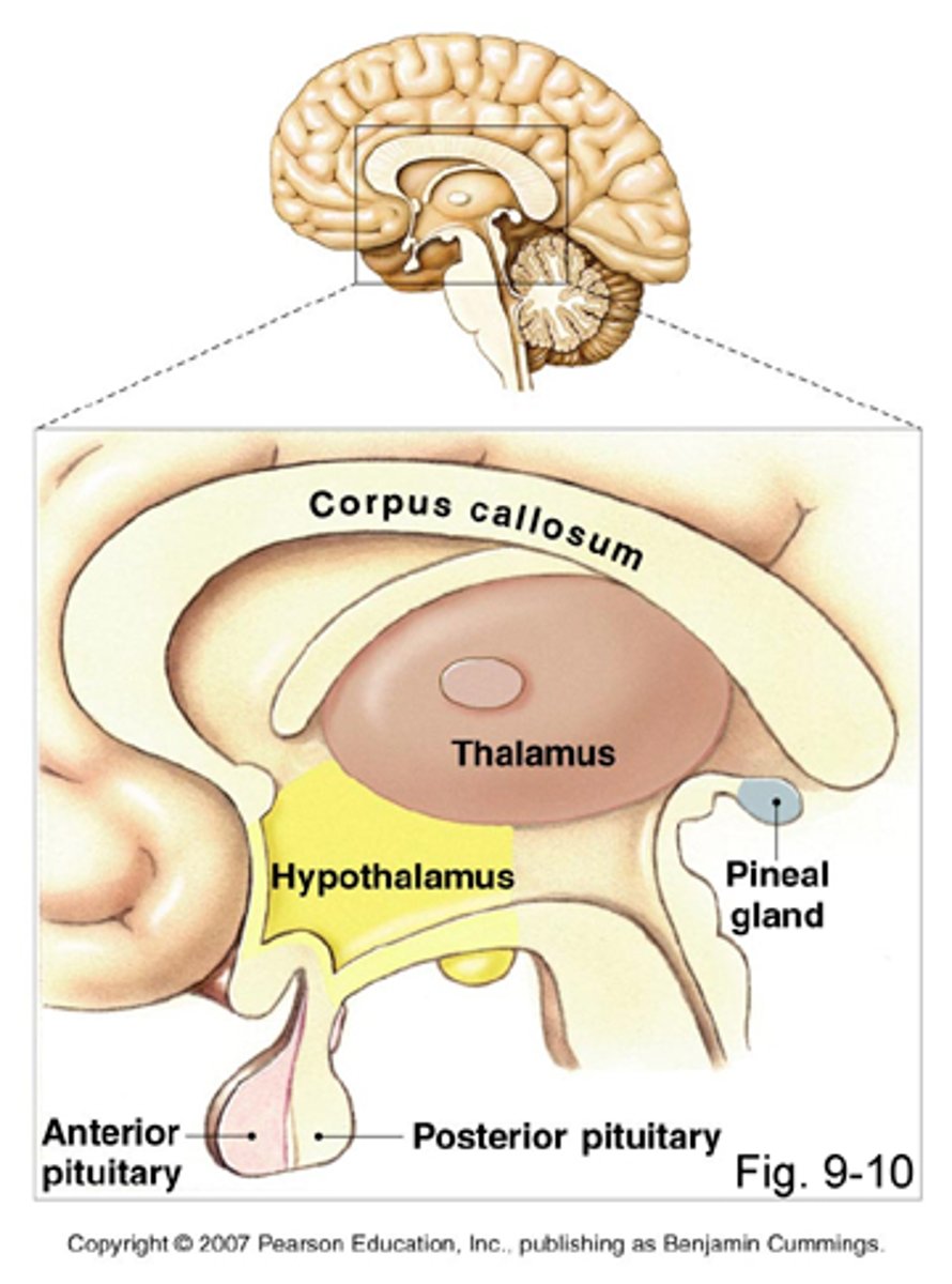
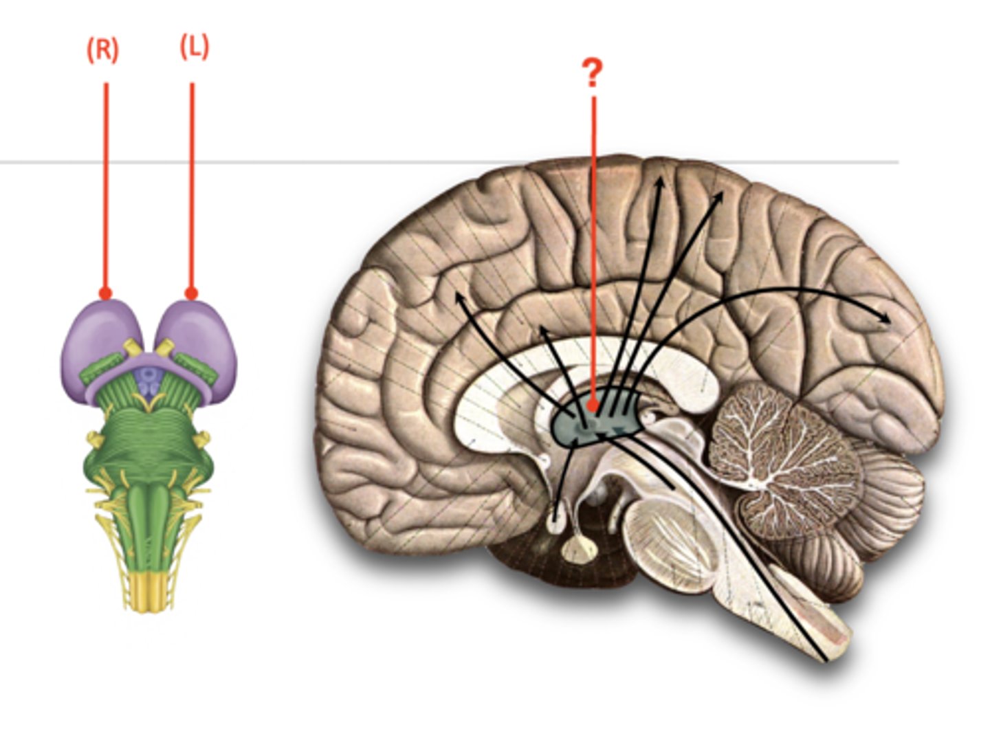
Thalamus
The brain's sensory relay center, located on top of the brainstem; directs messages to the sensory receiving areas in the cortex and transmits replies to the cerebellum and medulla.
It is one of three portions of the diencephalon.
Has left and right egg-shaped parts
Has nuclei that receive information from the spinal cord, brainstem, and eyes
Is relay center for all sensations except for olfaction (i.e. smell)
Receives, sorts, and relays sensory information to the appropriate area of the cortex capable of interpreting the sensation
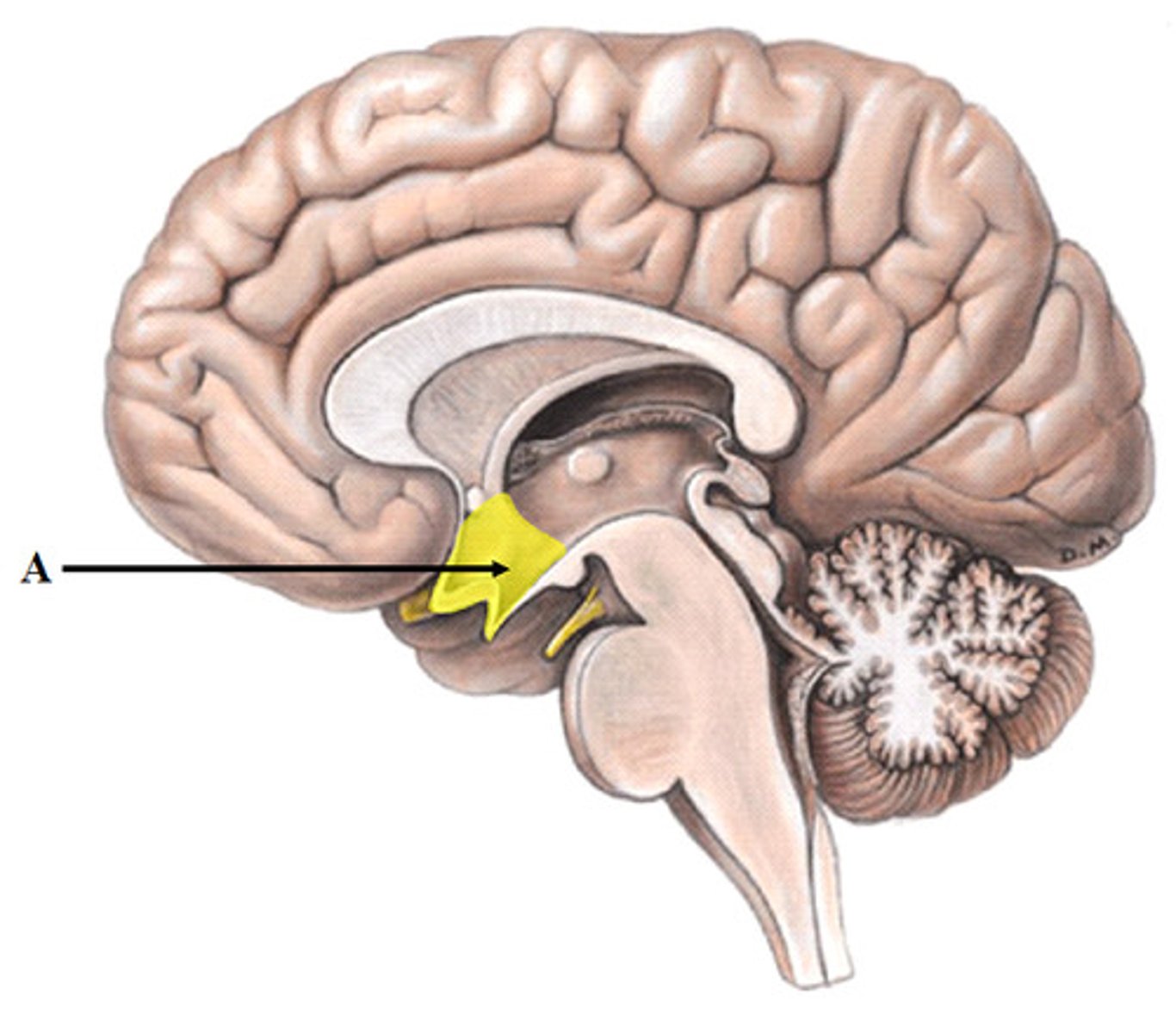
Hypothalamus
The portion of the diencephalon that is involved in the maintenance of homeostasis.
It is the link between the two systems used for communication: the nervous system and the endocrine system
It controls:
- Heart rate
- blood pressure
- food and water intake,
- A number of cyclical processes (e.g. sleep/wake, menstruation)
It is the “Command Center” of the endocrine system due to its effect on the release (or inhibition) of many hormones
It helps govern the endocrine system via the pituitary gland
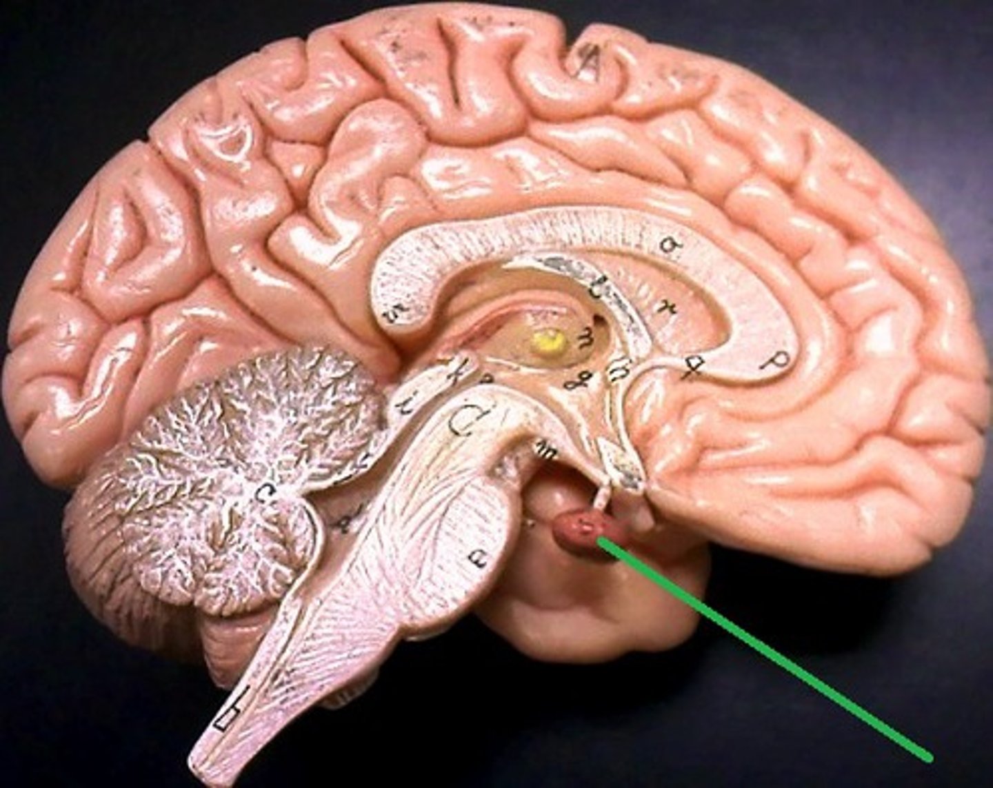
pituitary gland
Releases hormones in response to hormones from the hypothalamus
The endocrine system's most influential gland.
Under the influence of the hypothalamus, the pituitary regulates growth and controls other endocrine glands.
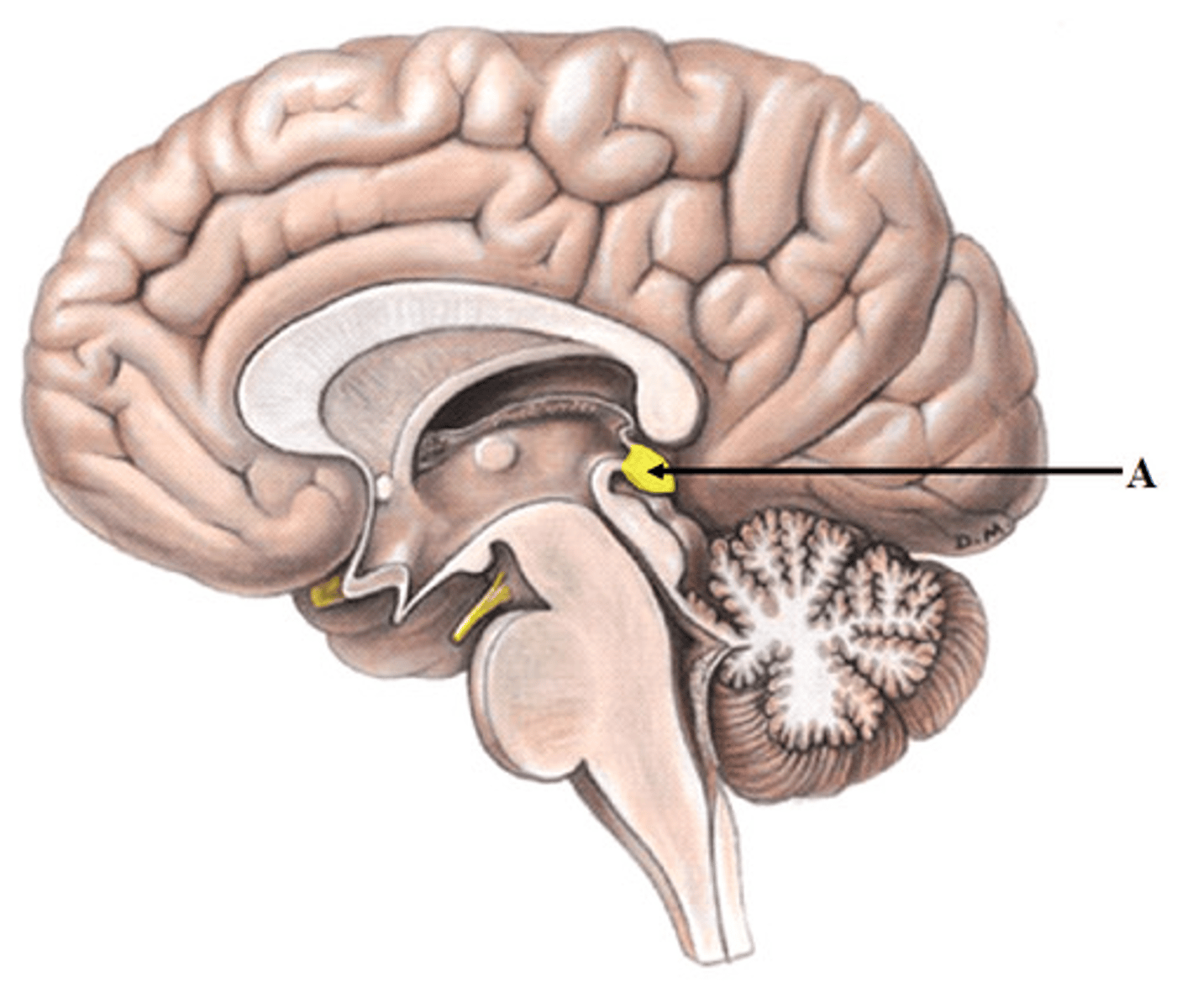
Epithalamus
Above and posterior to the thalamus.
It has a distinct feature, the pineal gland.
The Pineal gland produces melatonin in response to darkness and helps regulate a 24-hour circadian rhythm.
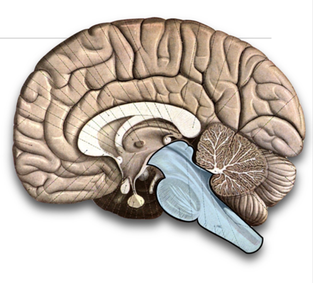
Brainstem
Essential to life.
Controls autonomic functions like breathing, heart beating
A continuation of the spinal cord; immediately superior to the foramen magnum
It serves as a highway for neurons traveling between the spinal cord & the brain
It connects many of the 12 cranial nerves connect to the CNS.
Has 3 subdivisions: midbrain, pons, and medulla oblongata
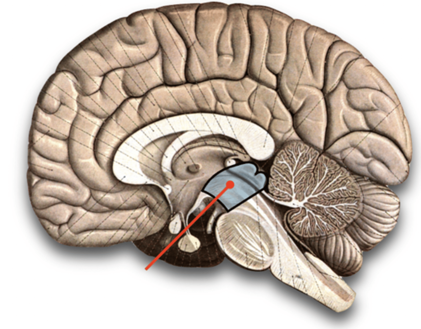
midbrain (mesencephalon)
The superior most section of the brainstem found just inferior to the thalamus.
It serves as a relay station for hearing and visual sensory input to the thalamus
It is involved in:
- Visual Reflexes
- Auditory Reflexes
Contains the cerebral aqueduct.
corpora quadrigemina
These 4 bodies are the 2 superior colliculi and 2 inferior colliculi and are located on the dorsal aspect of the midbrain
The superior colliculi are responsible for the visual reflex and the inferior colliculi are responsible for the auditory reflex.
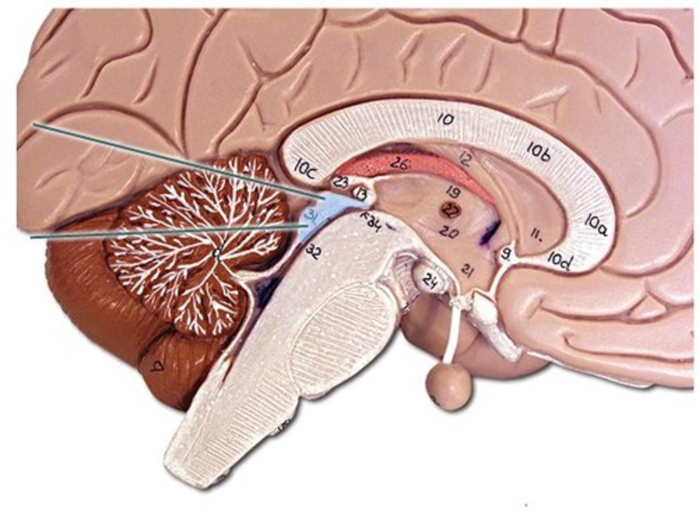
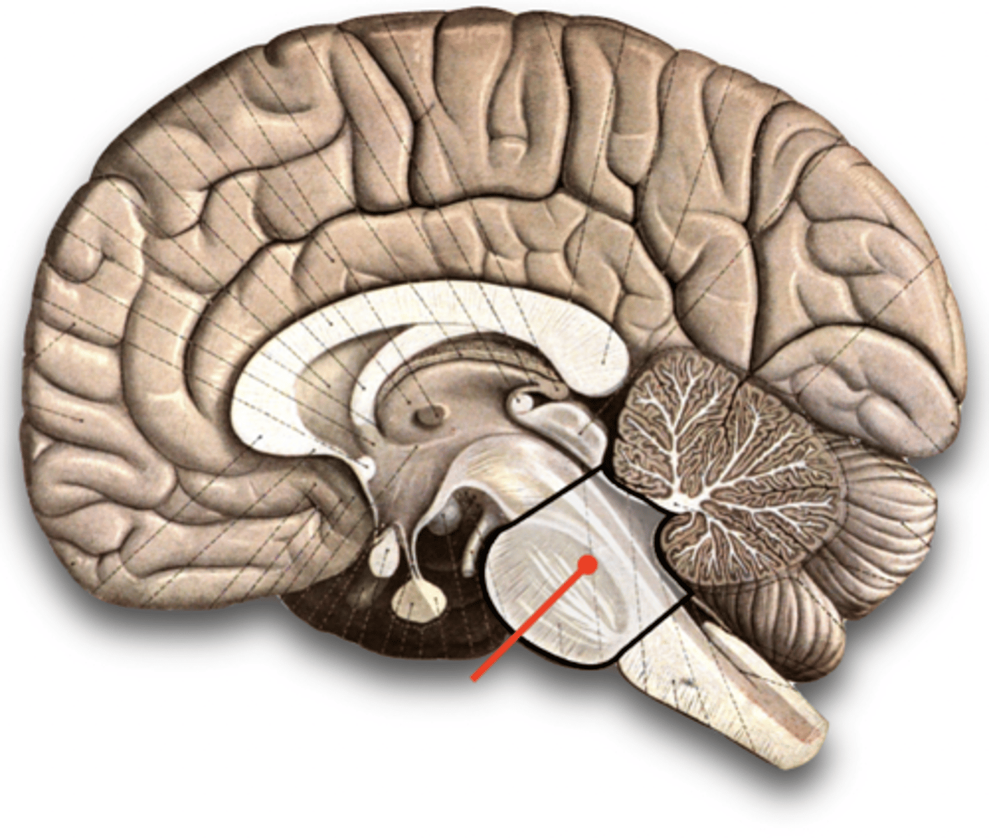
pons
Located between the midbrain and the medulla.
It has a great number of neurons that connect to the cerebellum and serves as a bridge between the cerebellum and the rest of the nervous system (pons means bridge).
Also serves as a bridge between the brain and spinal cord.
It is involved in many autonomic and sensory functions including arousal, respiratory processes, fine motor control, equilibrium, muscle tone, and the Circadian cycle (specifically regulating sleep).
Contains important centers that affect the rate and depth of inhalation/exhalation.
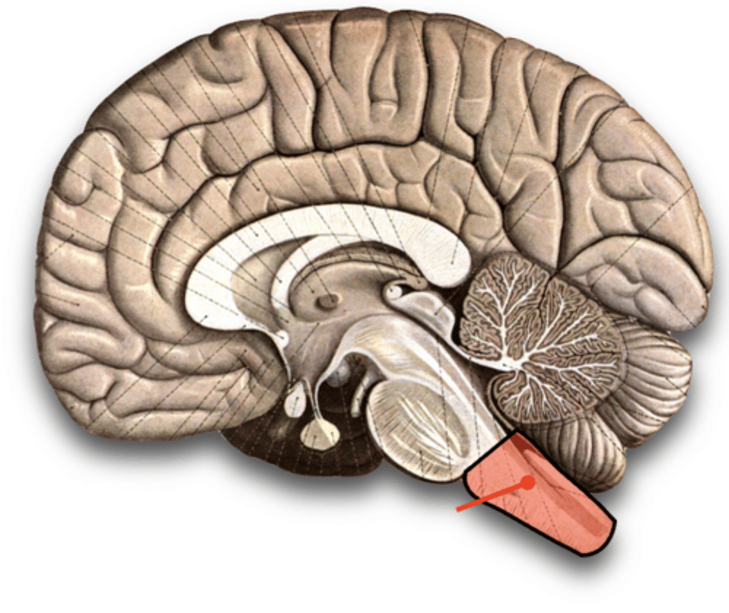
medulla oblongata
The inferior most section of the brainstem.
It is found just superior to the foramen magnum.
Has numerous nuclei that regulate vital bodily functions.
The respiratory, cardiovascular, vasomotor control centers in the medulla regulate breathing rates, heart rates, and blood pressure.
Also coordinates complex processes that involve several organs (e.g. vomiting, gagging, swallowing, and sneezing).
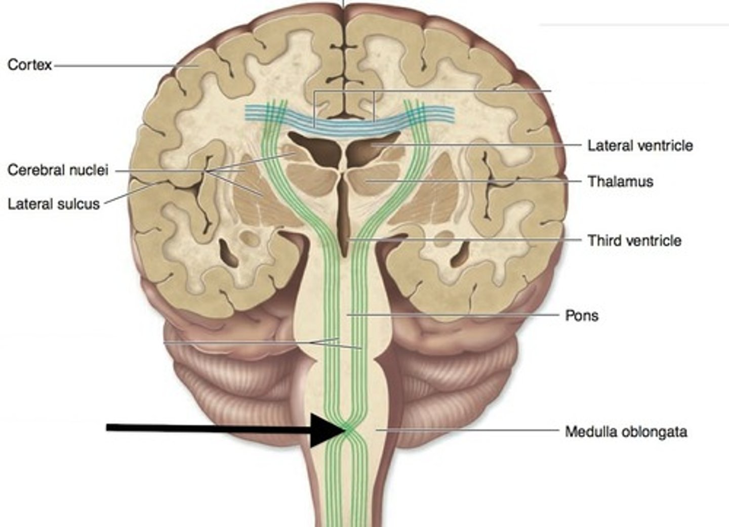
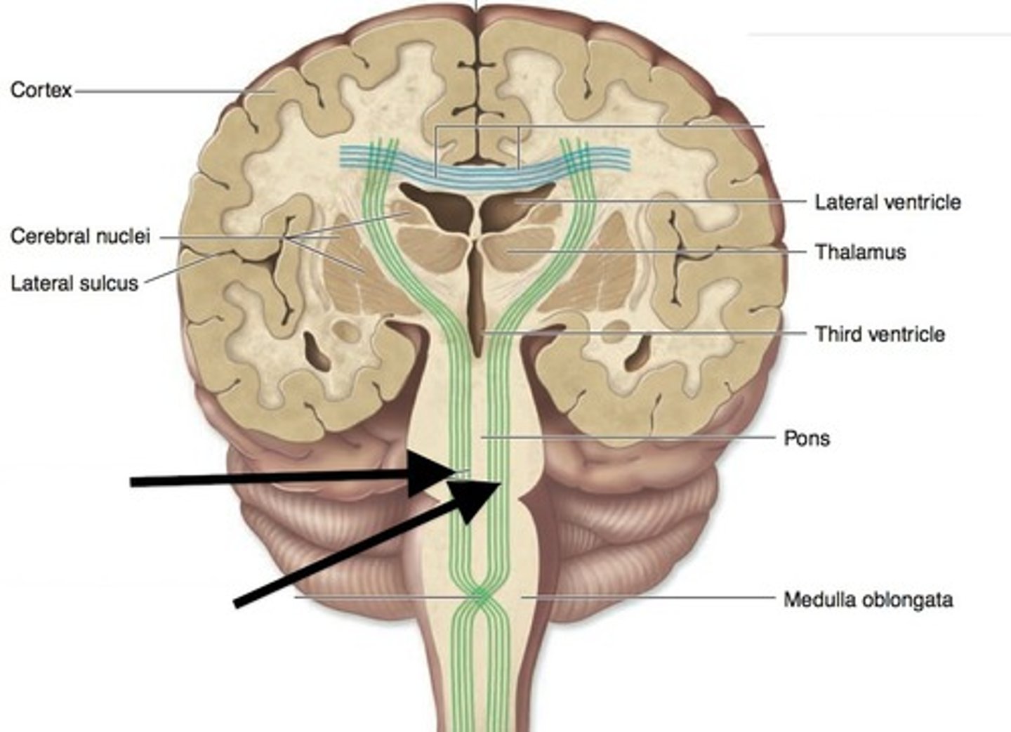
decussation of pyramids
cross over point for the tracts passing through the medulla
It occurs at the level of the medulla oblongata, which means that some of the motor fibers that originate in the CNS will cross to the opposite side of the brain.
This explains why when there is a stroke on one side of the brain, paralysis will result on the opposite side of the bod
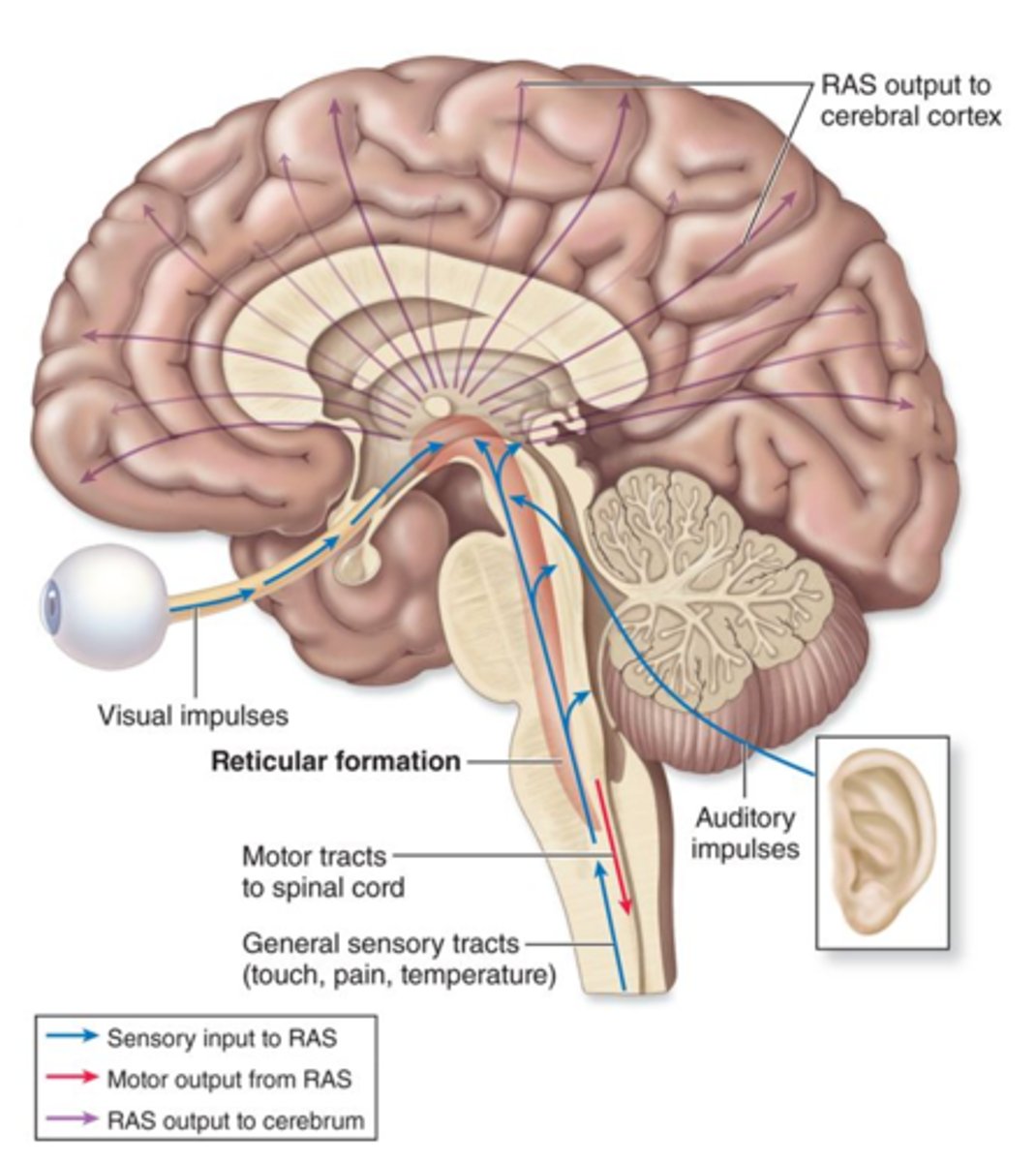
Reticular Formation (Reticular Activating System)
Neural network in brainstem (medulla and pons) and midbrain essential to the regulation of sleep, wakefulness, arousal, and attention
Maintains consciousness by connecting through diencephalon to cerebral cortex
It helps maintain muscle tone with the cerebellum, helps the pons and medulla oblongata to regulate respiration, heart rate and blood pressure as well as processing sensory stimuli such as touch, sight and sound.
It also helps to maintain consciousness and concentration.
Cerebrum
The "higher brain", involved in:
1. Personality
2. Thought
3. Language
4. Learning
5. Memory
6. Voluntary control of movement
Makes up 83% of the brain's volume.
Contains sensory, motor, and association areas associated with intellect, learning, memory, language, speech.
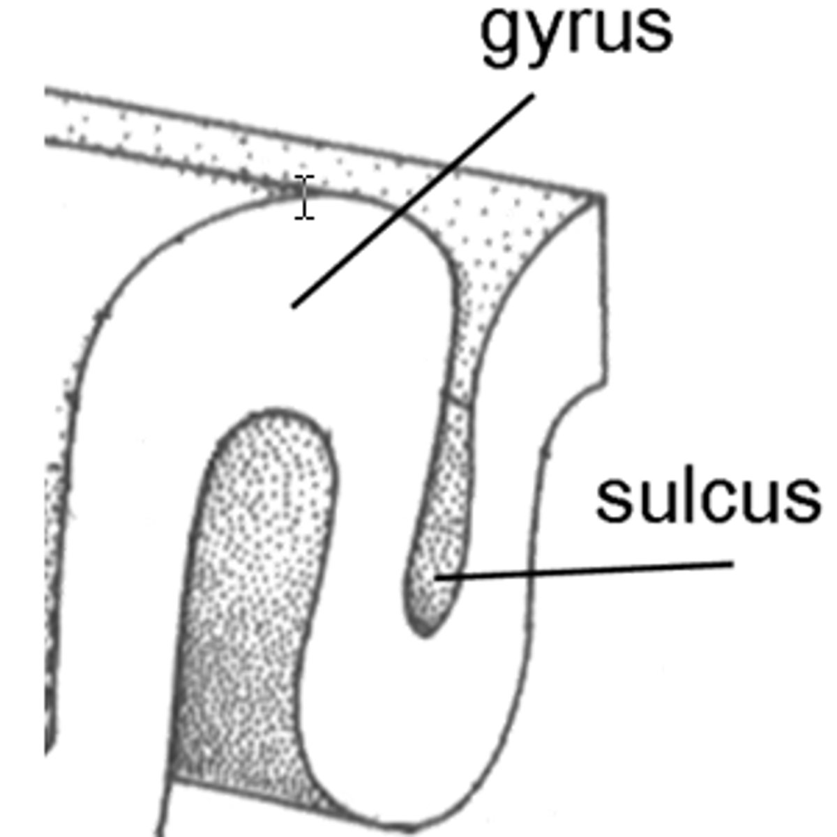
gyri
elevated ridges of brain tissue
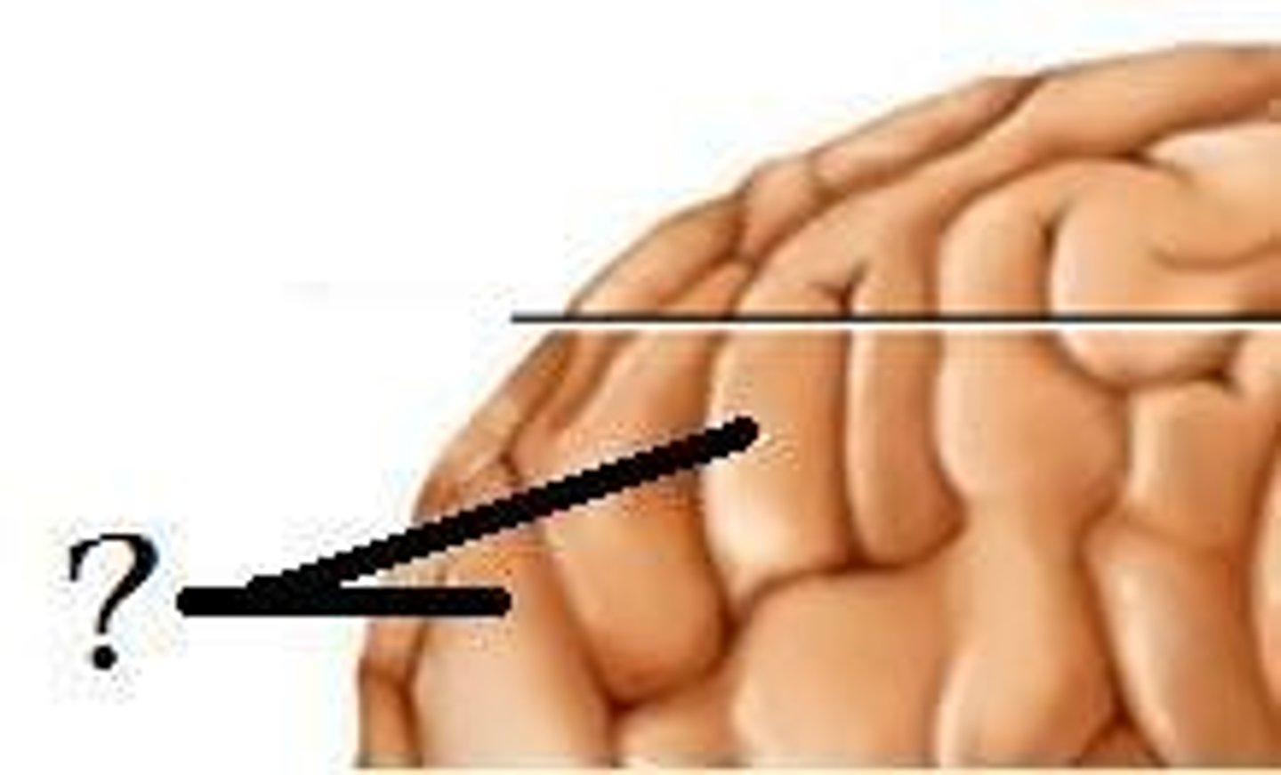
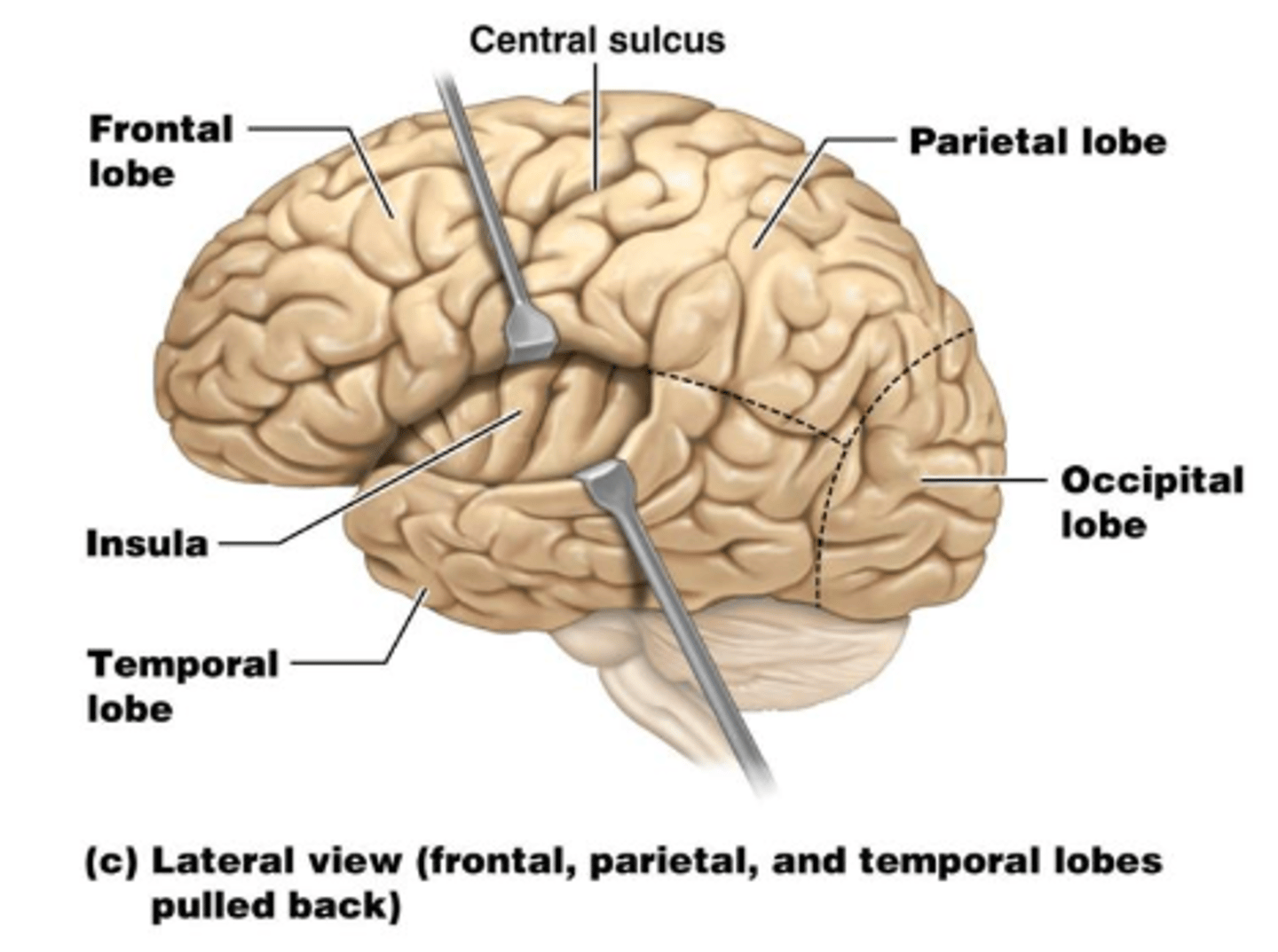
sulci
Shallow grooves that separate the gyri from one another.
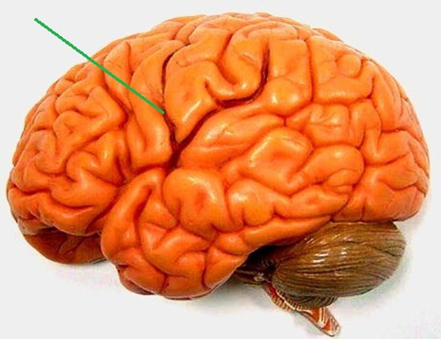
purpose of gyri and sulci
They serve to increase the surface area of the brain.
Two hemispheres of the cerebrum
1. The left hemisphere
2. The right hemisphere
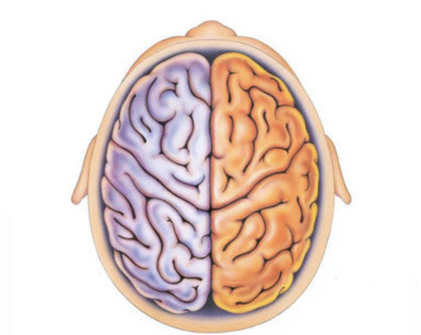
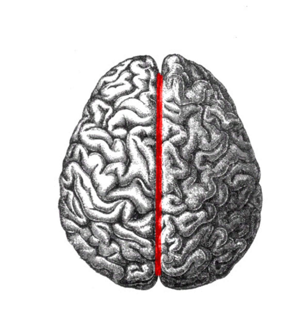
Longitudinal Fissure
Separates the left hemisphere from the right hemisphere.

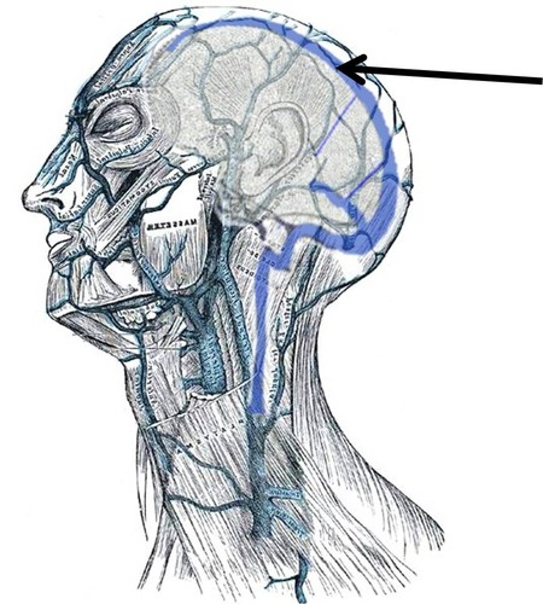
superior sagittal sinus
A vein that runs deep along the longitudinal fissure and receives cerebrospinal fluid and then returns that CSF into the venous circulation.
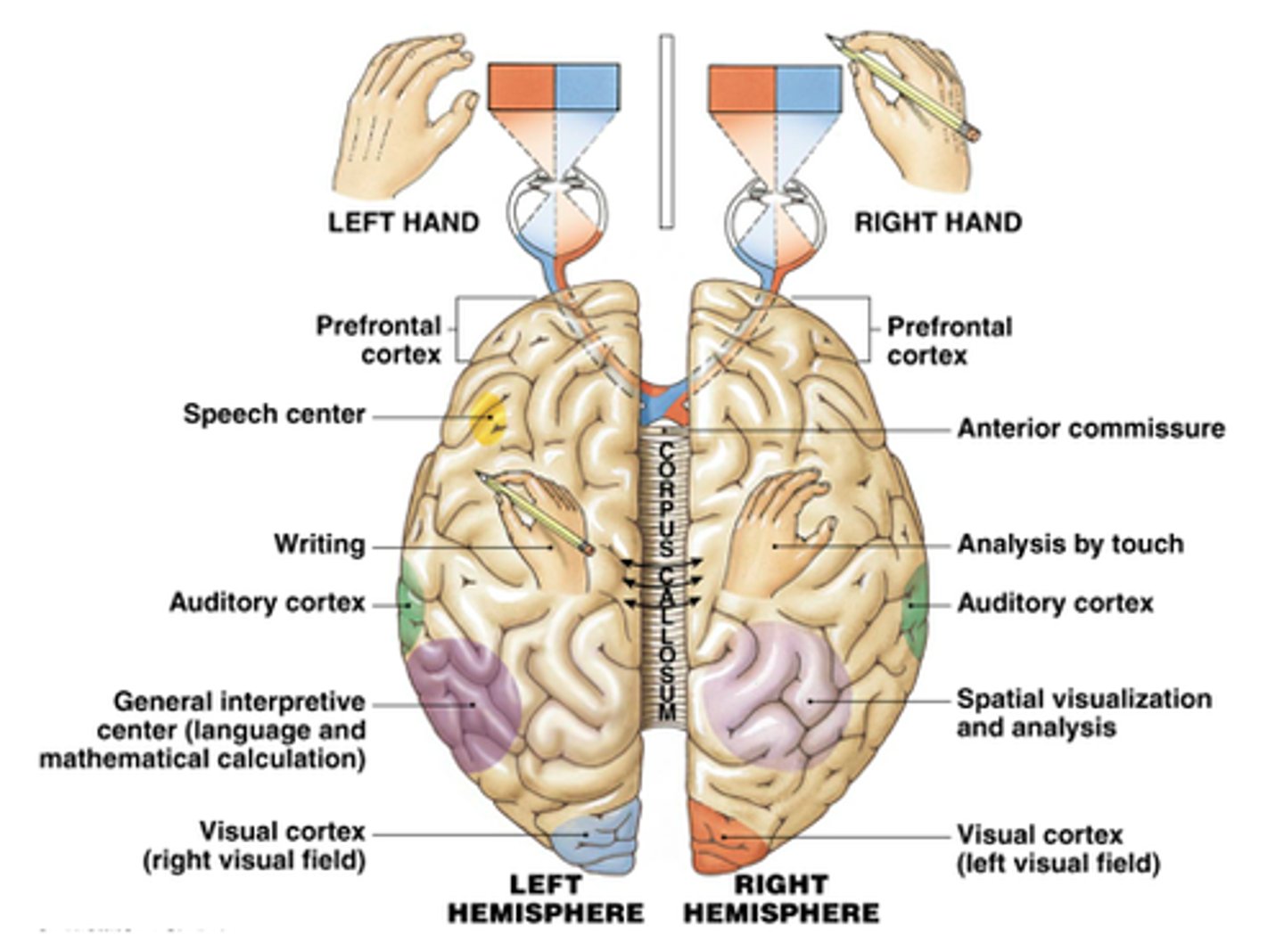
"Left Brain" vs. "Right Brain" (cerebral hemisphere differences)
Current research outlines that each hemisphere functions differently from one another.
The left cerebral hemisphere appears to handle functions such as language, math, music and logical and analytical thinking.
It is often considered to be the "rational" side of the brain or the more black and white thinking.
The right cerebral hemisphere is the more artistic hemisphere and functions more with visual-spatial perception, intuition, music and art appreciation, and emotional/holistic thinking.
It is often described as the more "big picture" or creative side of the cerebrum. The transverse fissure separates the cerebrum from the cerebellum on the posterior, inferior aspect of the brain.
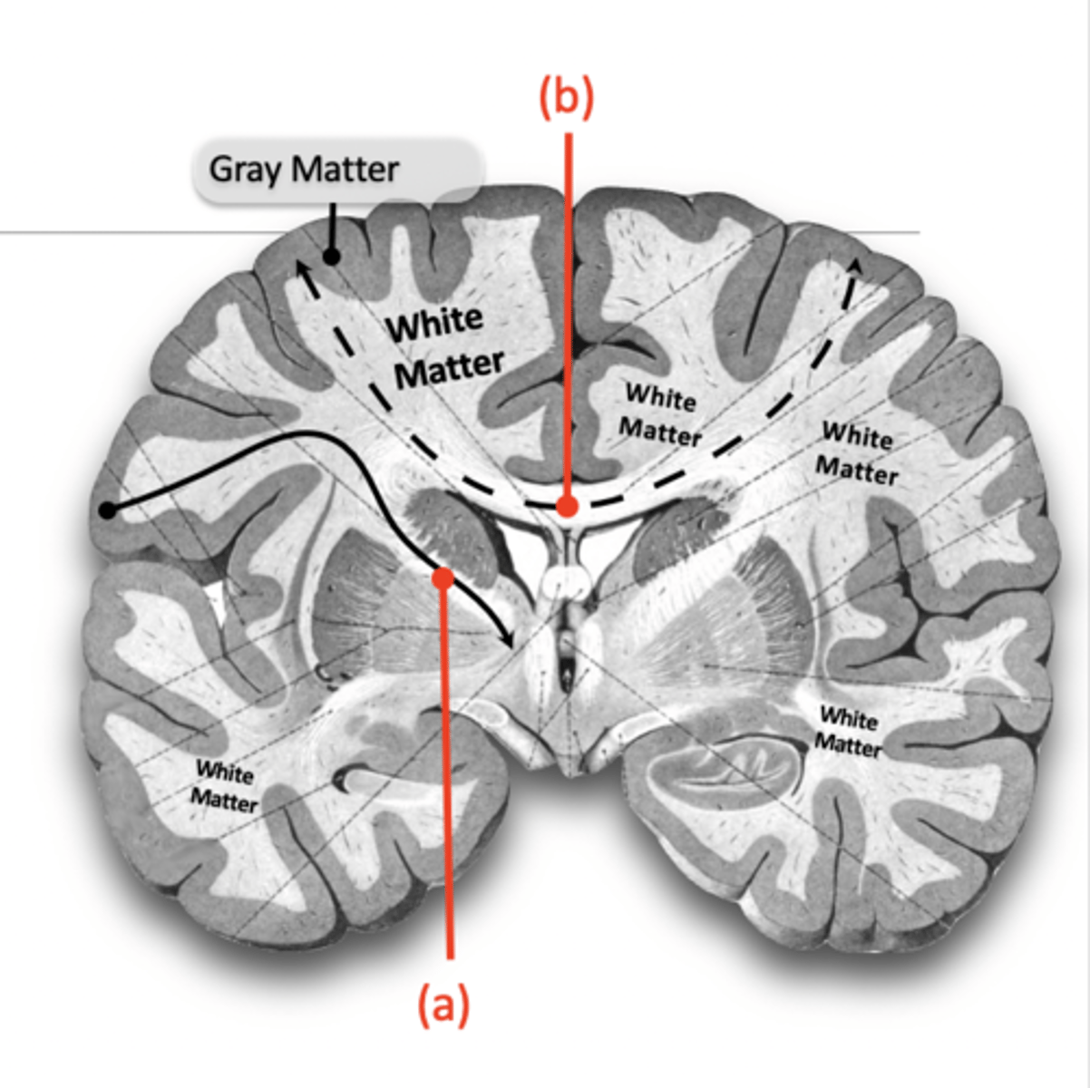
grey matter
Contains Cell bodies & dendrites of motor neurons and interneurons.
In the cerebrum and the cerebellum, the gray matter is the superficial layer (surface) of the brain.
This outer sheet of gray matter is called the Cortex.
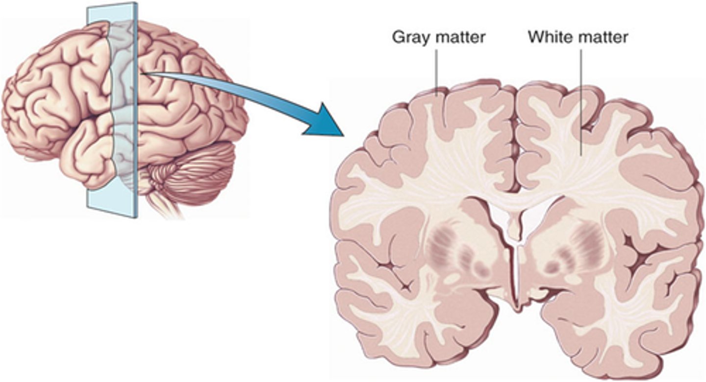
white matter
Is the location of:
- Myelinated axons (also known as nerve fibers)
- Myelin covering = lighter colored tissue
White matter is found deep to gray matter of the brain
These nerve fibers travel from gray matter to gray matter, for example:
- to and from brainstem or spinal cord
- between different areas of the cortex.
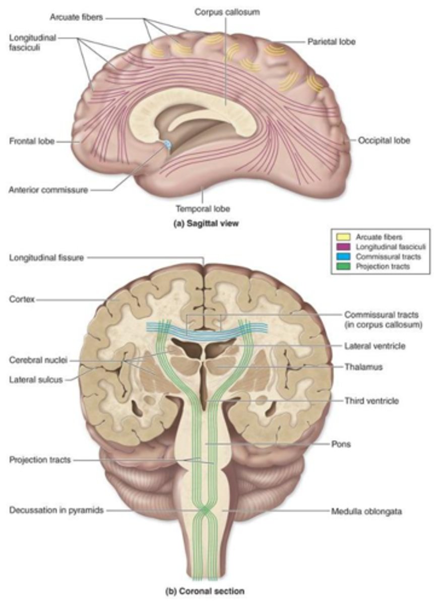
tracts
Specific collections of nerve fibers that often share common function.
Three classifications:
- Association tracts
- Commissural tracts
- Projection tracts
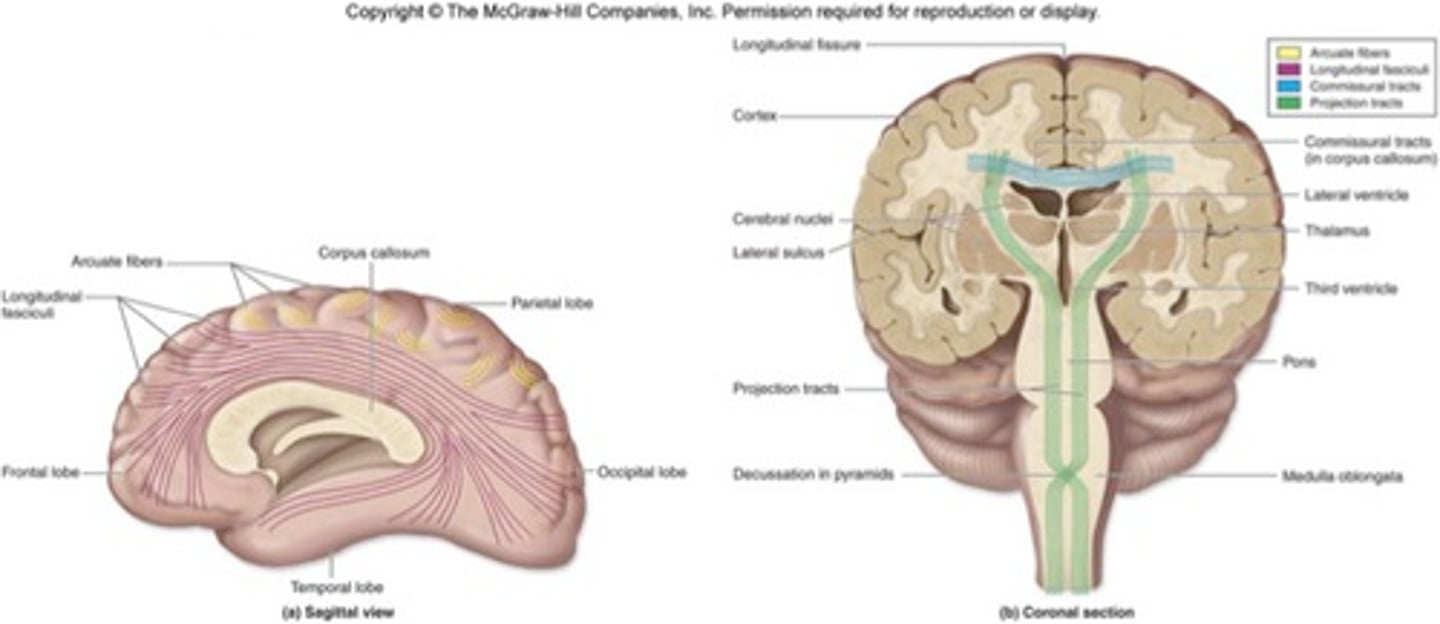
Association Tracts
Tracts of white matter that connect different areas of gray matter of the same hemisphere.
Connect nearby gyri of the same lobe or gyri of different lobes.
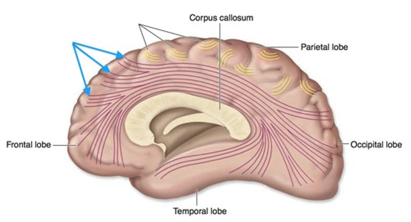
Commissural Tracts
Tracts of white matter that connect areas of gray matter between hemispheres (joins right and left hemispheres)
E.g. Corpus callosum
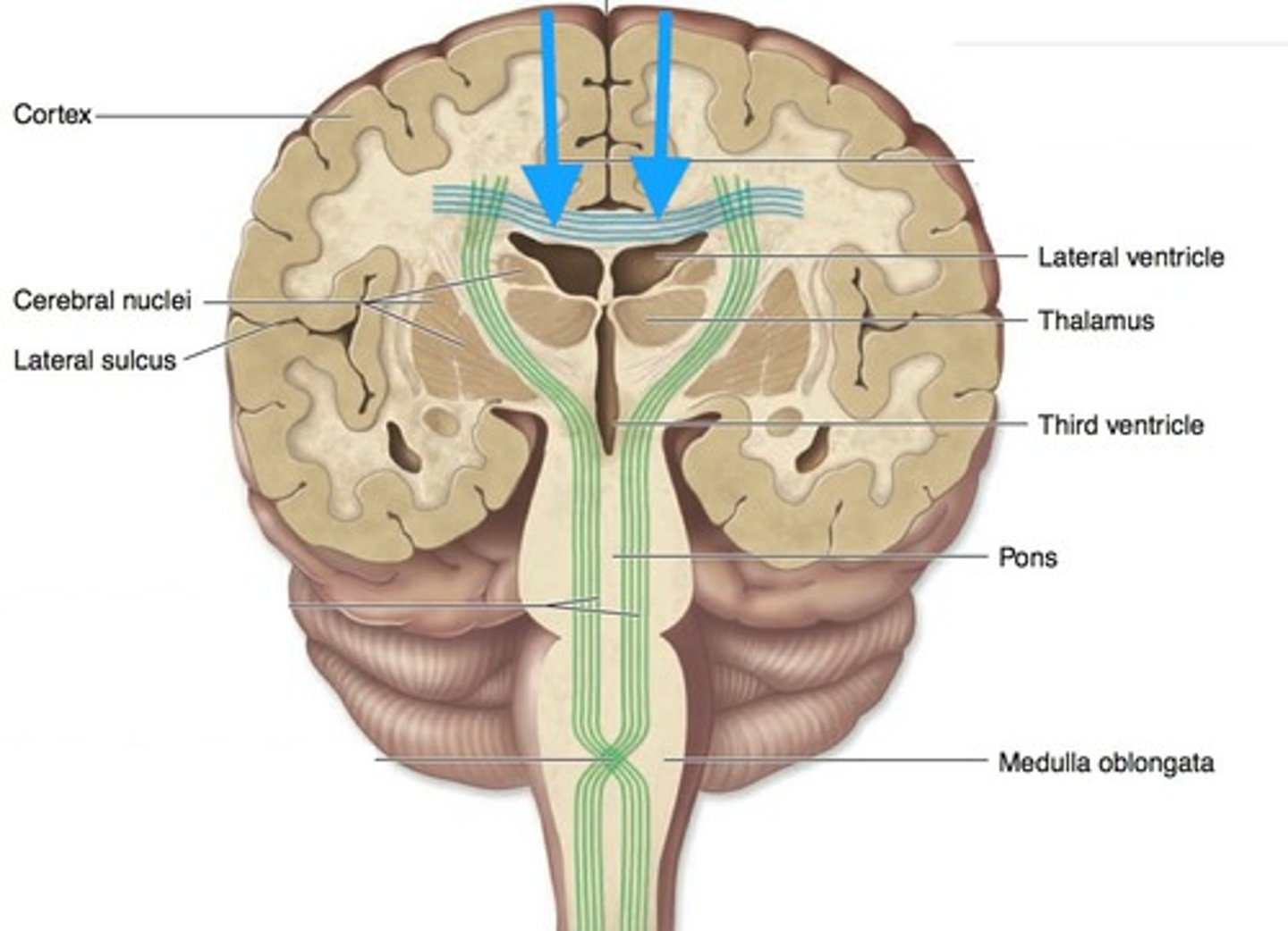
Projection tracts
Tracts of white matter that connect the cerebrum to other portions of brain or spinal cord.
fissure
A deep groove in the brain.
transverse fissure
Separates the cerebrum from the cerebellum.
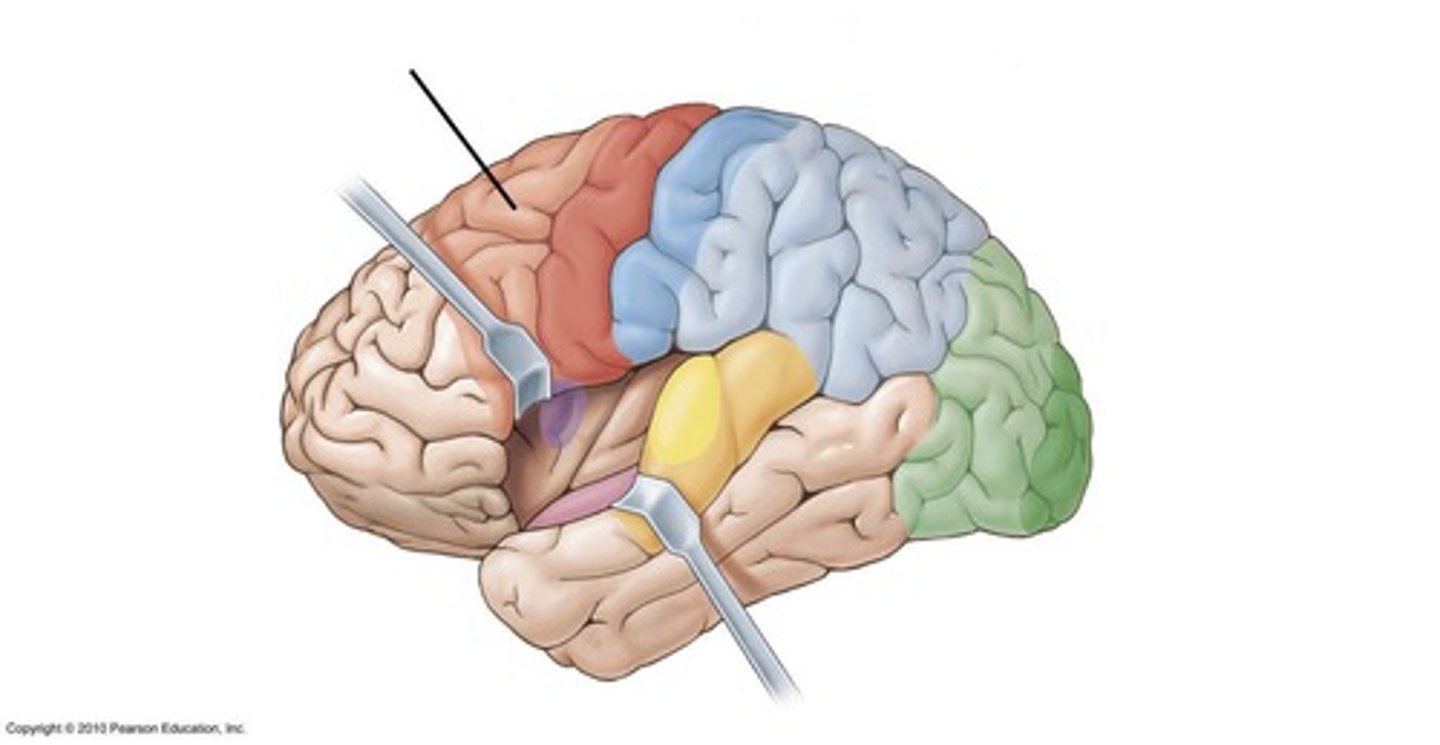
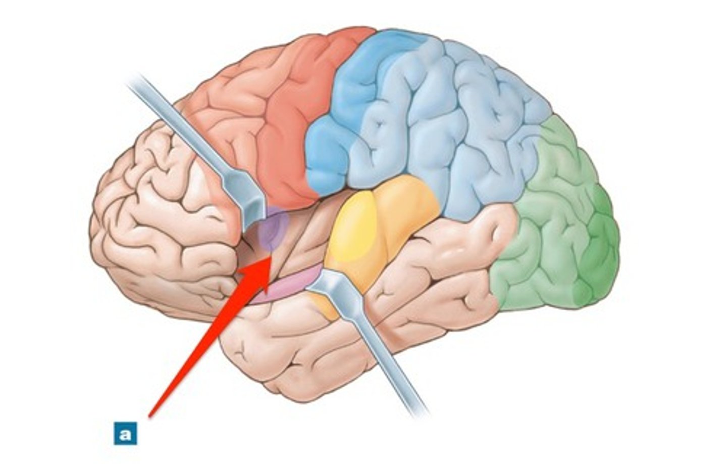
five lobes of the cerebrum
Four of them are superficial and are named for their overlying bones: Frontal, Parietal, Temporal, & Occipital
The fifth lobe, called the Insula, is found deep in the cerebrum.
What are association areas?
Areas of the cerebral cortex that are not involved in primary motor or sensory functions; rather, they are involved in higher mental functions such as learning, remembering, thinking, and speaking
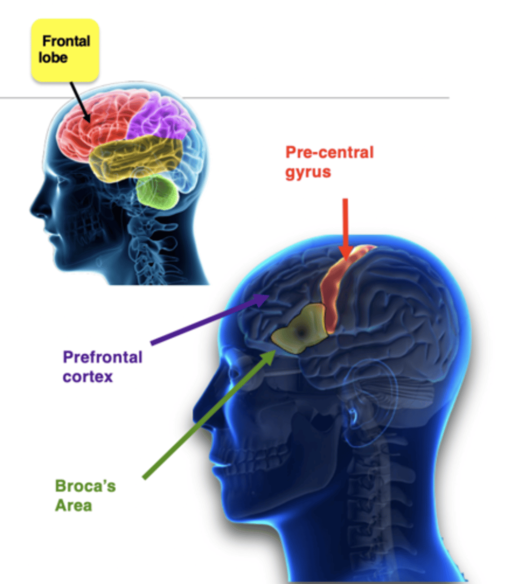
Frontal Lobes of the Cerebrum
Involved in:
- Thinking
- reasoning
- planning
- personality and emotions (prefrontal cortex)
- Initiating voluntary movements (pre-central gyrus)
- Producing speech (Broca's area)
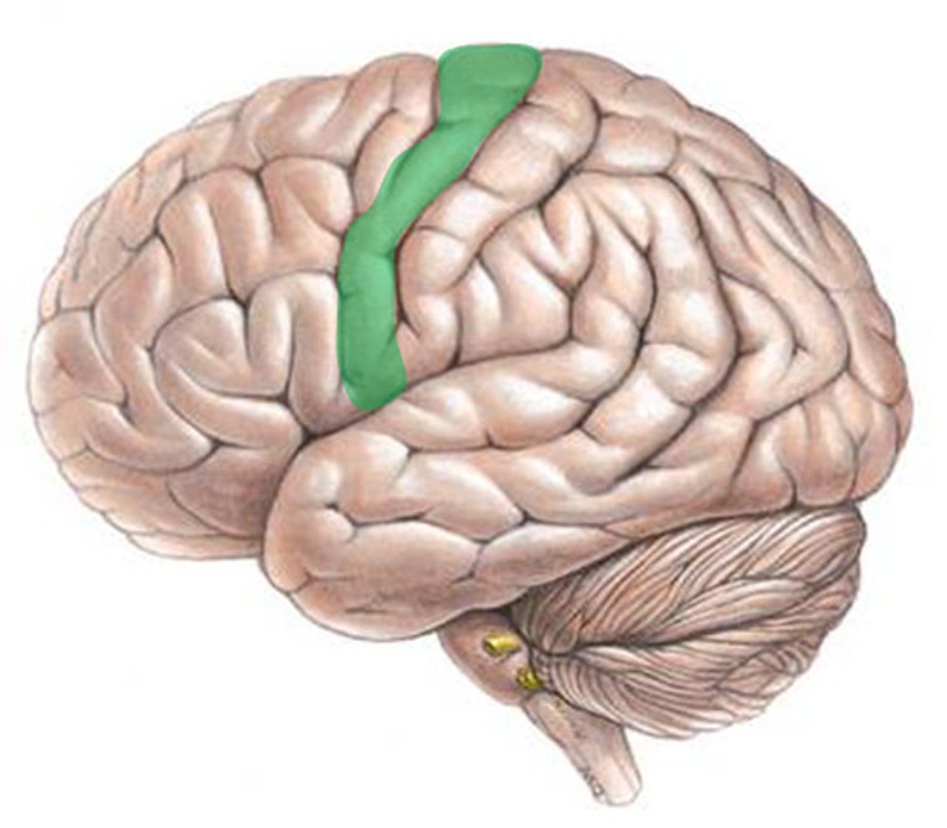
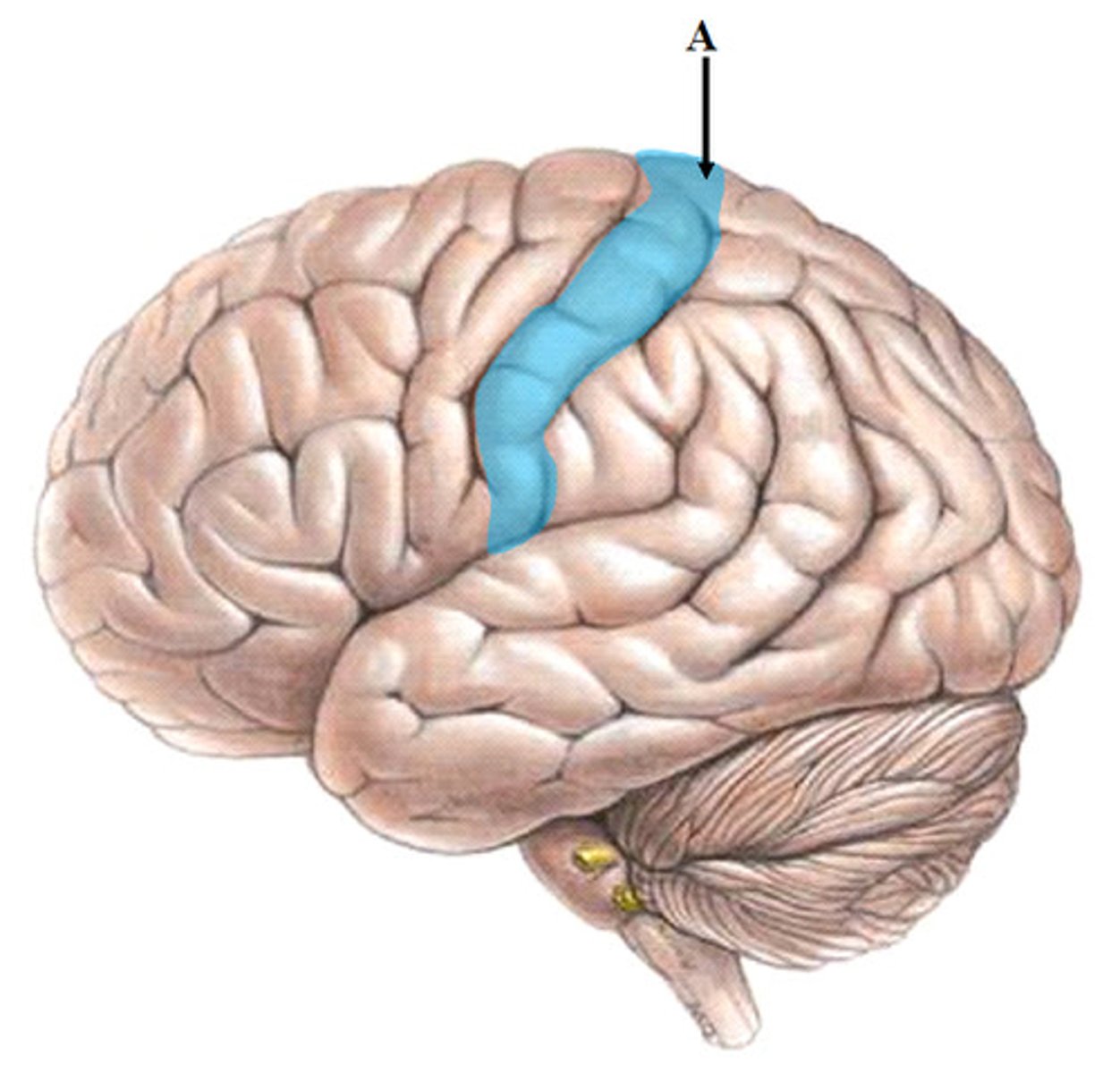
primary motor area
Precentral gyrus of frontal lobe
Sends impulses to skeletal muscles
Premotor area (motor association area)
Communicates with the primary motor area and thalamus to coordinate complex learned movements.
It is located anterior to the primary motor area in the frontal lobe.
Frontal eye field area
Area of the frontal cortex that controls voluntary scanning movements of the eyes (E.g. reading).
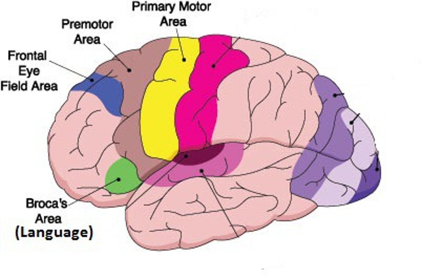
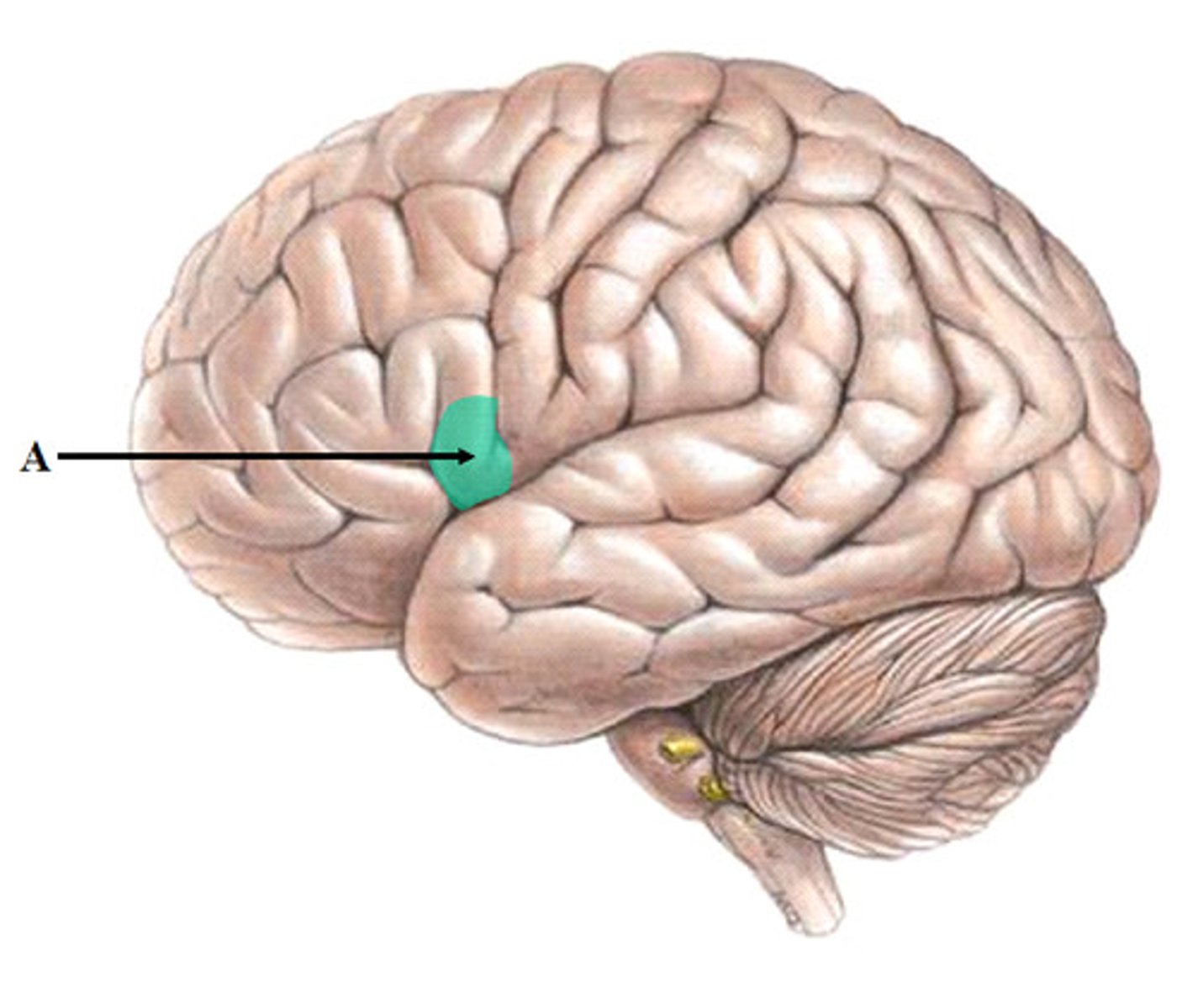
Broca's motor speech area
In the frontal lobe; controls muscle movements for speech
Usually in left lobe only
Receives impulses from Wernicke's sensory speech area (parietal lobe) formulates spoken response in concert with Prefrontal cortex (think about what you say!)
Relays impulses to premotor area and finally motor cortex.
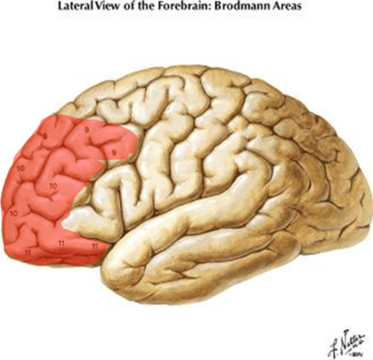
prefrontal cortex
The front-most portion of the frontal lobes; involved in planning and reasoning; one of the last areas of the brain to mature.
-primary association area (personality, intellect, cognition, etc.)
- Motivation and regulation of emotional behavior and mood.
- Goal setting
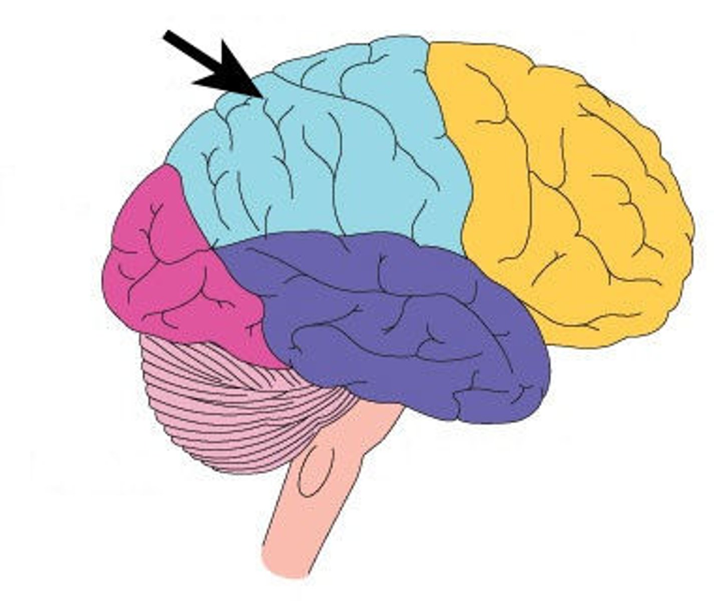
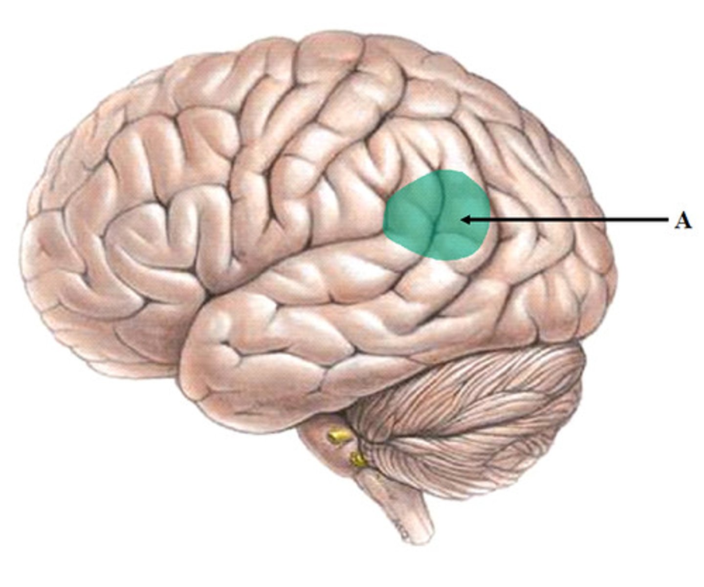
Parietal lobes of the Cerebrum
Involved in:
- Processing somatosensory information, including fine touch, pressure, temperature, pain (somatosensory cortex is in the post-central gyrus)
- Allowing us to understand our physical environment and how our body parts relate to each other
- Processing of some taste information
post central gyrus
primary somatosensory cortex (parietal lobe)
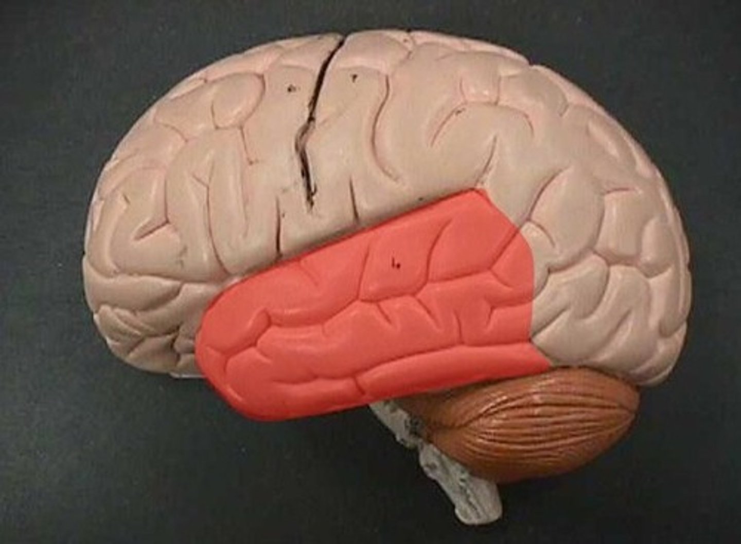
Temporal lobes of the Cerebrum
Involved in:
- Processing information on hearing and sound (primary auditory area - sensory for hearing).
- Word recognition and comprehension (Wernicke's area) = understanding speech and language. Contains the auditory association area. Will then send impulses to Broca's motor speech area
- Processing of some taste information
- Primary olfactory area - sensory for smell
- Memory
Wernicke's area
A brain area involved in language comprehension and expression; usually in the left temporal lobe
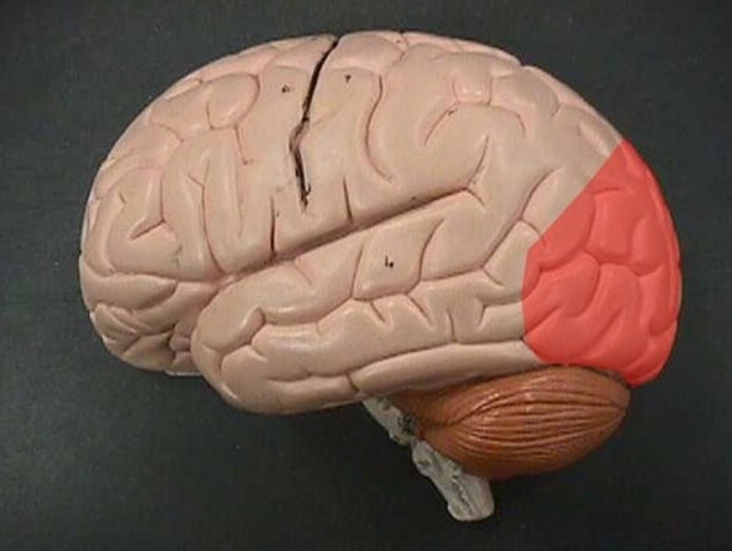
Occipital lobes of the Cerebrum
Involved in: Processing information on vision
Contains primary visual areas and visual association areas
Insula lobes of the Cerebrum
Found deep in the cerebrum
Also known as the limbic lobe
Involved in emotional responses and the behaviors and memories associated with them
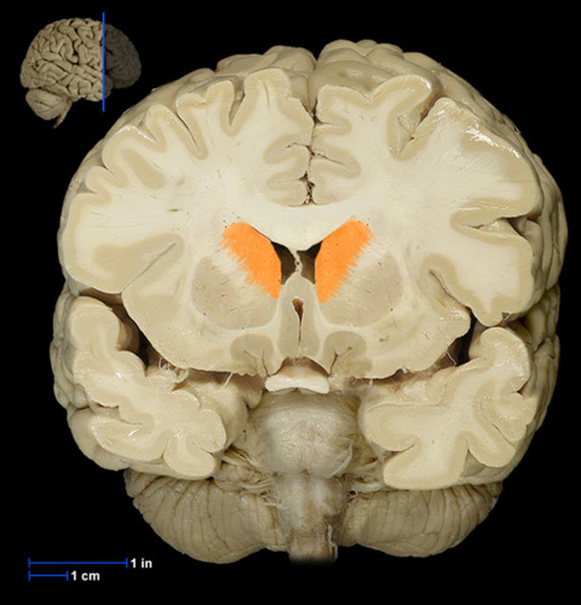
basal nuclei (basal ganglia)
Masses of gray matter found deep within the white matter of the cerebrum.
They integrate motor control by working with the motor cortex of the cerebrum to allow movements to start.
They also inhibit movements that are not necessary for what needs to be done.
Problems in the basal nuclei manifest with resting tremors (Parkinson's Disease)