Transcranial US Eval
 Call Kai
 Learn
Learn Practice Test
 Spaced Repetition
 Match
Match Flashcards
 Knowt Play
1/77

There's no tags or description
Looks like no tags are added yet.
Name | Mastery | Learn | Test | Matching | Spaced | Call with Kai | Chat |
|---|
No analytics yet
Send a link to your students to track their progress
78 Terms
TCD
Uses 2MHz non-imaging probe
0 degree angle
Vessel velocity, direction and pulsatility and turbulence assessed
Time averaged maximum velocity (TAMV) most commonly used for TCD evaluation
Microbubble contrast can improve exam
TCI/TCCD
Uses 1.8-2.5MHz phased array to produce B-mode, color and Doppler images
Can offer ability to angle correct for PSV instead of TAMV
Vertebrobasilar system and Circle of Willis evaluated with PW Doppler
Eval ACA, PCA, MCA, siphon and verts and basilar
Uses 5-10mm sample gate
Use small field of view
NEVER INVERT PW
Cervical ICA
Extracranial segment that originates at the carotid bifurcation and extends to the petrous portion of the temporal bone
Evaluated on extracranial Duplex exam
Petrous ICA
Intracranial segment that courses along the petrous portion of the temporal bone
Not able to evaluate using 2D imaging, requires Doppler evaluation
Cavernous ICA
Tortuous intracranial segment that extends from the petrous portion of the circle of Willis
Distal end of the cavernous segment is called the supraclinoid segment
Ophthalmic artery is the first branch of the ICA and originates at the cavernous segment
ICA terminates when it splits to MCA and ACA
MCA
Receives majority of blood from the ICA and is a larger vessel than the ACA
Supplies most of the temporal lobe, anterolateral frontal lobe, and parietal lobe
Each MCA divided into 3 segments
M1 (sphenoidal or horizontal segment, first portion)
M2 (Sylvian or insular segment, located in the sylvian fissure)
M3 (cortical segment, extends superiorly and inferiorly around the outer cerebrum and gives rise to numerous penetrating branches)
Terminal MCA connects to terminal ACA and PCA
Symptoms of MCA disease
Dysphasia, behavioral changes, severe hemiparesis in contralateral face/arms
MCA disease is related to Wernicke Aphasia -
Totally incomprehensive, unable to understand speech, speak, or follow directions
ACA
Supplies frontal lobe and parietal bones and corpus callosum
Each ACA is divided into two segments
A1 (Located between ICA and AcoA)
A2 (Located distal to the AcoA origin)
Symptoms of ACA disease
Loss of coordination, incontinence, severe leg hemiparesis or hemiplegia in contralateral leg, facial drooping or asymmetry
PCA
Both originate at the top of the basilar
Curve laterally, posteriorly and superiorly around the midbrain
Supply parts of midbrain, thalamus, temporal lobe, and occipital lobe
2 segments:
P1 (located between basilar and PcoA)
P2 (located distal to PcoA)
Vertebral arteries
Two vessels
Course from the subclavian arteries through vertebral pedicles and enter the cranium through the foramen magnum
Merge together in the cranium to form the single basilar artery
Proximal segments seen on extracranial evaluation, distal segments seen on TCI
Basilar artery
Formed at the junction of the two vertebral arteries inside the cranium
Supplies brainstem and cerebellum
Bifurcates into 2 PCA after superior cerebellar arteries branch off
Vertebrobasilar symptoms
Vertigo, ataxia, bilateral visual blurring or double vision, bilateral homonymous hemianopia, bilateral paresthesia, drop attack
Most common congenital anomaly of circle of Willis is
Absence of one or more communicating arteries
Most common location for intracranial aneurysm
Anterior communicating artery
AcoA
Single vessel that connects the two ACA branches
Potential for collateral flow between right and left anterior circulatory systems
Not normally directly evaluated in a TDC exam, but can be indirectly assessed by evaluating the flow direction of the main arteries of the Circle of Willis
PcoA
Two branches
Branches from the distal portion of the ICA before it terminates at the MCA/ACA bifurcation
Connects the PCA and MCA on each side
Potential for collateral flow
Not normally directly evaluated on TCD but can be indirectly assessed by evaluating the flow direction of the main renal arteries of the Circle of Willis
Most common indication for TDA/TCI
Assess vasospasm/vasoconstriction caused by subarachnoid hemorrhage
Most commonly occluded vessel with an acute stroke
MCA
Indications for TDC/TCI
Stroke, TIA, intracranial stenosis/occlusion, collateral flow due to occlusion, AV malformation (increased systolic and diastolic velocities), Vasospasm (increased PSV), Monitoring during procedures, microemboli detection, right to left shunt detection, patent foramen ovale, pediatric eval for sickle cell, suspected brain death
Contraindication for TCD/TCI
Recent eye surgery
Limitations of TDC/TCI
Hyperostosis - thickening of the cranial bones reduces sound penetration
Extensive time to pt and sonographer, incorrect vessel identification can be limited to position
Vasospasm can be mistaken for stenosis
Cannot evaluate the cerebral circulation for aneurysm formation
Cannot eval for aneurysm formation
Most common limitation of just TCD
Inaccurate vessel identification due to non-imaging technologist
Normal ACA/MCA velocity ratio
<1.2
Normal MCA/ICA velocity ratio
1.1-3.0
Normal basilar/vertebral velocity ratio
<2.0
Stenosis is most common in
The MCA, siphon and terminal ICA
MCA depth
30-60mm
MCA flow
Angle anteriorly and flow is anterior
MCA velocity
55 +- 12cm/s
ACA depth
60-80mm
ACA flow
Angle anteriorly, flow is away
PCA (P1 and P2) depth
60-70mm
PCA P1 flow
Angle posterior and caudal, flow is toward
PCA P1 Velocity
39 +- 10cm/s
PCA P2 flow
Angle posterior, flow is away
PCA P2 velocity
40 +- 10cm/s
Distal ICA depth
55-65mm
Distal ICA flow
Angle posterior and caudal, bi-directional flow
Distal ICA velocity
39 +- 9cm/s
Vertebral depth
60-90mm
Vertebral flow
Angle laterally, flow is away
Vertebral velocity
34 +- 8cm/s
Basilar depth
80-120mm
Basilar flow
Angle midline, flow is away
Basilar velocity
41 +- 10cm/s
Siphon depth
60-80mm
Siphon flow
Angle slightly lateral, flow can be away or bidirectional
Siphon velocity
41 +- 11cm/s
Ophthalmic depth
40-60mm
Ophthalmic flow
Angle medially, flow is toward
Ophthalmic velocity
21 +- 5cm/s
Abnormal intracranial vessel PSVs in TCI
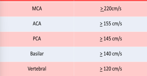
Most common collateral pathway intracranially
AcoA
Anterior communicating artery collateral
Provides collateral flow between the right and left anterior systems for high grade ipsilateral ACA or ICA stenosis
Contralateral ACA will demonstrate high velocities
Flow reversal is the A1 segment of the ipsilateral ACA
Posterior communicating artery collateral
Provides collateral flow between the carotid and basilar arteries for ipsilateral ICA stenosis or occlusion
Increased velocity in the ipsilateral P1 segment
Decreased velocity in the ipsilateral P2 segment
ECA to ICA collateralization
Occurs with ipsilateral ICA stenosis/occlusion
Reversal of flow in the ipsilateral ophthalmic artery
Increased systolic and diastolic velocities in contralateral ophthalmic artery
ECA > superficial temporal artery > supraorbital artery > ophthalmic artery > ICA
ECA > superficial temporal artery > frontal artery > ophthalmic artery > ICA
ECA > facial artery > angular artery > frontal artery > nasal artery
Peri-orbital Doppler
CW Doppler of the frontal artery (branch of ophthalmic)
Pt supine with 8-10MHz probe
Normal direction is towards probe
Compression of the infraorbital, facial and superficial temporal arteries is performed on both sides
FLOW AWAY from probe = IPSILATERAL ICA Occlusion
Determines if collaterals are being used to redirect flow
Intracranial AVM
Emerge during fetal development
Complications include subarachnoid hemorrhage, cerebral ischemia, cerebral aneurysm, brain damage or stroke
Vein of Galen defect
Wyburn-Mason syndrome - presents with unilateral vascular AVMs that involve brain, orbits and facial structures
US not normally used
Vasospasm
Vasoconstriction of intracranial arteries
Occurs within 14 days of the start of a subarachnoid hemorrhage
Flow velocities increase in MCA, ACA, and PCA
Always perform the repeat exams using the same depth as the initial eval
Treated with vasodilator
Lindegaard ratio → >3 = mildly abnormal, >6 = severely abnormal
Record highest velocities
Vasospasm is seen as a complication in
Pts with subarachnoid hemorrhage, usually a slow bleed
Patent foramen ovale
HITS are seen in the MCA a few seconds of injection of microbubbles
Sickle Cell anemia
Abnormally shaped RBCs produced by spleen
Can cause increased PSV in cranial vessels and increased risk for stroke
>200cm/s is abnormal for terminal ICA and MCA and indicate increased risk of stroke in children
Intraoperative monitoring
Most studies use depth of 50-55mm to eval M1
Can be performed at short intervals or continuously with a headband
Eval for microemboli from procedure
Noninvasive
TCD monitoring is used during:
Endarterectomy
Open heart surgery with bypass
ICU monitoring
Brian death - All waveforms have to and fro pattern and high increased resistance
Fistula and aneurysm
Intracranial arteriovenous malformations are most common in
Men
ICA terminates into
MCA and ACA
What vessels originate from the basilar
PCAs
The anterior communicating artery allows for
Flow between the right and left side of the circle of Willis
The posterior communicating arteries connect the
Anterior and posterior cerebral circulation systems
An acute stroke is most associated with blockage in:
MCAs
Limitations of TCD
Hyperstenosis
Incorrect vessel identification due to lack of imaging
Uncooperative pt
When evaluating the MCA from the temporal window, how do you move to assess the terminal ICA
Angle inferior
The transorbital window is typically used to eval the:
Ophthalmic artery
Which sign of laminar flow is not visualized in normal cerebral arteries
Spectral window
From the transtemporal window, the flow direction in a normal MCA should be:
Toward the transducer
What cardiac defect can be diagnosed using a TCD
Atrial septal defect