McGraw Hill Questions - Chapter 19
 Call Kai
 Learn
Learn Practice Test
 Spaced Repetition
 Match
Match Flashcards
 Knowt Play
1/116

There's no tags or description
Looks like no tags are added yet.
Name | Mastery | Learn | Test | Matching | Spaced | Call with Kai | Chat |
|---|
No analytics yet
Send a link to your students to track their progress
117 Terms
a common error is to describe arteries as the vessels that always transport ______ blood instead of describing it as a vessel that takes blood ___ from the heart
oxygenated; away
Which receive blood from veins?
Atria
The cardiovascular system consists of two circulations which are the _____ circulation and the _____ circulation
pulmonary; systemic
The space between the parietal layer and visceral layer of the pericardium contains _____.
serous fluid
The heart is a relatively small, conical organ approximately the size of a person's ______.
clenched fist
The fibrous pericardium is attached to both the _____ and the base of the _____ vessels
diaphragm; great
The wrinkled flap-like extensions visible in the anterior view of the heart are the ____.
auricles
The thin space between the parietal and visceral layers of the serous pericardium is the _____
pericardial
The heart is surrounded by a visceral and parietal ____.
pericardium
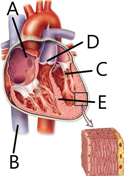
Match each structure with correct letter (Interior Heart)
A → fossa ovalis
B → inferior vena cava
C → chordae tendineae
D → pulmonary semilunar valve
E → interventricular septum
The _____ atrium and ventricle are prominent when viewing the heart from the anterior view.
Right
The position of the interventricular septum is deep to the _____________ located on the heart's superficial surface.
interventricular sulci
The outermost heart layer of the heart wall is the ______ and is also known as the ______ layer of the serous pericardium.
epicardium, visceral
The interatrial septum forms a thin wall between the right and
_______ ________.
left atria
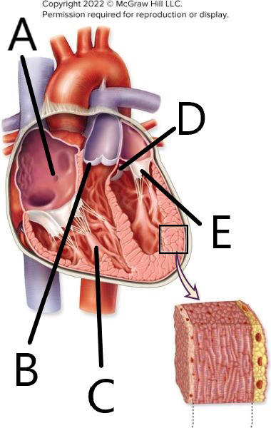
Match each structure with the indicator letter (heart interior)
A → opening of the coronary sinus
B → pulmonary semilunar valve
C → papillary muscle
D → aortic semilunar valve
E → left atrioventricular valve
Which side of the heart is more visible from an anterior view?
right
The right atrium receives venous blood from the ______.
superior and inferior vena cava
The right and left ventricles are separated by the ______
inter ventricular septum
There is an oval depression in the interatrial septum called the ______.
fossa ovalis
The posterior atrial wall is smooth, but the auricle and anterior wall exhibit obvious muscular ridges, called _______.
pectinate muscles
The _____ are smaller than the _____.
atria; ventricles
The internal wall surface of each ventricle displays characteristic large, smooth, irregular muscular ridges, called the ______.
trabeculae carneae
Identify the three major vessels that empty into the right atrium
superior vena cava
inferior vena cava
coronary sinus
Openings of ______ are visible in the left atrium.
pulmonary veins
During ventricular relaxation, blood is allowed to flow through the right ______ valve into the right ventricle.
atrioventricular
Like the right atrium, the left atrium also has ______ in its auricle.
pectinate muscles
Correctly match the valve with its structure
right atrioventricular → three triangular-shaped cusps of dense connective tissue
semilunar → three half-moon-shaped cusps of dense connective tissue
left atrioventricular → two triangular-shaped cusps of dense connective tissue
The heart valves consist of _______-lined fibrous connective tissue flaps called _________.
endothelium, cusps
The left atrioventricular valve has ______ cusps.
2
The right _____ valve is forced closed when the right ventricle begins to contract, preventing blood from flowing back into the right atrium
atrioventricular (AV)
The ______ delivers oxygen-rich blood to the thick heart wall while the ______ carries the deoxygenated blood away from the heart wall.
coronary arteries, coronary veins
Match the valve with its correct function
Right atrioventricular → prevents backflow of blood into the right atrium
Pulmonary semilunar → prevents backflow of blood into the right ventricle
Left atrioventricular → prevents backflow of blood into the left atrium
Aortic semilunar → prevents backflow of blood into the left ventricle
True or false: Papillary muscles are associated with semilunar valves.
False
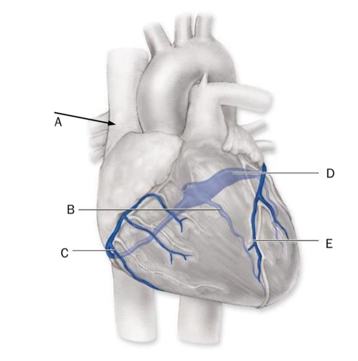
Match the coronary vein with the indicator letter in the diagram
A → superior vena cava
B → middle cardiac vein
C → small cardiac vein
D → coronary sinus
E → great cardiac vein
The right coronary artery typically branches into the right ______ artery, which supplies the lateral wall of the right ventricle, and the posterior ______ artery, which supplies the posterior surface of both the left and right ventricles
marginal, inter ventricular
True or false: The heart myocardium is nourished primarily through the diffusion of nutrients from the blood in the heart chambers
False; the walls do not absorb nutrients or oxygen by diffusion and are dependent on the coronary arteries to deliver them
The left AV valve is forced ______ when the left ventricle begins to contract, ______ blood backflow into the left atrium.
closed, preventing
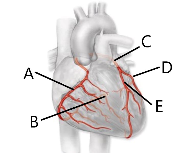
Match the coronary artery with the indicator letter in the diagram
A → right coronary artery
B → posterior interventricular artery
C → left coronary artery
D → circumflex artery
E → anterior interventricular artery
The left coronary artery typically branches into the ______ interventricular artery and the ______ artery.
anterior, circumflex
An arterial ________ forms when more than one artery is providing blood to a given tissue.
anastomosis
The anterior interventricular artery (LAD) supplies the ______ wall of the left ventricle and ______ the interventricular septum.
anterior; most of
Coronary arteries are considered functional end arteries and not anastomoses because ______.
if one becomes blocked there is insufficient blood flow to the tissue
The ____ lies within the anterior interventricular sulcus
great cardiac vein
Blood from the coronary circulation returns to the right atrium through one of several _______ _______.
coronary veins
The circumflex artery supplies blood to the ______ ventricle.
left
The opening for the coronary sinus is located in the wall of the ______.
right atrium
The ______ cardiac vein runs alongside the ______ interventricular artery
middle; posterior
The ______ cardiac vein travels close to the right ______ artery.
small, marginal
True or False: The sarcoplasmic reticulum is more extensive in smooth muscle fibers than in skeletal muscle fibers
False
Cardiac muscle can use fatty acids, ________, lactate, amino acids, and ________ bodies as fuel sources.
glucose, ketone
The ______ lies within the anterior interventricular sulcus.
great cardiac vein
The heartbeat is initiated by the specialized cardiac muscle cells of the ____, which are located in the posterior wall of the right atrium, adjacent to the entrance of the ____.
sinoatrial node; superior vena cava
The ______ cardiac vein travels alongside the ______ interventricular artery
great, anterior
Choose all that are part of the cardiac muscle cells
sarcolemma
T tubules
myofibrils
The AV node is located in the floor of the ____.
right ventricle
Select all that contribute to the ability of a cardiac muscle to meet its energy demands
numerous mitochondria
an extensive blood supply
myoglobin
True or False: The cells of the cardiac conduction system do not contract
False; they initiate and conduct electrical singal’s but they do not contract
After initiation the muscle impulse travels from the SA node to the ____ ____.
AV node
Parasympathetic and the sympathetic axons extend from the ____ to the heart
cardiac center
Parasympathetic innervation ____ the heart rate, but generally tends to have no effect on the force of contractions
decreases
The cardiac center is within the ____
medulla oblongata
True or false: The innervation by autonomic centers in the brainstem does not initiate a heartbeat, but it can increase or decrease the rate of the heartbeat.
True
True or False: Sympathetic fibers arise from the cardioinhibitory center
False; they arise from cardioacceleratory center
Parasympathetic innervation of the heart comes from the ____ via the left and right vagus nerves
cardioinhibitory center
In the heart, action potentials spread across the ____ of cardiac muscle cells, causing them to contract
sarcolemma
What is the SA node are the pacemaker cells that initiate a heartbeat?
nodal cells
Sympathetic fibers cause ____ of coronary arteries
dilation
True or False: All Neurons have a pacemaker potential
False; SA nodal cells do have pacemaker potential because they exhibit autorhythmicity
Put the sequence of events in the correct order to describe the initia
SA node fires action potential: The sinoatrial (SA) node acts as the heart's natural pacemaker and generates the initial electrical impulse
Action potential reaches AV node via gap junctions: The electrical wave spreads across the atria and arrives at the atrioventricular (AV) node
Action potential is delayed at the AV node: The signal pauses briefly here to let the atria finish contracting and empty blood into the ventricles
Action potential travels down the bundle branches: The impulse moves from the AV bundle (bundle of His) down the right and left bundle branches through the interventricular septum
Action potential travels up the Purkinje fibers: The Purkinje fibers rapidly conduct the signal upward through the ventricular walls to trigger a full contraction
Nodal cells in the SA initiate a heartbeat by spontaneously ____
to generate an action potential.
depolarization
Cardiac muscle cells contain Ca2+ pumps that keep more Ca2+ ____
the cell.
outside
The ability to reach threshold without stimulation is called the ____ ____
of the SA node.
Pacemaker; potential
Place in the correct order the sequence of events resulting in the action potential for cardiac muscle cells.
Na+ ions enter causing depolarization.
Depolarization causes opening of voltage-gated K+ channels.
Slow voltage gated-Ca2+ channels open causing a Ca2-induced Ca2 release
Plateau occurs
Voltage-gated Ca2+ channels close
Repolarization occurs returning to the resting membrane potential
The purpose for the delay in the conduction of the action potential from the AV node to the bundle branches of the ventricles is to allow ______, before the ventricles contract
the atria to complete their contraction
The sarcolemma of the cardiac muscle cells has fast voltage-gated ____ channels that participate in the depolarization of the cell membrane
sodium, Na+, or Na
Which shorten as a result of sarcomeres shortening?
Both cardiac and skeletal muscle cells
The movement of ______ is responsible for the depolarization phase of the action potential in cardiac muscle cells.
Na+
The complex of an ECG that represents the electrical changes that are associated with ventricular depolarization is the ____ complex.
QRS
The time from the start of one heartbeat to the initiation of the next is called a(n) ____ cycle.
cardiac cycle
Which cells cannot exhibit tetany?
cardiac muscle
When the ______ contract, blood is forced into the ______ through the open AV valves.
atria; ventricles
During atrial relaxation, blood is entering the ______ atrium via superior and inferior vena cavae, and blood is entering the ______ atrium via the pulmonary veins
right; left
The wave of an ECG that reflects the electrical changes of atrial depolarization is the ____ wave
p
When the ventricles contract, the ______ close as blood pushes against the cusps of the AV valves, and their edges meet to form a seal.
atrioventricular valves
During the cardiac cycle, blood flows from veins into the atria under ______
low pressure
As the cardiac cycle begins, the semilunar valves are closed because the pressure exerted by the blood in the ventricles is ..................
than the pressure the blood exerts in the great arteries.
Blank 1: lower, low, less, or lesser
Toward the end of ventricular filling, the left and right ______ contract simultaneously.
atria
Papillary muscles and the ______ prevent the AV valve cusps from ______.
chordae tendineae, everting
During the cardiac cycle, blood only passes from the atria into the ventricles if the ____ valves are open.
AV
The amount of blood in the ventricles immediately prior to initiation of contraction is called ____ ____volume.
Blank 1: end
Blank 2: diastolic
Passive filling of the ventricles through the open ....................
valves occurs at the beginning of the cardiac cycle while the ventricles are in ................
Blank 1: AV or atrioventricular
Blank 2: diastole or relaxation
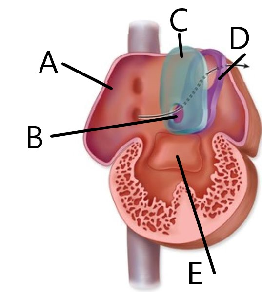
Match
A → right atrium
B → foramen ovale
C → septum secundum
D → septum primum
E → endocardian cushion
Electrical activity is started at the SA node, which causes an action potential to spread through the heart through the ______.
conduction system
Choose all that atrial contraction causes
Blood is pushed into ventricles
Openings of the great veins are compressed
The amount of blood that leaves the heart during the ventricular contraction is called the ____ ____
stroke volume
During isovolumetric relaxation, the ____ valves close to prevent blood backflow into the ventricles
semilunar
Choose all events that occur during the atrial concentration and ventricular filling phase of the cardiac cycle
The ventricles have reached their EDV
Blood flow into the atria from the veins is restricted
SA node initiates atrial contraction
Prolonged pumping of unequal amounts of blood by the left and right ventricles can lead to ____
edema
The contraction phase of the cardiac cycle that includes closure of the AV and semilunar valves even though the Purkinje fibers have initiated ventricular contraction is the __________ phase.
isovolumetric
The amount of blood in the ventricles immediately prior to initiation of contraction is called ___________ __________ volume.
end diastolic