UNIT 12. CATALASE-POSITIVE, GRAM-POSITIVE COCCI
 Call Kai
 Learn
Learn Practice Test
 Spaced Repetition
 Match
Match Flashcards
 Knowt Play
1/158

There's no tags or description
Looks like no tags are added yet.
Name | Mastery | Learn | Test | Matching | Spaced | Call with Kai | Chat |
|---|
No analytics yet
Send a link to your students to track their progress
159 Terms
Staphylococci
Micrococci
CATALASE-POSITIVE, GRAM-POSITIVE COCCI usually refers to the two genera
ability of microorganism to produce catalase to breakdown hydrogen peroxide into water and oxygen = EFFERVESCENCE
principle involved in Catalase test
“staphle” = bunches of grapes
etymology of “Staphylococci”
capablity of producing coagulase to form clumping or clot in the presence of plasma
principle of Coagulase test
Slide Method = detect clumping factor (bound coagulase)
Tube Method = detect free coagulase
Slide Method = detect _
Tube Method = detect _
Staphylococcus aureus
Couagulase test is usually specific for what specie?
Staphylococcus delphini
Staphylococcus intermedius
Staphylococcus pseudintermidius
ENUMERATE the Staphylococcus species (seen in animals) other the S. aureus that may give a positive coagulase test
Staphylococcus aureus
What is/are the catalase-positive, coagulase-positive, gram-positive staphylococci
Staphylococcus epidermidis
Staphylococcus haemolyticus
Staphylococcus saprophyticus
Staphylococcus lugdunensis
Staphylococcus schleiferi
Staphylococcus capitis
Staphylococcus caprae
Staphylococcus warneri
Staphylococcus auricularis
Staphylococcus hominis
Staphylococcus nohnii
Staphylococcus xylosus
Staphylococcus simulans
Micrococcus spp. and related genera
Alloiococcus
What is/are the catalase-positive, coagulase-negative, gram-positive staphylococci
Staphylococcaceae
Family of Staphylococcus
aerobic or facultative anaerobic;
S. aureus subsp. anaerobius;
S. sacchrolyticus;
obligate anaerobes
Staph are generally ___ (O2 requirement) except ___ and ___ which are ___
F
They are nonmotile, non spore-forming
T or F
Staph are motile and non spore forming
spherical
0.5-1.5 um
singly, in pair, in clusters
Shape, size, and arrangement of Staph
skin
mucous membranes
intestines
normal location of Staph
community-acquired
Staphylococcus aureus nature of infection is _
nosocomial infection
Other Staphylococci (coagulase-negative) causes _
medium sized (4-8 mm)
cream-colored, white, or rarely light gold,
“buttery-looking”
other spp. may have gray colonies
some may be B-hemolytic (S. aureus)
Describe the appearance and morphology of Staph colonies grown in BAP
fermenter
Staph result in OF medium
oxidizer
Micrococci result in OF medium
Staphylococcus aureus
true coagulase positive and most virulent species of staphylococci
NA and TSB
S. aureus grow well on most routine media like __ and __
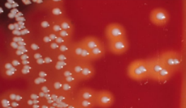
golden yellow color
B-hemolytic
describe S. aureus grown in BAP
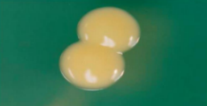
round,
smooth,
opaque,
butyrous
describe S. aureus grown in solid media
Protein A
Peptidoglycan
Teichoic Acid
Clumping Factor
Capsular Polysaccharide
[PePeT Cum Cum]
ENUMERATE
(Virulent factors) Antigenic Structure of S. aureus
Peptidoglycan
Teichoic Acid
(VIRULENT FACTORS) ANTIGENIC STRUCTURE OF S. AUREUS
it protects the bacteria from lysis and probably aids in adherence
Clumping Factor
(VIRULENT FACTORS) ANTIGENIC STRUCTURE OF S. AUREUS
component on cell wall responsible for clumping of the whole Staphylococci in the presence of plasma
Protein A
(VIRULENT FACTORS) ANTIGENIC STRUCTURE OF S. AUREUS
group specific antigen unique to S. aureus
Capsular Polysaccharide
(VIRULENT FACTORS) ANTIGENIC STRUCTURE OF S. AUREUS
protects for phagocytosis
COAGULASE (STAPHYLOCOAGULASE)
HYALURONIDASE (SPREADING FACTOR)
STAPHYLOKINASE (FIBRINOLYSIN)
LIPASE (FAT-SPLITTING ENZYME)
DEOXYRIBONUCLEASE (DNASE) AND PHOSPHATASE
PROTEASE
GELATINASE
B-LACTAMASE
ENUMERATE
(VIRULENT FACTORS) ENZYMES OF S. AUREUS
COAGULASE (STAPHYLOCOAGULASE)
(VIRULENT FACTORS) ENZYMES OF S. AUREUS
coagulates fibrinogen in the plasma
promotes fibrin layer formation around the staphylococcal abscess protecting the bacteria from phagocytosis
Cell-bound Coagulase of Clumping Factor
Unbound or Free Coagulase
ENUMERATE
2 TYPES OF STAPHYLOCOAGULASE
Cell-bound Coagulase of Clumping Factor
2 TYPES OF STAPHYLOCOAGULASE
bound the cell wall and clots human, rabbit, or pig plasma
Unbound or Free Coagulase
2 TYPES OF STAPHYLOCOAGULASE
extracellular enzymes not bound to the cell wall and cause clot formation when bacterial cells are incubated with plasma
HYALURONIDASE (SPREADING FACTOR)
(VIRULENT FACTORS) ENZYMES OF S. AUREUS
hydrolyzes hyaluronic acid present in the intracellular ground substance, permitting the spread of infection
STAPHYLOKINASE (FIBRINOLYSIN)
(VIRULENT FACTORS) ENZYMES OF S. AUREUS
fibrinolytic activities by dissolving fibrin clot
LIPASE (Fat-Splitting Enzyme)
(VIRULENT FACTORS) ENZYMES OF S. AUREUS
act on lipids present on the surface of the skin, particularly fats and il secreted by the sebaceous glands
LIPASE (Fat-Splitting Enzyme)
(VIRULENT FACTORS) ENZYMES OF S. AUREUS
important in the formation of furuncles, carbuncles, and boils
T
T or F
LIPASE (Fat-Splitting Enzyme) are produced by both coagulase (+) and coagulase (-) staphylococci
DEOXYRIBONUCLEASE (DNASE) AND PHOSPHATASE
(VIRULENT FACTORS) ENZYMES OF S. AUREUS
lowers viscosity of exudates giving the pathogen more mobility
destroys DNA
B-LACTAMASE
(VIRULENT FACTORS) ENZYMES OF S. AUREUS
breaks down Penicillin and B-lactam drugs
CYTOTOXIC TOXINS
Alpha (α)-hemolysin
Beta (β)-hemolysin
Delta (δ)-hemolysin
Gamma (γ)-hemolysin
Staphylococcal (Panton-Valentine) Leukocidin
ENTEROTOXINS
TOXIC SHOCK SYNDROME TOXIN-1 (TSST-1)
EXFOLIATIVE TOXIN
ENUMERATE
(VIRULENT FACTORS) TOXINS OF S. AUREUS
Alpha (α)-hemolysin
Beta (β)-hemolysin
Delta (δ)-hemolysin
Gamma (γ)-hemolysin
ENUMERATE
4 Types of Hemolysins
Alpha (α)-hemolysin
4 Types of Hemolysins
damage RBC, platelets, and macrophages and cause severe tissue damage
Alpha (α)-hemolysin
4 Types of Hemolysins
predominant hemolysin of S. aureus
Beta (β)-hemolysin / Sphingomyelinase C
4 Types of Hemolysins
acts on sphingomyelin in the plasma membrane of RBC
Beta (β)-hemolysin / Sphingomyelinase C
4 Types of Hemolysins
aka “hot-cold” lysine
enhanced hemolytic activity in incubation at 37 C (heat labile) and subsequent exposure to cold (4 C)
Delta (δ)-hemolysin
4 Types of Hemolysins
produced by all S. aureus strain that cause RBC injury in culture and produce edematous lesions
less toxic to cells than either a-hemolysin of B-hemolysin
Gamma (γ)-hemolysin
4 Types of Hemolysins
associated with Panton-Valentine Leukocidin (PVL)
Staphylococcal Leukocidin / Panton-Valentine Leukocidin
exotoxin lethal to polymorphonuclear leukocytes ;
associated with community-acquired staphylococcal infections
Staphylococcal Leukocidin / Panton-Valentine Leukocidin
pore forming exotoxin that suppress phagocytosis and associated with severe cutaneous infections and necrotizing pneumonia
100° C
30 minutes
ENTEROTOXINS are heat-stable exotoxin, able to grown at __ for _
gastric and jejunal enzymes
ENTEROTOXINS resistant to hydrolysis by _
ENTEROTOXINS
act as neurotoxins that stimulate vomiting through the vagus nerve
30-5
ENTEROTOXINS are produced by _% of S. aureus isolates
ENTEROTOXINS A, B, and D
Staphylococcal food poisoning
Usually causes nausea, vomiting, abdominal cramps, and diarrhea.
Enterotoxins B and C (sometimes G and I)
Enterotoxins that causes Toxic Shock Syndrome (TSS)
Enterotoxin B
causes Staphylococcal Pseudomembranous Enterocolitis (from contaminated milk products)
ENTEROTOXIN F
PYROGENIC EXOTOXIN C
TOXIC SHOCK SYNDROME TOXIN-1 (TSST-1) aka?
TOXIC SHOCK SYNDROME TOXIN-1 (TSST-1)
menstruating-associated TSS = TSS associated with tampon use
chromosomal-mediated toxin
SUPERANTIGEN;
T-cell
cytokines
TSST-1 acts as a ___ stimulating __ proliferation and production of a large amount of _
EPIDERMOLYTIC TOXIN A and B
EXFOLIATIN serotypes A and B
EXFOLIATIVE TOXIN aka?
EXFOLIATIVE TOXIN
serine protease that divides the intracellular bridges of the epidermis and causes excessive sloughing of the epidermis (stratum granulosum)
Staphylococcal Scalded Skin Syndrome (Ritter’s Disease);
Bullous Impetigo
Exfoliative toxin causes ____ (___) and is implicated in ___
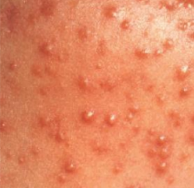
Folliculitis
mild inflammation of a hair follicle or oil gland; infected area is raised and red
raised, domed pustules form around hair follicles
Furuncles (Boils)
focal suppurative lesions which has resulted from an infection (folliculitis) that extend into subcutaneous tissue; large, raised, superficial abscesses
large abscess forms around a hair follicle
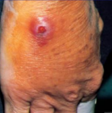
Carbuncles
larger, more invasive lesions develop from multiple furuncles, which can progress into deeper tissues; present with fever and chills, indicating systemic infection
multilocular abscess around several hair follicles

Bullous Impetigo
larger pustules surrounded by a small zone of erythema
highly contagious infection that spread by direct contact, fomites, or autoinoculation
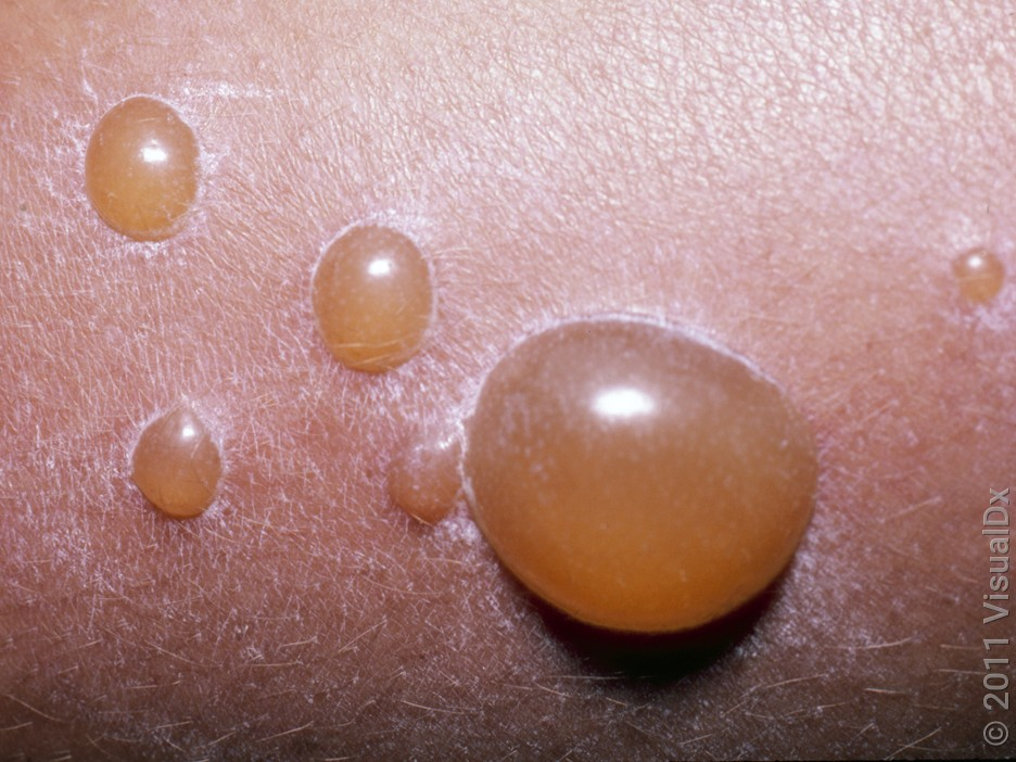
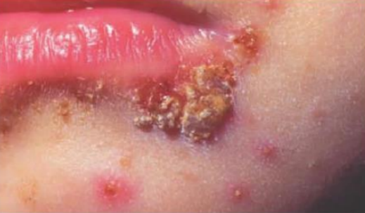
Impetigo
superficial cutaneous infection commonly seen in newborns and young children characterized by the formation of encrusted pustules surrounded by red border
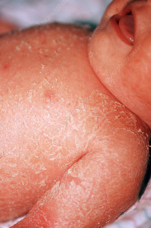
Scalded Skin Syndrome
bullous exfoliative dermatitis that occurs primarily in newborns and previously healthy young children
causes the skin to peel off in sheets
likely to occur in children under age 2.
few blisters
pemphigus neonatorum
Ritter disease
Localized form of SSS
cutaneous erythema
profuse peeling of the epidermis
Generalized form of SSS
Toxic Epidermal Necrolysis (TEN)
clinical manifestation with multiple causes;
symptoms are due to hypersensitivity reaction
TOXIC SHOCK SYNDROME
rare but potentially fatal, multisystem disease characterized by sudden onset of fever, chills, vomiting, diarrhea, muscle aches, and rash, which can quickly progress to hypotension and shock
FOOD POISONING
intoxication resulting from ingestion of a toxin formed outside the body
symptoms appear rapidly (2 to 8 hours after ingestion) and resolve within 24 to 48 hours
nausea, vomiting, abdominal pain, and severe cramping, diarrhea
Enterotoxin A (78%)
Enterotoxin D (38%)
Enterotoxin B (10%)
enterotoxins causing food poisoning
FOOD POISONING
perfuse and watery diarrhea due to water and electrolyte loss
food won’t appear or taste tainted
Enterotoxin A
LukE
LukP
Virulence factors causing ENTEROCOLITIS
STAPHYLOCOCCAL BACTEREMIA
leads to secondary pneumonia and endocarditis observed among IV drug users
STAPHYLOCOCCAL OSTEOMYELITIS
secondary to bacteremia
STAPHYLOCOCCAL PNEUMONIA
secondary to influenza virus infection
multiple abscesses and focal lesions in the pulmonary parenchyma
SEPTIC ARTHRITIS
frequent in children and occur in patients with a history of rheumatoid arthritis or IV drug abuse
Pus
Purulent Fluids
Sputum
Urine
Blood
Specimens used for S. aureus diagnosis
Gram (+) cocci in irregular clusters
Gram stain reaction of S. areus
Columbia Colistin-Nalidixic Acid (CNA)
purulent exudates of S. aureus should be grown in what type of medium?
MSA and PEA
heavily contaminated specimens of S. aureus should be grown in what type of medium?
CHROM Agar
growth medium for selective-differential for MRSA
Coagulase Test
best single criterion of recognition and pathogenicity of S. aureus
Slide Method
Coagulase test methods:
rapid screening test
detects cell-bound coagulase of clumping factor
Tube Method
Coagulase test methods:
sensitive but definitive
confirm all slide negative results
detects extracellular or free coagulase
1-4 hours
35-37 C
Tube Method: incubate for ___ at ___
room temperature
20 hours
Tube Method: incubate for 1-4 hours at 35 C - 37 C; if no clot forms after 4 hours reincubate at ___ for additional ___
4 hours
false (-)
FIBRINOLYSIN
Coagulase test result should be read within ___ to prevent ___ reaction due to ___ which lyses the clot formed
Causes False (+) result for Pseudomonas and Enterococci
use citrate and release calcium forming clot in the absence of coagulase
Why is coagulase plasma with citrate not suitable?
mannitol
(7.5-10 %)
S. aureus can ferment ___ and can tolerate high salt concentrations ___
positive result: yellow-colored colonies surrounded by a yellow halo
Mannitol Fermentation Test results for S. aureus
JET-BLACK COLONIES
S. aureus reduce tellurite (in Tellurite Glycine Agar) producing ___
S. aureus = resistant
other Staphylococci = susceptible
Polymyxin B Sensitivity results
S. aureus = sensitive
Micrococci = resistant
Lysostaphin Sensitivity Test (2 ug/mL) Results
S. intermedius
Voges-Proskauer (VP) test differentiate S. aureus (+) from _ (-)
positive
S. aureus = DNAse _