Snells Ch 8 - The Structure and Functional Localiation of the Cerebral Cortex
1/90
There's no tags or description
Looks like no tags are added yet.
Name | Mastery | Learn | Test | Matching | Spaced |
|---|
No study sessions yet.
91 Terms
Cerebral Cortex is made of which kind of matter?
grey matter
The cerebral cortex forms a complete covering of the
cerebral hemisphere.
The thickness of the cortex varies from 1.5 to 4.5 mm. The cortex is thickest over the crest of a ___ and thinnest in the depth of a ____
gyrus
sulcus
The cerebral cortex, like gray matter elsewhere in the central nervous system, consists of ....
mixture of nerve cells, nerve fibers, neuroglia, and blood vessels.
The following types of nerve cells are present in the cerebral cortex:
(1) pyramidal cells
(2) stellate cells, (3) fusiform cells, (4) horizontal cells of Cajal, and
(5) cells of Martinotti
giant pyramidal cells, also known
betz cells
The apices of the pyramidal cells are oriented toward the pial surface of the ...
cortex
Each dendrite in the pyramidal cell possesses numerous dendritic spines for ____ _____ with axons of other neurons
synaptic junctions
stellate cells
small star-shaped cortical interneurons
fusiform cells
characteristics
- long axis vertical to surface
- mostly in deepest cortical laters
- dendrites from each pole of the body
- The inferior dendrite branches within the same cellular layer
- the superficial dendrite ascends toward the surface of the cortex and branches in the superficial layers
- axon from inferior part (enters white matter)
horizontal cells of cajal
- small
- fusiform
- horizontally oriented cells found in the most superficial layers of the cortex.
- dendrite from each layer of cell
- axon is parallel to surface of cortex
cells of Martinotti
- small
- multipolar
- present in all layers of cortex
- short dendrites
- axon is directed toward the pial surface of the cortex
- ends in most superficial layer
Radial Fibers
_____ fibers
- enter what kind of fibers?
- terminate in the cortex, and the axons of pyramidal, stellate, and fusiform cells, which leave the cortex to become projection, association, and commissural fibers of the _____ matter of the cerebral hemisphere.
afferent fibers
- enter projection, association, and commissural fibers,
- terminate in the cortex, and the axons of pyramidal, stellate, and fusiform cells, which leave the cortex to become projec- tion, association, and commissural fibers of the white matter of the cerebral hemisphere.
The _____ fibers run parallel to the cortical surface and are, for the most part, collateral and terminal branches of _____ fibers
tangential
afferent
in which layers are tangential fibers are most concentrated in?
layers 4 and 5, where they are referred to as the outer and inner bands of Baillarger
where are the bands of Baillarger well developed?
in the sensory areas due to the high concentration of the terminal parts of the thalamocortical fibers
In the visual cortex, the outer band of Baillarger, which is so thick that it can be seen with the naked eye, is known as the ... Because of this obvious band, or stria, the visual cortex in the walls of the _____ sulcus is sometimes called the striate cortex.
stria of Gennari
calcarine
what are the layers which divide the cerebral cortex
1. Molecular layer (plexiform layer)
2. external granular layer
3. . External pyramidal layer
4. . Internal granular layer.
5. . Ganglionic layer (internal pyramidal layer)
6. Multiform layer (layer of polymorphic cells)
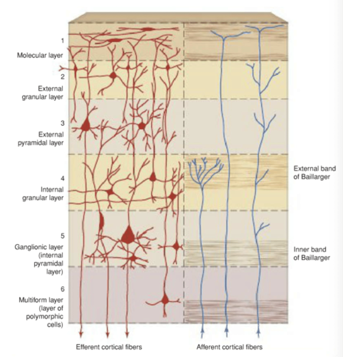
which is the most superficial layer?
molecular layer (plexiform layer)
which fibers are derived from the apical dendrites of the pyramidal cells and fusiform cells, the axons of the stellate cells, and the cells of Martinotti? in which layer can this be found?
tangentially oriented nerve fibers
molecular layer
Afferent fibers originating in the thalamus and in association with commissural fibers also are present in which layer?
molecular layer
true or false
This most superficial layer ( the external granular layer) of the cortex clearly is where large numbers of synapses between different neurons occur
false
- molecular layer
true or false
External granular layer. This layer contains large numbers of small pyramidal cells and stellate cells.
true
In the external granular layer:
The dendrites of these cells terminate in the ____ layer, and the axons enter deeper layers, where they terminate or pass on to enter the ___ matter of the cerebral hemisphere.
molecular
white
External pyramidal layer.
This layer is composed of what kind of cell?
. The apical dendrites pass into the ______ layer, and the axons enter the white matter as projection, association, or commissural fibers.
pyramidal cells,
molecular
which layer does this describe
This layer is composed of closely packed stellate cells with a high concentra- tion of horizontally arranged fibers known collec- tively as the external band of Baillarger.
a. external pyramidal layer
b. internal granular layer
c. molecular layer
d. ganglionic layer
B
the ganglionic layer is also known as the
internal pyramidal layer
in the ganglionic layer
Scattered among the pyramidal cells are ____ cells and cells of ____
`stellate
martinotti
in which layer is the inner band of the baillarger formed
in the ganglionic layer
Ganglionic Layer -
In the ___ cortex of the ____ gyrus, the pyramidal cells of this layer are very large and are known as ___ cells.
These cells account for about 3% of the projection fibers of the corticospinal or pyramidal tract.
motor
precentral
Betz
Multiform layer (layer of polymorphic cells). Although the majority of the cells are ____, many of the cells are modified pyramidal cells, whose cell bodies are triangular or ovoid. The cells of ____ also are con- spicuous in this layer. Many nerve fibers are present that are entering or are leaving the underlying white matter.
fusiform
Martinotti
Those areas of the cortex in which the basic six layers cannot be recognized are referred to as ____,
as opposed to the majority which possess 6 layers are refered to as?
heterotypical
homotypical
Two heterotypical areas are described as :
the granular and the agranular type.
Characteristics of the Granular Type
(which layers are well developed and poorly developed)
- well developed layers
- densely packed stellate cells
- layers 2 and 4 are well developed
- layers 3 and 5 poorly developed
- layers 2 through 5 merge into a single layer of predominantly granular cells. - These cells receive thalamocortical fibers.
- The granular type of cortex is found in the postcentral gyrus, in the superior temporal gyrus, and in parts of the hippocampal gyrus.
Characteristics of the Agranular Type
the granular layers are poorly developed, so layers 2 and 4 are practically absent. The pyramidal cells in layers 3 and 5 are densely packed and are very large
where is the agranular type of cortex found
in the precentral gyrus and other areas in the frontal lobe
true or false
An afferent fiber may synapse directly with an efferent neuron or may involve vertical chains of internuncial neurons
true
The ______ area is situated in the precentral gyrus
precentral
The precentral area may be divided into
posterior and anterior regions
pre central area posterior region
The posterior region, which is referred to as the motor area, primary motor area, or Brodmann area 4, occupies the precentral gyrus extend- ing over the superior border into the paracentral lob- ule
pre central area anterior region
The anterior region is known as the premotor area, secondary motor area, or Brodmann area 6 and parts of areas 8, 44, and 45. It occupies the anterior part of the precentral gyrus and the posterior parts of the superior, middle, and inferior frontal gyri.
Somatosensory (most to contralateral side of body; oral to same side; pharynx, larynx, and perineum bilateral)
origin
cortical area
destination
sensory or motor
Ventral posterior lateral
and ventral posterior medial nuclei of thalamus
Primary somesthetic area (B3, 1, and 2), posterior central gyrus
Secondary somesthetic area; primary motor area
sensory
vision
origin
cortical area
destination
sensory or motor
- lateral geniculate body
- Primary visual area (B17)
- Secondary visual area (B18 and 19)
sensory
auditory
origin
cortical area
destination
sensory or motor
- medial geniculate body
- Primary auditory area (B41 and 42)
- Secondary auditory area (B22)
- sensory
taste
sensory or motor
origin
cortical area
destination
- nucleus soliatrius
- posterior central gyrus (b43)
- no destination
- sensory
smell
sensory or motor
origin
cortical area
destination
- olfactory bulb
- Primary olfactory area; periamygdaloid and prepiriform areas
- Secondary olfactory area (B28)
- sensory
Fine movements (most to contralateral side of body; extraocular muscles, upper face, tongue, mandible, larynx, bilateral)
sensory or motor
origin
cortical area
destination
motor
Thalamus from cerebellum, basal ganglia; somatosensory area; premotor area
Primary motor area (B4)
Motor nuclei of brainstem and anterior horn cells of spinal cord; corpus striatum
which area is able to carry out the individual movements of different parts of the body.
the function of the primary motor area
The _______ motor area is situated in the medial frontal gyrus on the medial surface of the hemisphere and anterior to the paracentral lobule. Stimulation of this area results in movements of the contralateral limbs, but a stronger stimulus is necessary than when the primary motor area is stimulated. Removal of the supplementary motor area produces no permanent loss of movement.
supplementary
The ___ ____ field extends forward from the facial area of the precentral gyrus into the middle frontal gyrus (parts of Brodmann areas 6, 8, and 9).
frontal eye
true or field
Electrical stimulation of the frontal eye field causes conjugate movements of the eyes, especially toward the opposite side.
true
The motor speech area of Broca is located in the
inferior frontal gyrus
true or false
In those individuals in whom the right hemisphere is dominant, the area on the left side is of importance.
false
right side is more important
The ____ ____ ____ brings about the formation of words by its connections with the adjacent primary motor areas; the muscles of the larynx, mouth, tongue, soft palate, and the respiratory muscles are appropriately stimulated.
Broca speech area
The _____ cortex is an extensive area that lies anterior to the precentral area. It includes the greater parts of the superior, middle, and inferior frontal gyri; the orbital gyri; most of the medial frontal gyrus; and the anterior half of the cingulate gyrus
prefrontal
what connects the prefrontal area with other areas of the cerebral cortex, the thalamus, the hypothalamus, and the corpus striatum
Large numbers of afferent and efferent pathways
The prefrontal area is concerned with the makeup of
an individuals personality
The primary somesthetic area
occupies the postcentral gyrus on the lateral surface of the hemisphere and the posterior part of the paracentral lobule on the medial surface (Brodmann areas 3, 1, and 2).
The primary somesthetic areas of the cerebral cortex receive projection fibers from
the ventral posterior lateral and ventral posterior medial nuclei of the thalamus
what is represented in the most inferior part of the postcentral gyrus
The pharyngeal region, tongue, and jaws followed by the face, fingers, hand, arm, trunk, and thigh
The leg and the foot areas are found on the _____ surface of the hemisphere in the posterior part of the paracentral lobule.
medial
The ____ part of the postcentral gyrus situated in the central sulcus receives a large number of ____ fibers from muscle spindles, tendon organs, and joint receptors.
anterior
afferent
The ____ _____ area (secondary somatic sensory cortex 52) is in the superior lip of the posterior limb of the lateral fissure. The secondary sensory area is much smaller and less important than the primary sen- sory area.
secondary somesthetic
The ____ _____ area occupies the superior parietal lobule extending onto the medial surface of the hemisphere (Brodmann areas 5 and 7).
This area has many connections with other sensory areas of the cortex, and its main function is probably to receive and integrate different sensory modalities.
example of this area functioning
somesthetic association
In other words, it not only receives information concerning the size and shape of an object but also relates this to past sensory experiences; thus, the information may be interpreted, and recognition may occur. A quarter placed in the hand can be distinguished from a dime or a nickel by the size, shape, and feel of the coin without having to use one's eyes.
The ___ ____ area (Brodmann area 17) is situated in the walls of the posterior part of the _____ sulcus and occasionally extends around the occipital pole onto the lateral surface of the hemisphere
primary visual area
calcarine
The visual cortex receives afferent fibers from the
lateral geniculate body.
The fibers first pass forward in the _____ matter of the temporal lobe and then turn back to the primary visual cortex in the ______ lobe.
white
occipital
The visual cortex receives fibers from the temporal half of the _____ retina and the nasal half of the ____ retina.
ipsilateral
contralateral
The right half of the field of vision, therefore, is represented in the visual cortex of the ____ cerebral hemisphere and vice versa. Note that the superior retinal quadrants (inferior field of vision) pass to the superior wall of the ____ sulcus, while the inferior retinal quadrants (superior field of vision) pass to the inferior wall of the calcarine sulcus.
left
calcarine
The ____ ____, which is the central area of the retina and the area for most perfect vision, is repre- sented on the cortex in the posterior part of area 17 and accounts for one-third of the visual cortex. The visual impulses from the peripheral parts of the retina terminate in concentric circles anterior to the occipital pole in the anterior part of area 17.
macula lutea
The _____ _____ area (Brodmann areas 18 and 19) surrounds the primary visual area on the medial and lateral surfaces of the hemisphere. This area receives afferent fibers from area 17 and other cortical areas as well as from the thalamus. The function of the secondary visual area is to
secondary visual
relate the visual information received by the primary visual area to past visual experiences, thus enabling the individual to recognize and appreciate what he or she is seeing.
The occipital eye field is thought to exist in which area
secondary visual area
Stimulation of the occipital eye filed produces conjugate ____ of the eyes, especially to the oppo site side. The function of this eye field is believed to be reflex and associated with .......
The occipital eye fields of both hemispheres are connected by nervous pathways and also are thought to be connected to the superior colliculus. By contrast, the frontal eye field controls .___ ____ ____ of the eye and is independent of visual stimuli.
deviation
movements of the eye when it is following an object.
voluntary scanning movements
The primary auditory area includes the
gyrus of Heschl
Projection fibers to the auditory area arise principally in the medial geniculate body and form ....
the auditory radiation of the internal capsule.
medial geniculate body receives fibers mainly from the organ of ___ of the opposite side as well as some fibers from the same side.
Corti
secondary auditory area
important for interpretation of sounds
The sensory speech area of Wernicke is localized where
left hemisphere (anterior temporal gyrus)
Wernicke area is connected to the _____ area by a bundle of nerve fibers called the ____ fasciculus. lt receives fibers from the visual cortex in the occipital lobe and the ____ cortex in the superior temporal gyrus.
Broca
arcuate
auditory
The _____ area permits understanding of written and spoken language and enables a person to read a sentence, understand it, and say it out loud
Wernicke
where is the taste area situated
lower end of post central gyrus
superior wall of lateral sulcus
in the insula
vestibular area is found near what
maintains what
part of the postcentral gyrus concerned with sensations of the face. Its location lies opposite the auditory area in the superior temporal gyrus. The vestibular area and the vestibular part of the inner ear are concerned with appreciation of the positions and movements of the head in space.
posture and balance
the insula is important for
planning or coordinating the articulatory movements necessary for speech
The primary _____ areas with their ____ cortex and the primary ____ areas with their _____ cortex form only a small part of the total cortical surface area.
sensory
granular
motor
agranular
Three main association areas are recognized there are...
prefrontal, anterior temporal, and posterior parietal.
The ____ ____ cortex is thought to play a role in the storage of previous sensory experiences. Stimulation may cause the individual to recall objects seen or music heard in the past.
anterior temporal
In the _____ ____ cortex, visual information from the posterior occipital cortex and the sensory input of touch and pressure and ____- from the anterior parietal cortex is integrated into concepts of size, form, and texture. This ability is known as ____. A con- scious appreciation of the body image is also assembled in the posterior parietal cortex. The brain knows at all times where each part of the body is located in relation to its environment. This information is so important when performing _____ ______
posterior parietal
proprioception
stereognosis
body movements
true or false
The right side of the body is represented in the left hemisphere, and the left side of the body is represented in the right hemisphere.
true
which parts of the cerebral commissures provide a pathway for information that is received in one hemisphere to be transferred to the other
the cerebral commissures, especially the corpus callosum and the anterior commissure, provide a pathway for information that is received in one hemisphere to be transferred to the other.
are Handedness, perception of language, and speech are functional areas of behavior controlled by the dominant hemisphere or non dominant hemispheres in most individuals?
dominant
are spatial perception, recognition of faces, and music interpreted by the nondominant hemisphere or the dominant hemisphere?
non dominant