orbit and eyes
1/72
There's no tags or description
Looks like no tags are added yet.
Name | Mastery | Learn | Test | Matching | Spaced | Call with Kai | Chat |
|---|
No analytics yet
Send a link to your students to track their progress
73 Terms
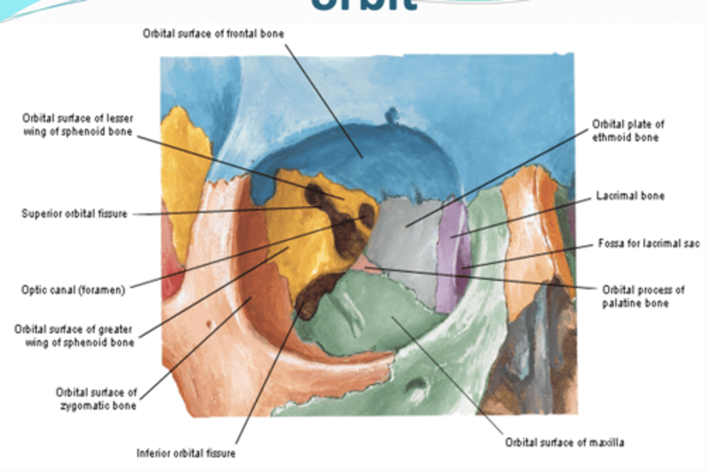
What is the orbit?
quadrangular pyramid, with base anteriorly, apex posteriorly, and 4 walls
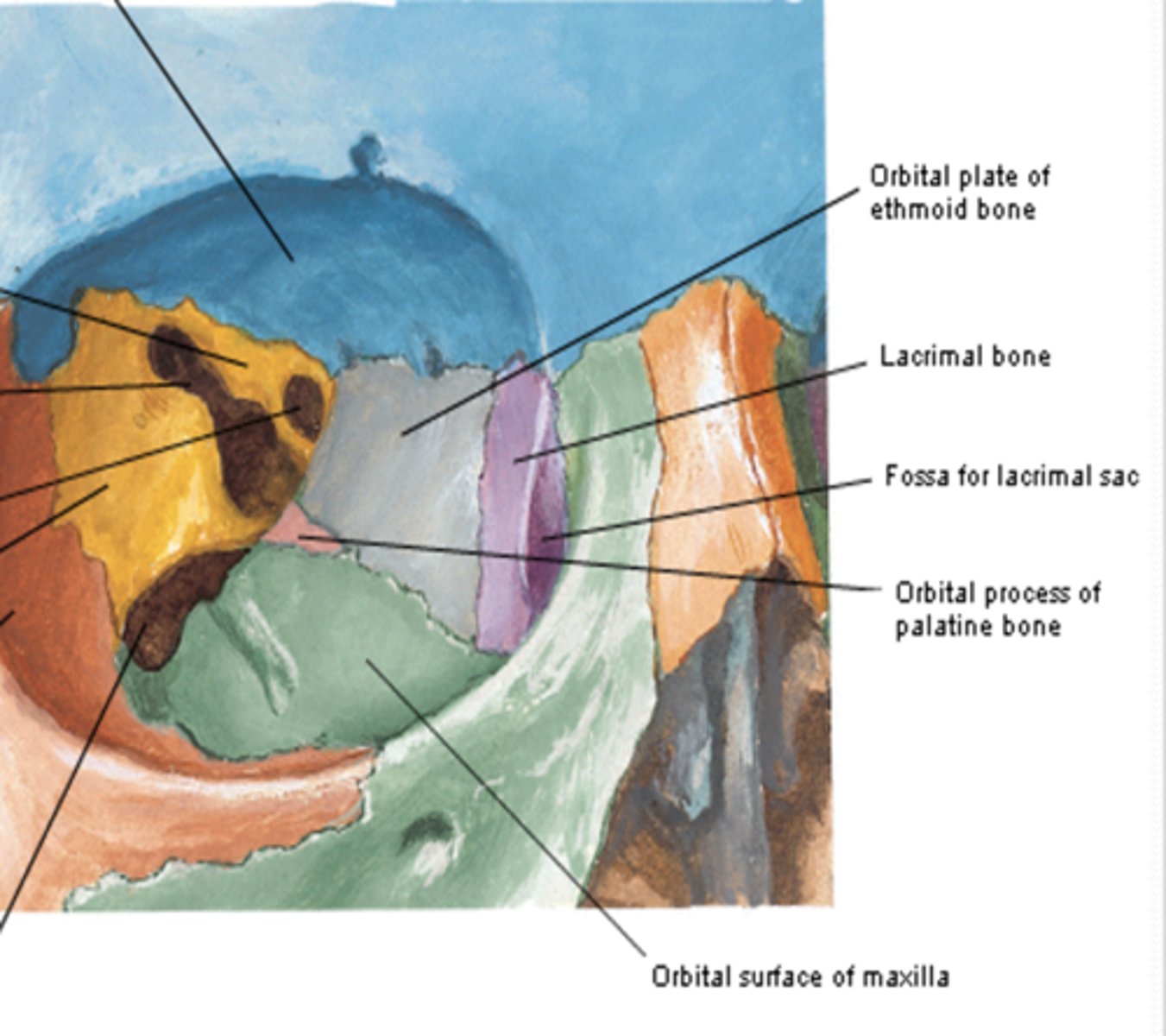
What contributes to the medial wall of the orbit? what fossa does it contain?
1. Lacrimal bone contains fossa for lacrimal sac
2. Orbital plate of ethmoid bone (lamina papyracea): posterior to the lacrimal bone
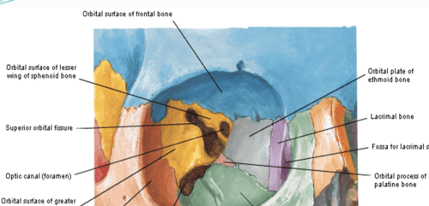
What contributes to the superior wall of the orbit?
1. Frontal Bone
2. Lesser wing of sphenoid bone (near the apex)
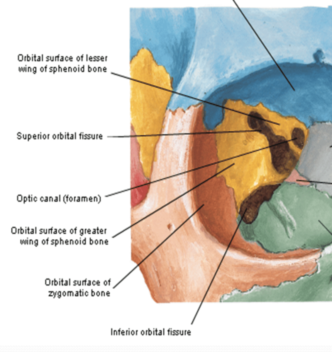
What contributes to the lateral wall of the orbit?
1. Greater wing of the sphenoid bone
2. Zygomatic bone (SIGNIFICANTLY)
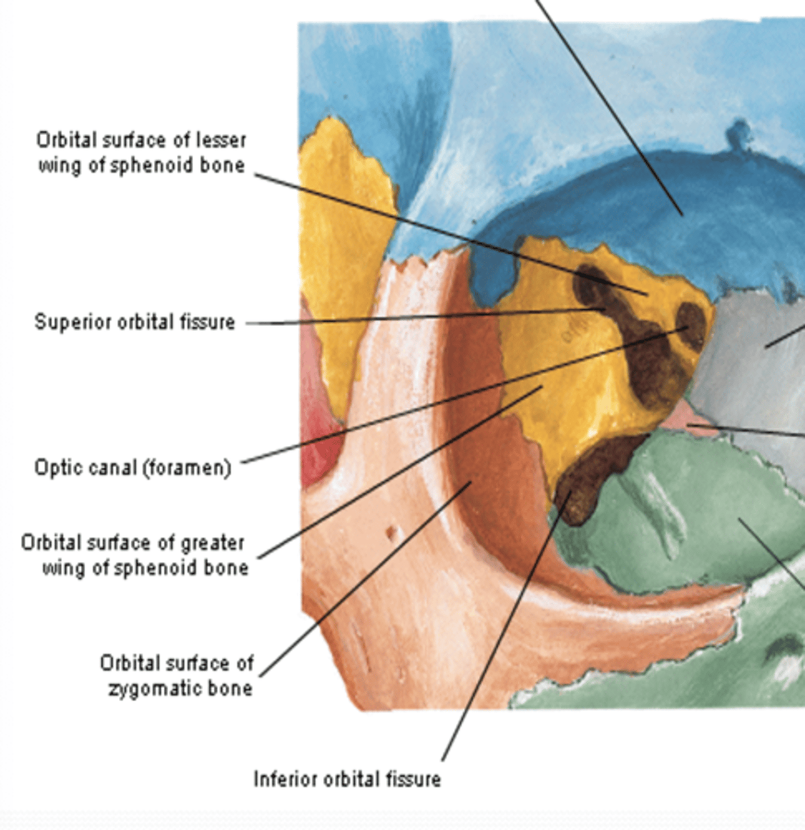
What does the optic canal in the orbit pass though?
passes though the lesser wing of sphenoid bone
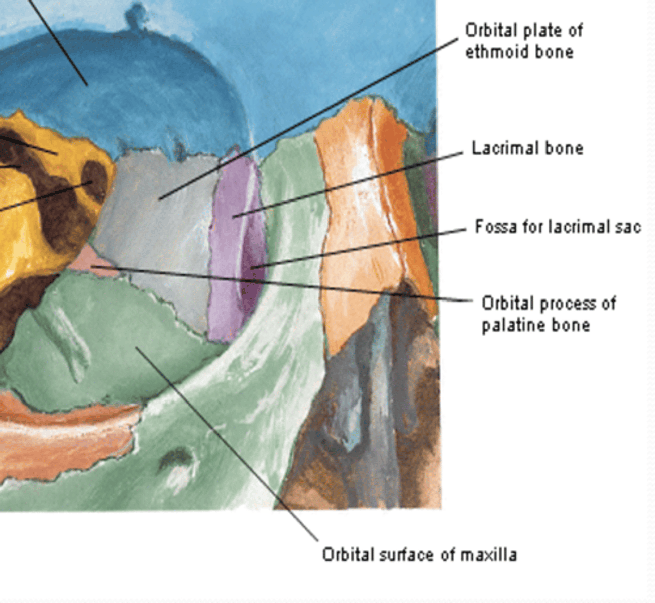
What bone contributes slightly to the apex of the orbit?
Orbital process of palatine bone
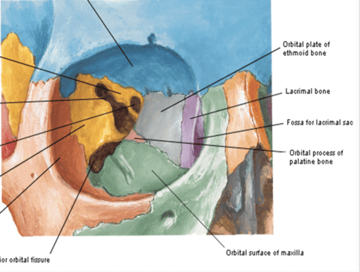
What contributes to the inferior wall of the orbit?
Maxilla
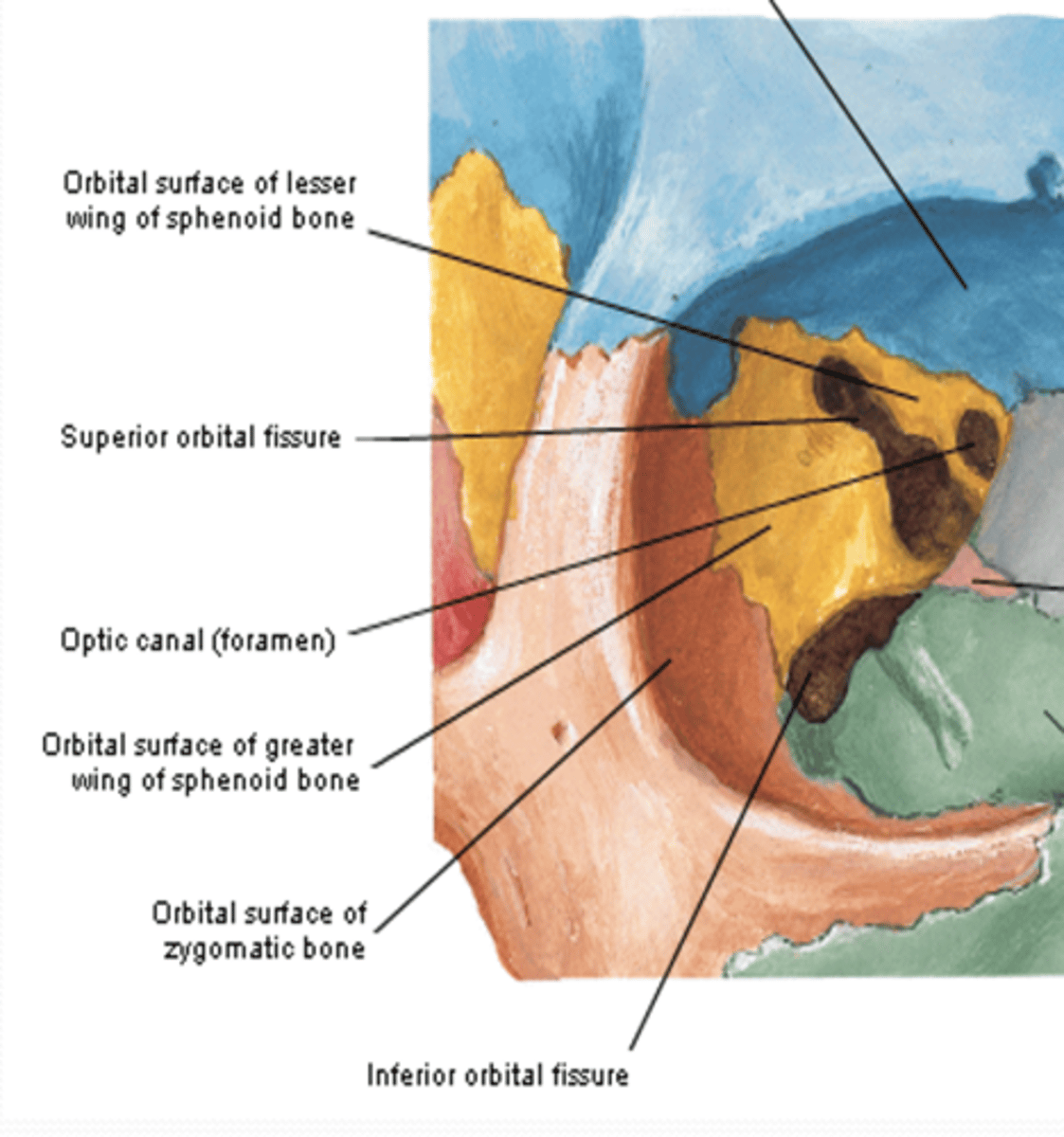
The superior orbital fissure of the orbit is between what bones?
located between the greater and lesser wings of the sphenoid
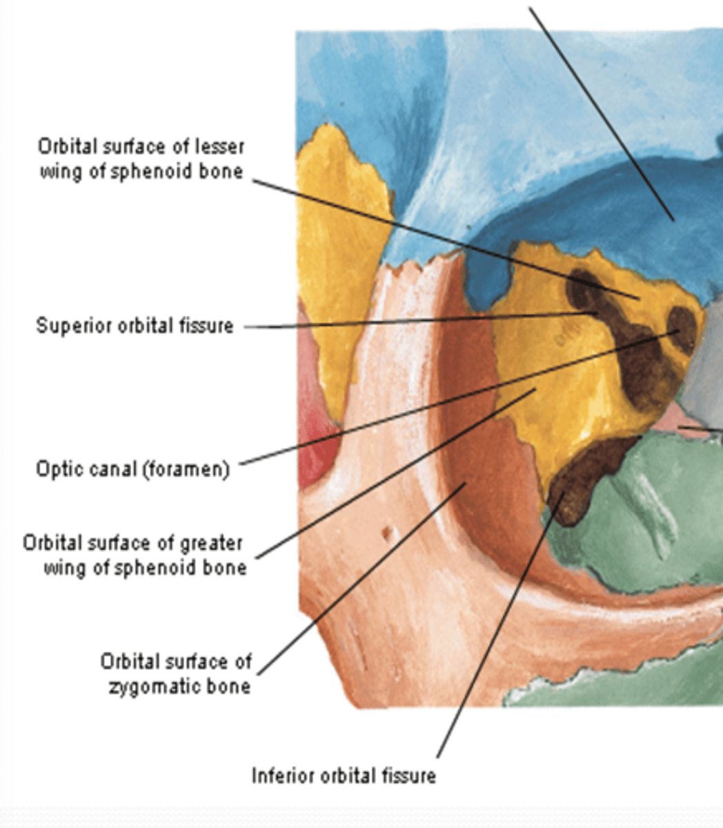
The inferior orbital fissure of the orbit is between what bones?
between maxilla, zygomatic, and greater wing of the sphenoid
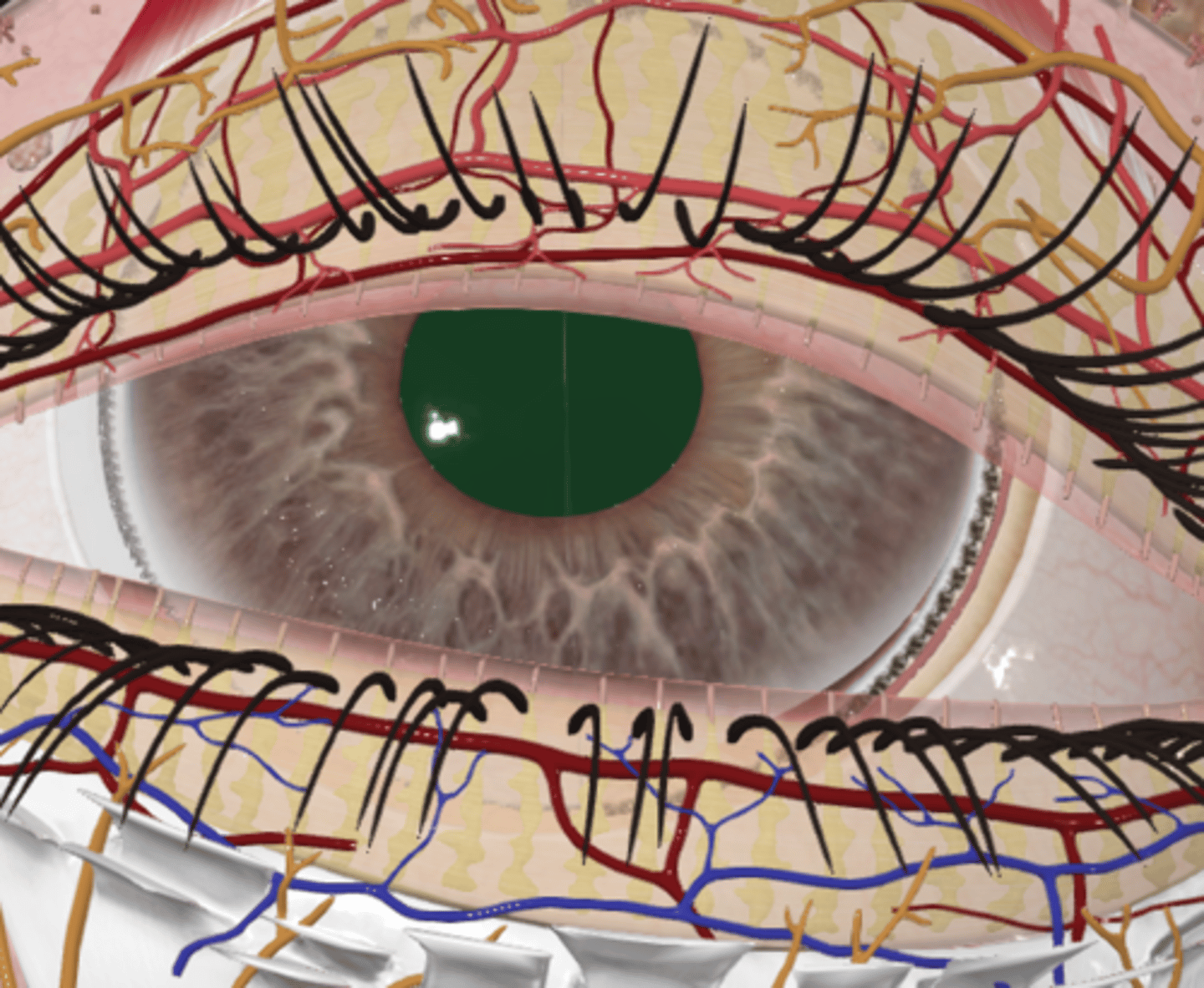
What structure bounds the orbit anteriorly and limits the exposure of the anterior eyeball?
eyelids
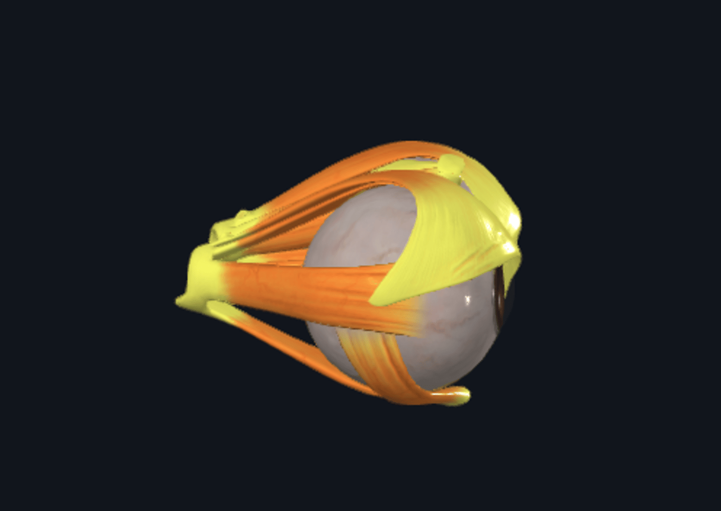
What is the function of the extra-ocular muscles of the eyes?
They position the eyeball and raise the superior eyelid
What surrounds the eyeball and muscles within the orbit?
Orbital fascia
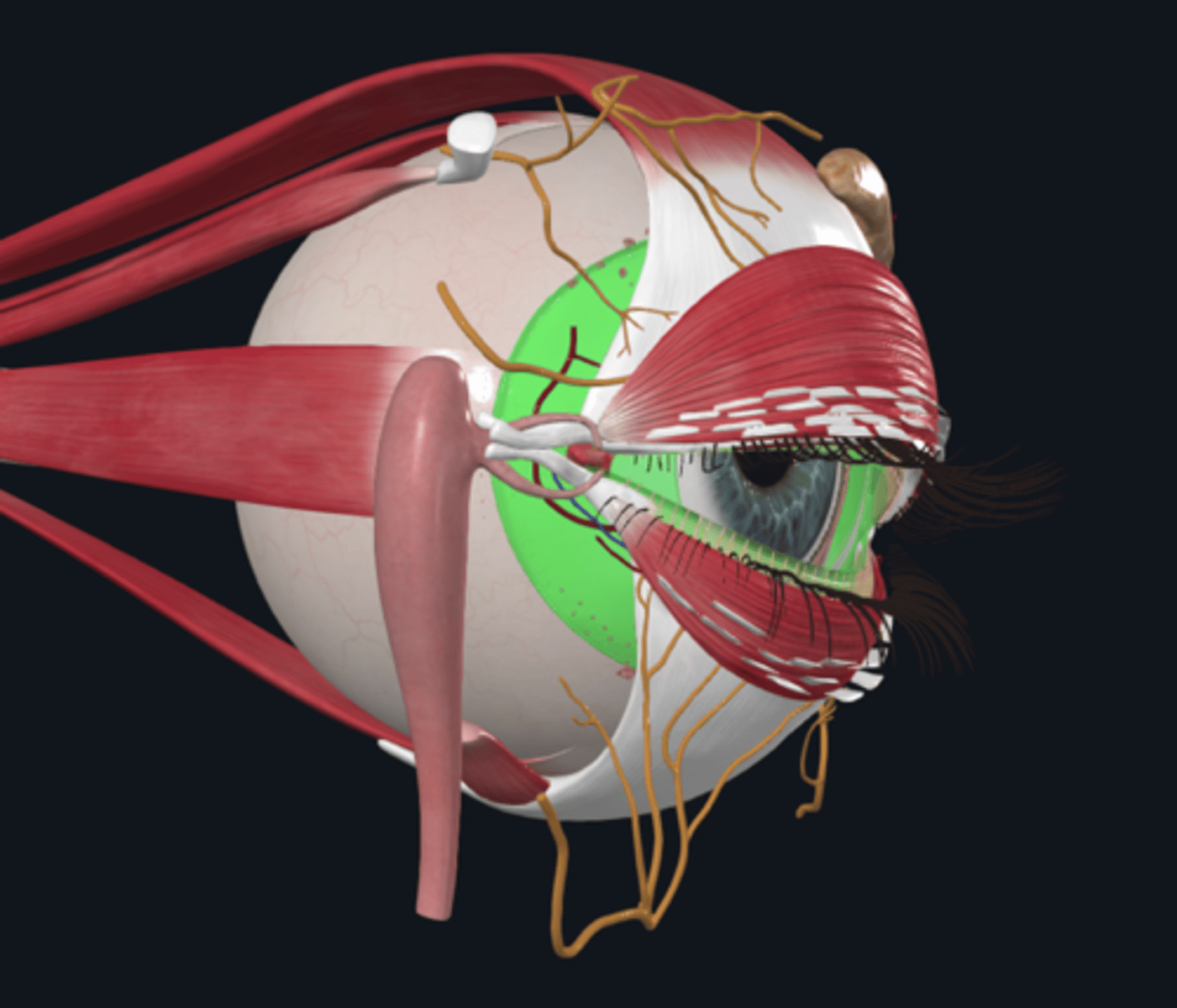
What is the role of the mucous membrane (conjunctiva) in the eye?
It lines the anterior aspect of the eyeballs, the interior aspect of the eyelids, and most of the lacrimal apparatus.
What occupies the rest of the orbital space, embedding various structures?
Orbital fat
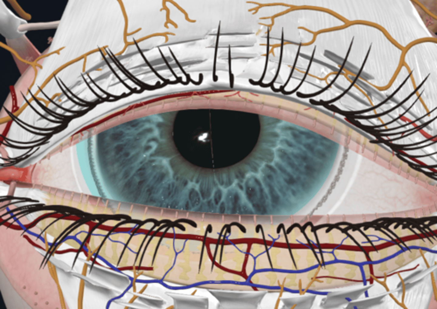
What is the primary function of the eyelids?
The eyelids are movable folds that cover and protect the eyeball anteriorly and keep the cornea moist by spreading lacrimal fluid
What are the components of the eyelids?
1. Skin (external layer)
2. Connective tissue (contains eyelashes)
3. Skeletal muscle (orbicularis oculi muscle)
4. Tarsofascial layer (contains tarsi and orbital septum)
5. Conjunctiva (inner layer)
What is the function of the connective tissue in the eyelids?
It lies immediately deep to the skin and contains the eyelashes.
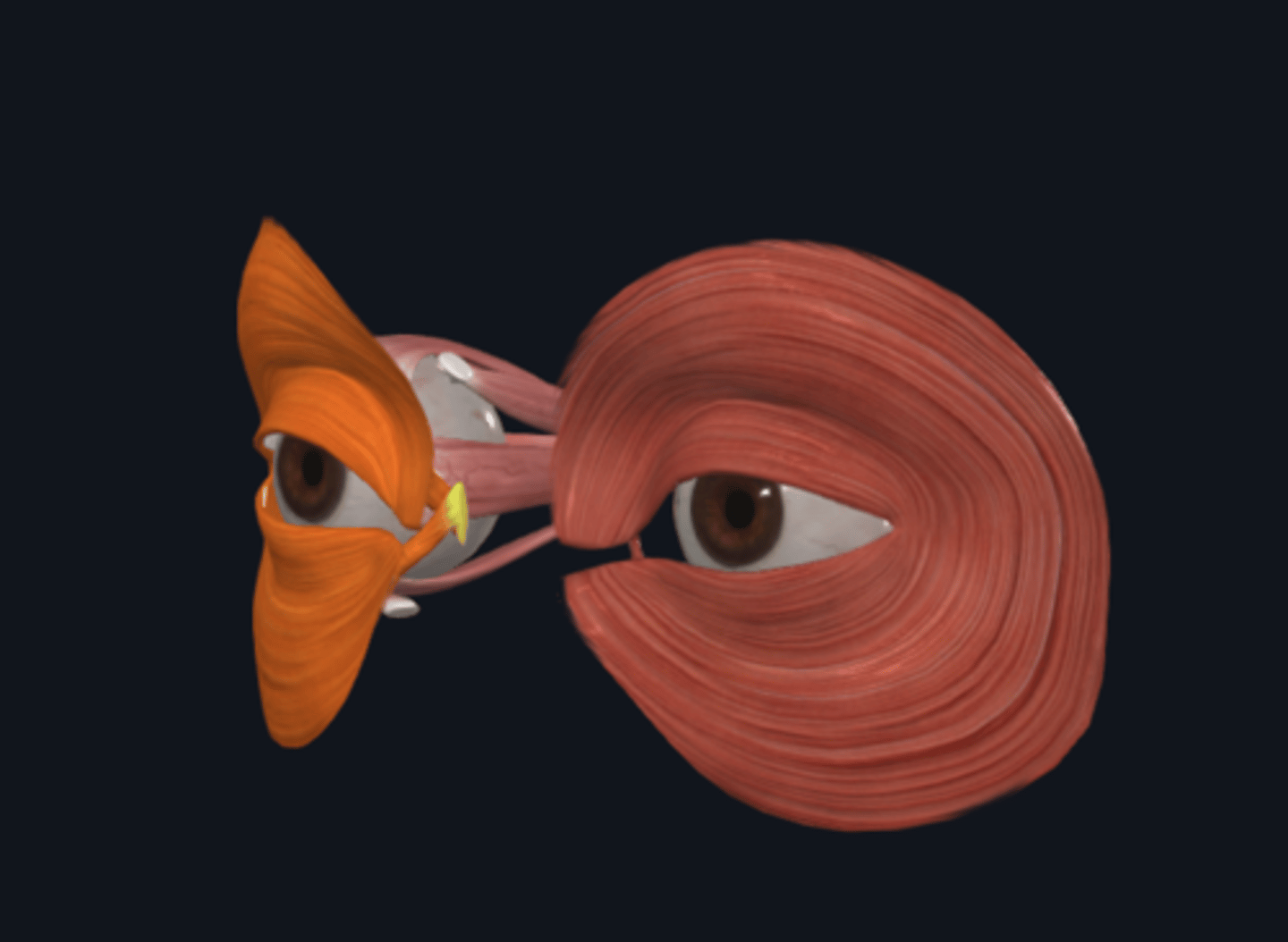
What muscle acts as the sphincter of the eye, located in the eyelid?
The orbicularis oculi muscle
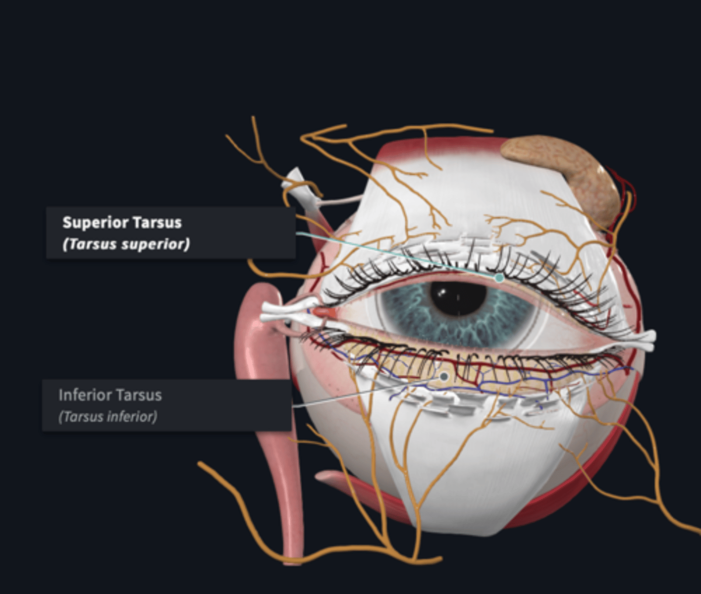
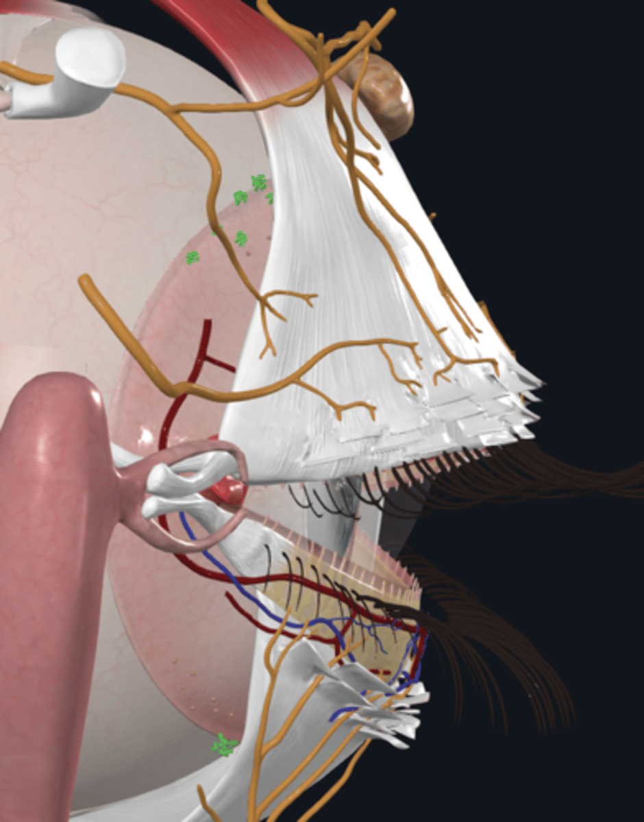
What is the tarsofascial layer and what are its components?
The tarsofascial layer consists of:
Superior and inferior tarsi: Dense bands of connective tissue that strengthen the eyelids
Orbital septum: A continuation of the periosteum of the surrounding orbital bones, attaching to the tarsi
What is the function of the tarsal glands (Tarsofascial layer) in the eyelids?
Tarsal glands are sebaceous glands that secrete lipids to resist tear overflow

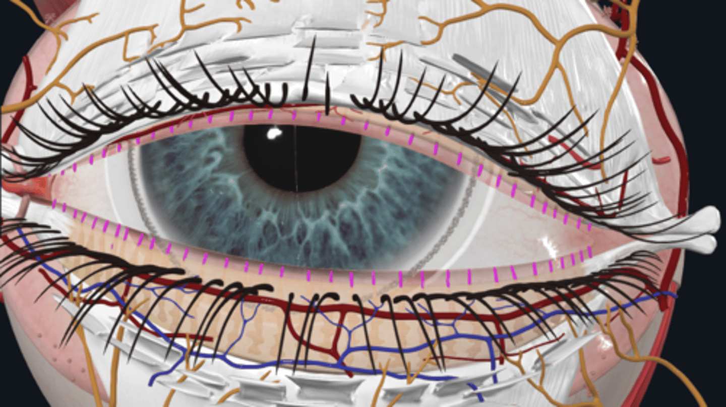
What are the two parts of the conjunctiva and what are their differences?
1. Palpebral conjunctiva: Closest to the eyelid, it is opaque
2. Bulbar conjunctiva: Overlies the cornea, it is transparent and receives sensory innervation from the long ciliary nerve
What is the conjunctival sac?
It is the space bound by the bulbar and palpebral conjunctiva.
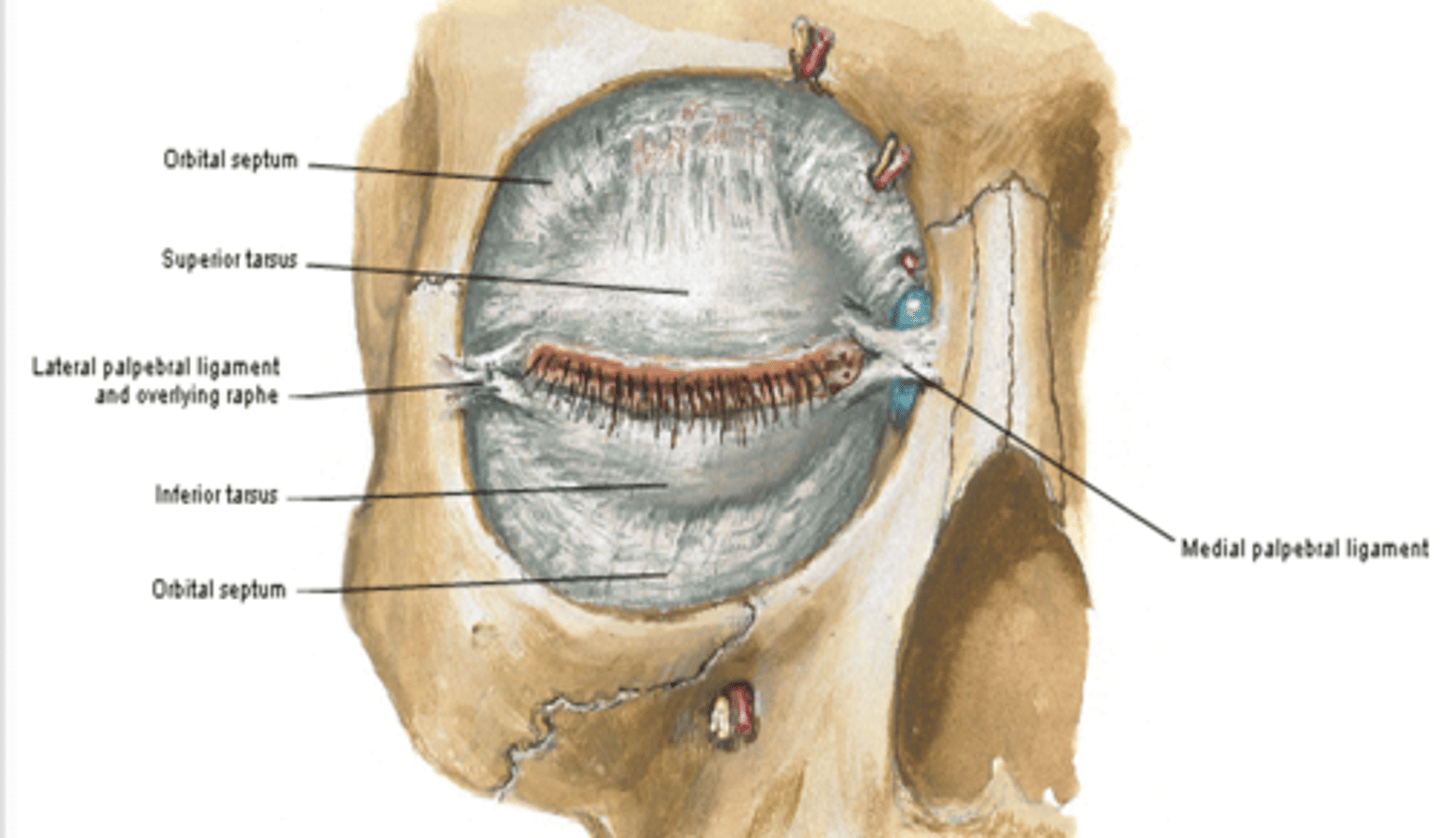
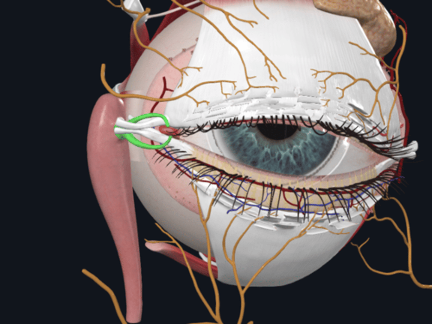
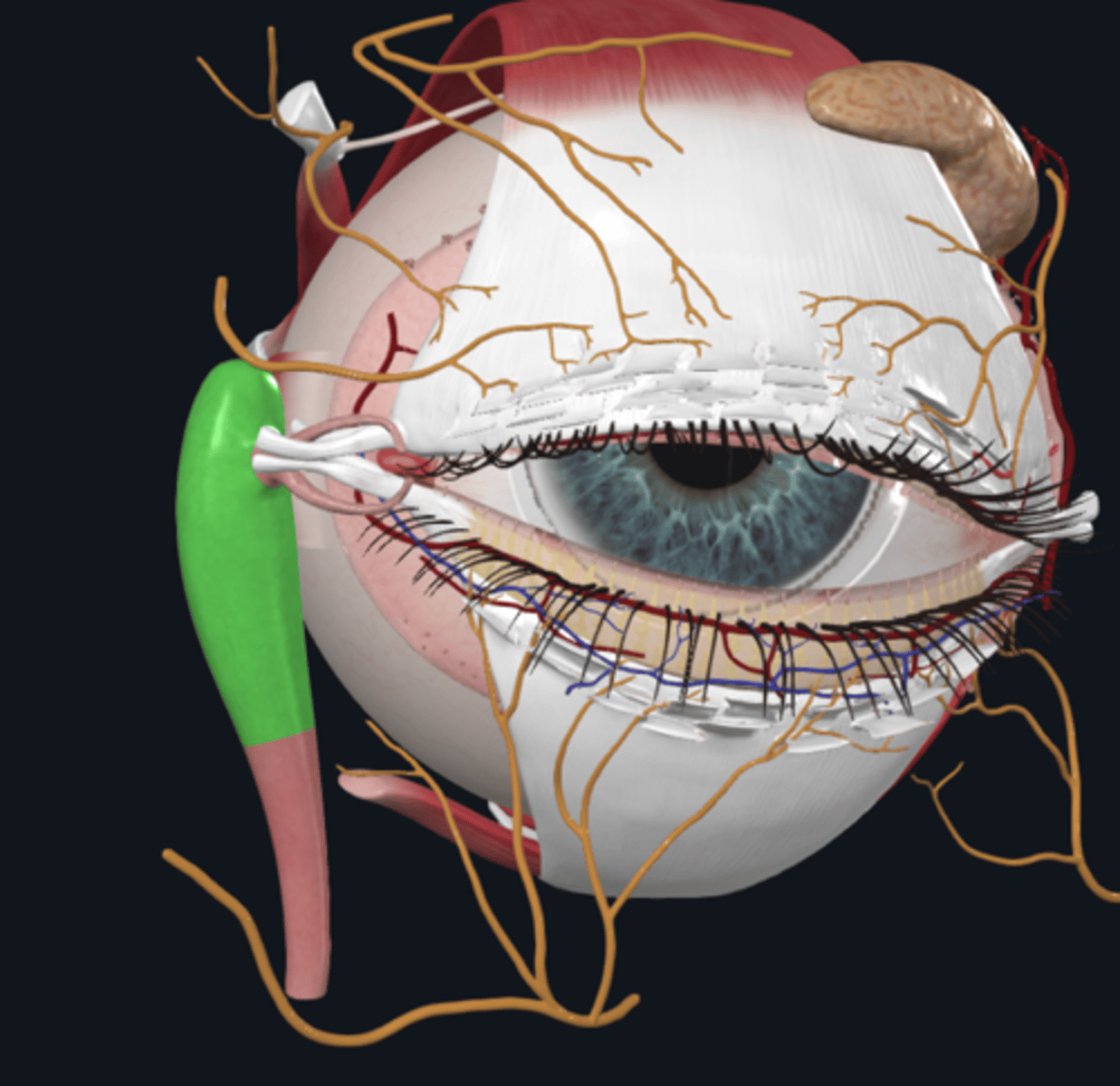
What are the medial and lateral palpebral commissures?
They are the junctions of the superior and inferior eyelids, forming the medial and lateral angles (canthi) of the eye.
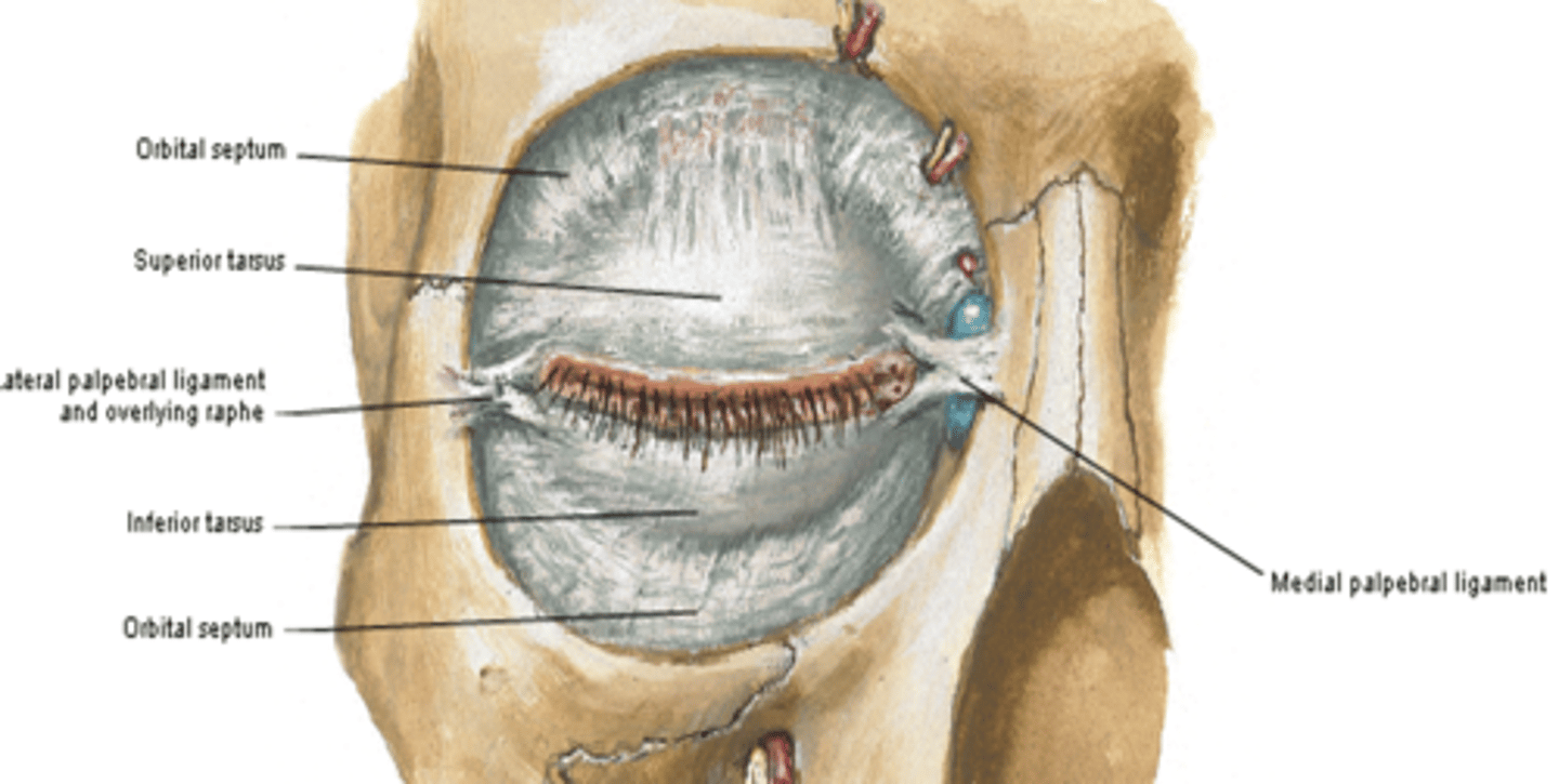
What is the function of the medial palpebral ligament?
It connects the tarsi to the medial margin of the orbit.
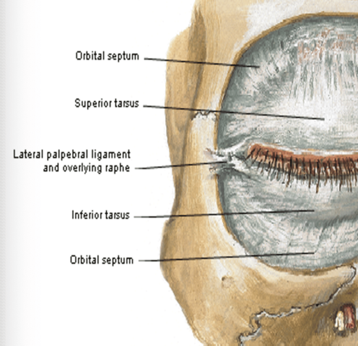
What is the function of the lateral palpebral ligament?
It connects the tarsi to the lateral margin of the orbit.
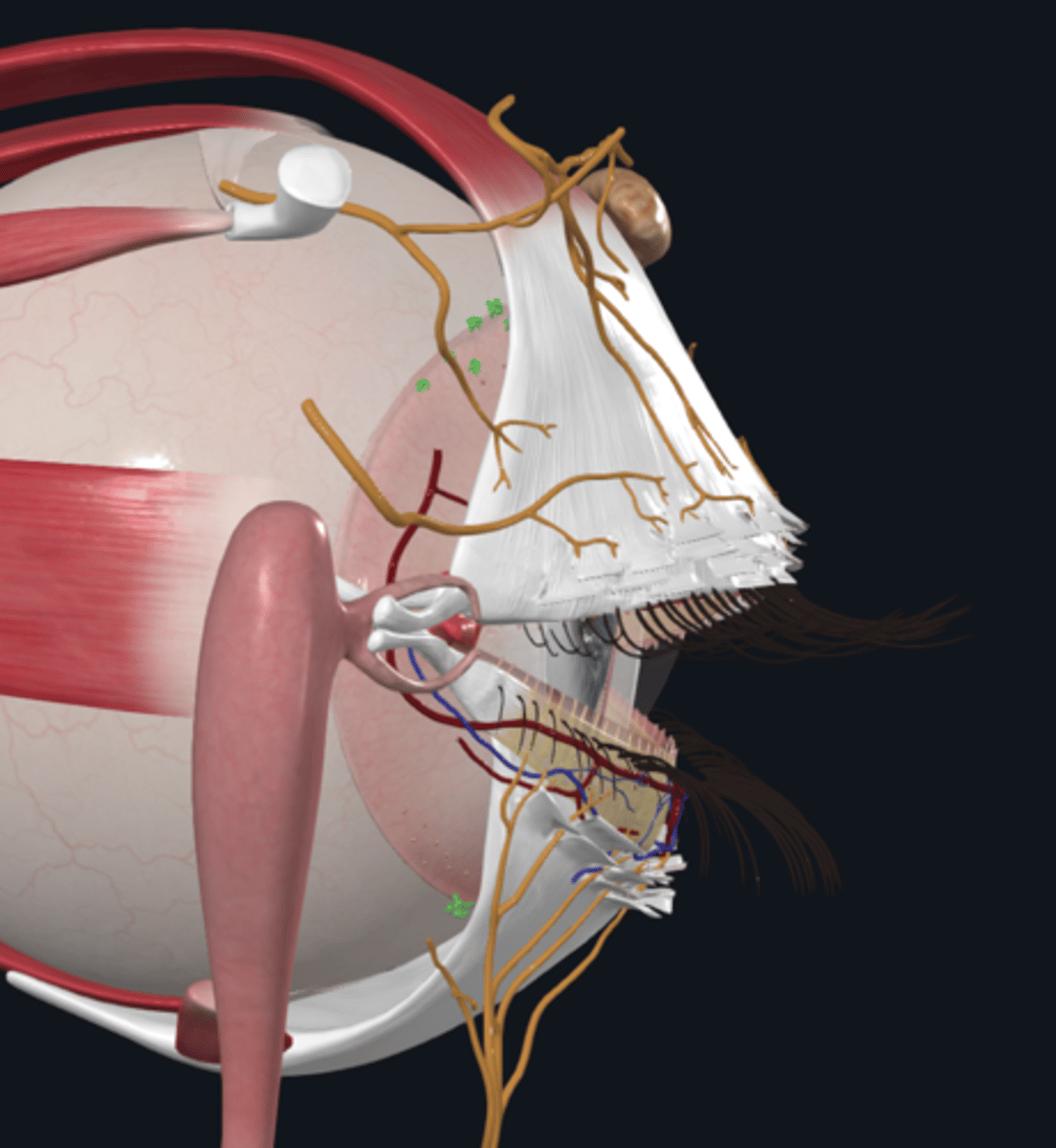
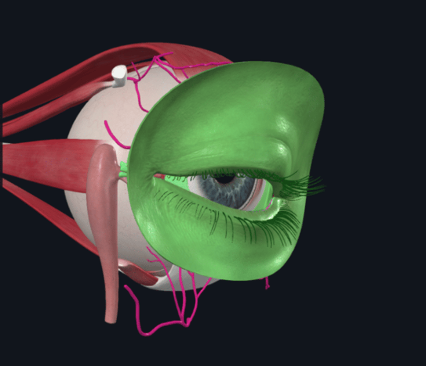
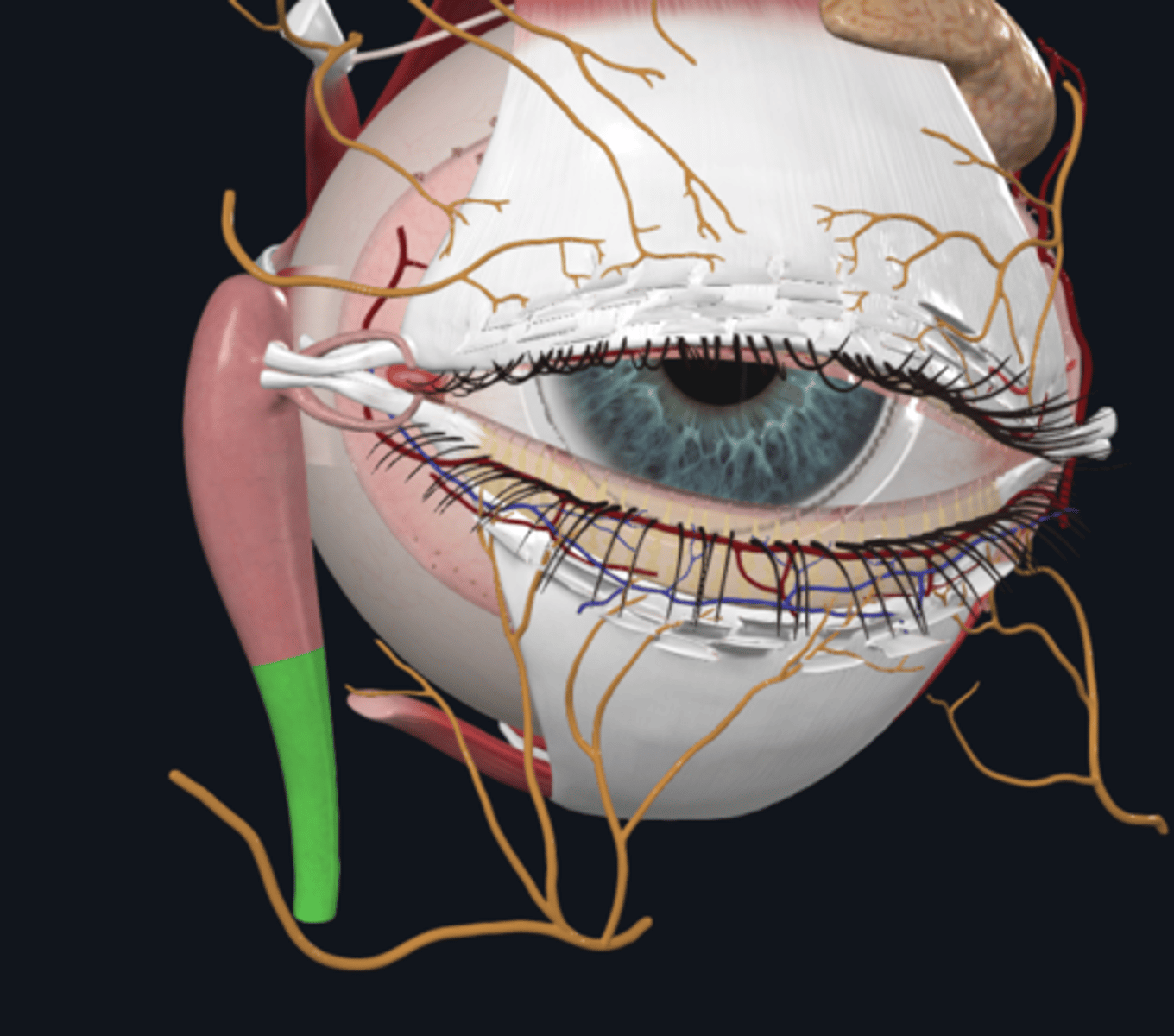
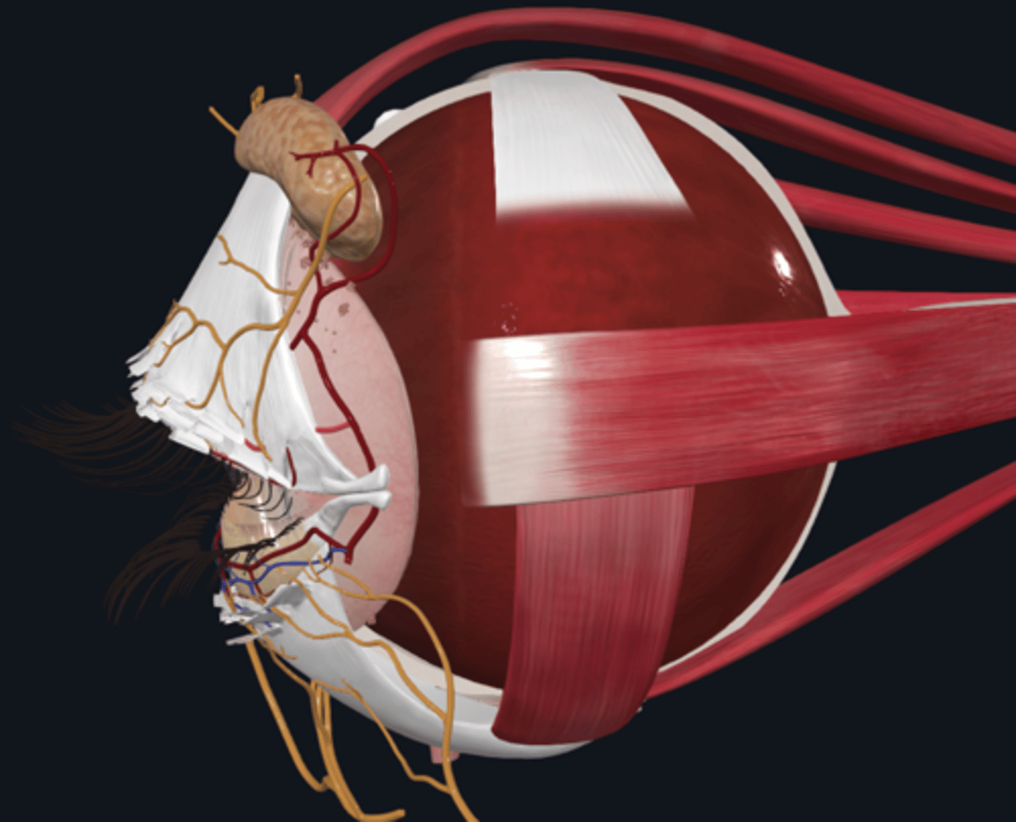
What is the function of the lacrimal glands?
The lacrimal glands secrete lacrimal fluid and are divided into superior (orbital) and inferior (palpebral) parts
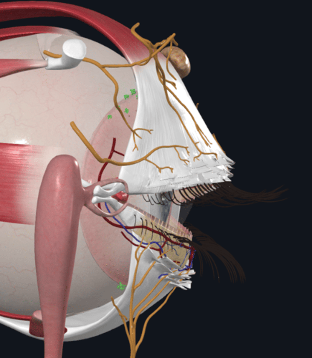
Where are the lacrimal glands located?
They are located in the superior lateral part of the orbit, in a small depression in the frontal bone called the lacrimal fossa (about 2 cm in diameter and oval-shaped).
What is lacrimal fluid and how is it secreted?
Lacrimal fluid is a watery physiological saline solution, secreted through 8-12 excretory ducts.
What is the function of the excretory ducts in the lacrimal apparatus?
The excretory ducts convey lacrimal fluid to the conjunctival sac
What are lacrimal canaliculi?
Lacrimal canaliculi are small canals that begin at the lacrimal punctum (opening) on the lacrimal papilla in the medial angle of the eye.
What is the lacrimal sac and where is it located?
The lacrimal sac is the dilated superior part of the nasolacrimal duct and lies in the medial wall of the orbit, in the depression of the lacrimal bone.
What is the function of the nasolacrimal duct?
The nasolacrimal duct conveys lacrimal fluid inferiorly to the inferior nasal meatus in the nasal cavity
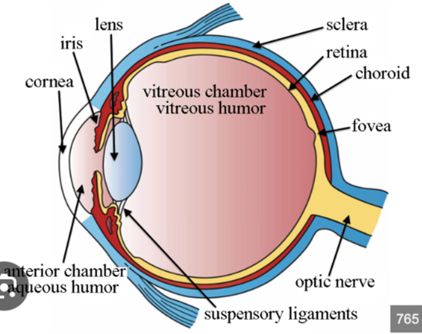
What does the eyeball contain and where is it located?
The eyeball contains the optic apparatus and occupies most of the anterior portion of the orbit.
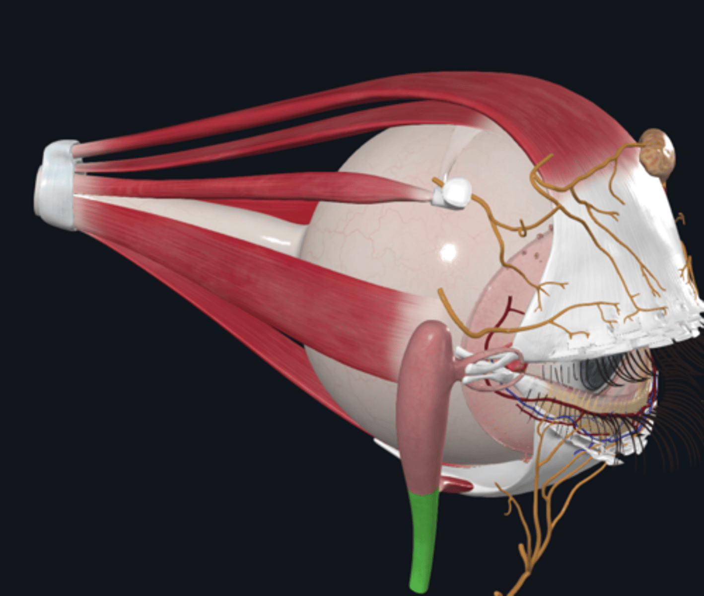
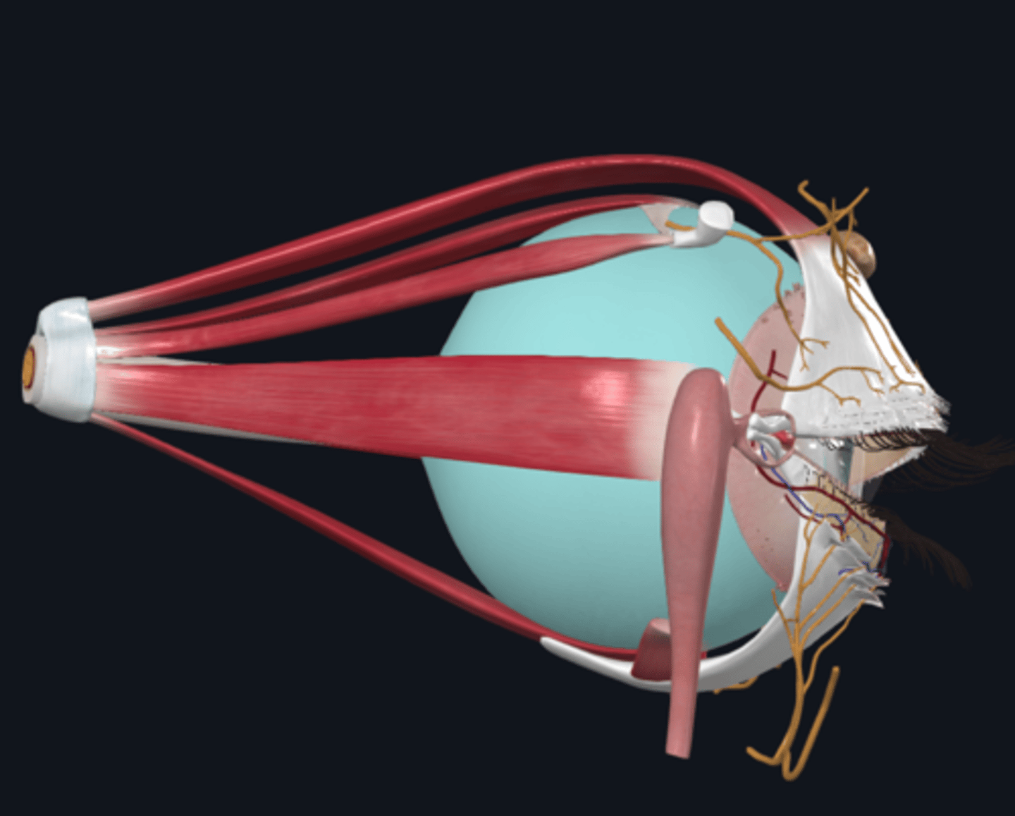
How is the eyeball suspended within the orbit?
It is suspended by 6 extrinsic muscles and a fascial suspensory apparatus.
What are the three layers of the eyeball?
1. Fibrous layer
2. Vascular layer
3. Neural layer
What is the fibrous layer of the eyeball and what is its function?
The fibrous layer is the external fibrous skeleton of the eyeball, providing shape and resistance.
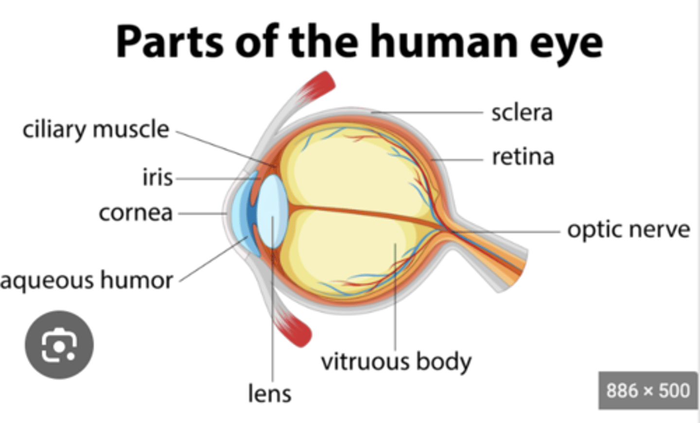
What is the sclera (fibrous layer) and where is it located?
The sclera is the tough, opaque part that covers the posterior 5/6th of the eyeball. It provides attachment for the eye muscles and is relatively avascular
What is the cornea (fibrous layer) and what are its characteristics
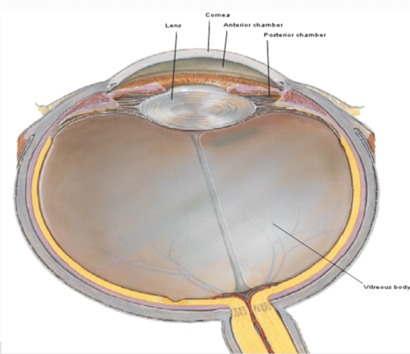
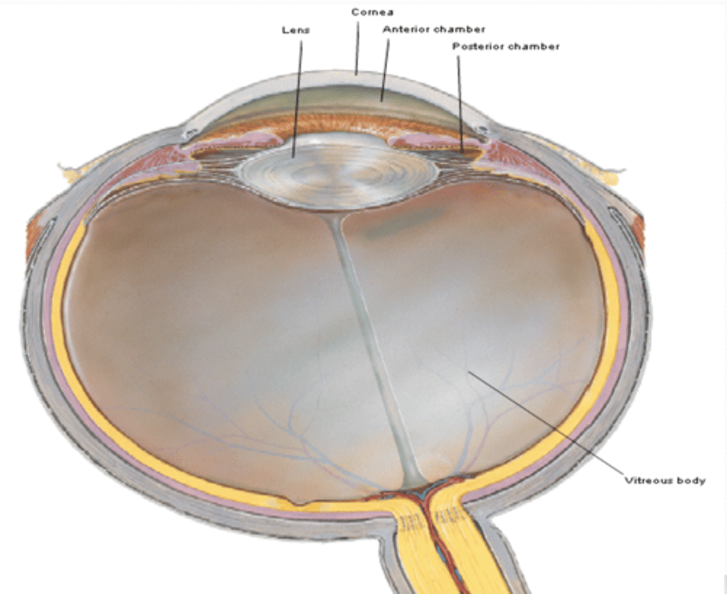
The cornea is the transparent part that covers the anterior 1/6th of the eyeball. It is completely avascular, receiving nourishment from lacrimal fluid and aqueous humor, and it is innervated by the ophthalmic nerve (CN V1).
What is the choroid (vascular layer) and what is its role in the eye?
The choroid is the dark reddish-brown, pigmented layer between the sclera and retina. It is the largest part of the vascular layer and is responsible for the “red-eye” reflection in flash photography.
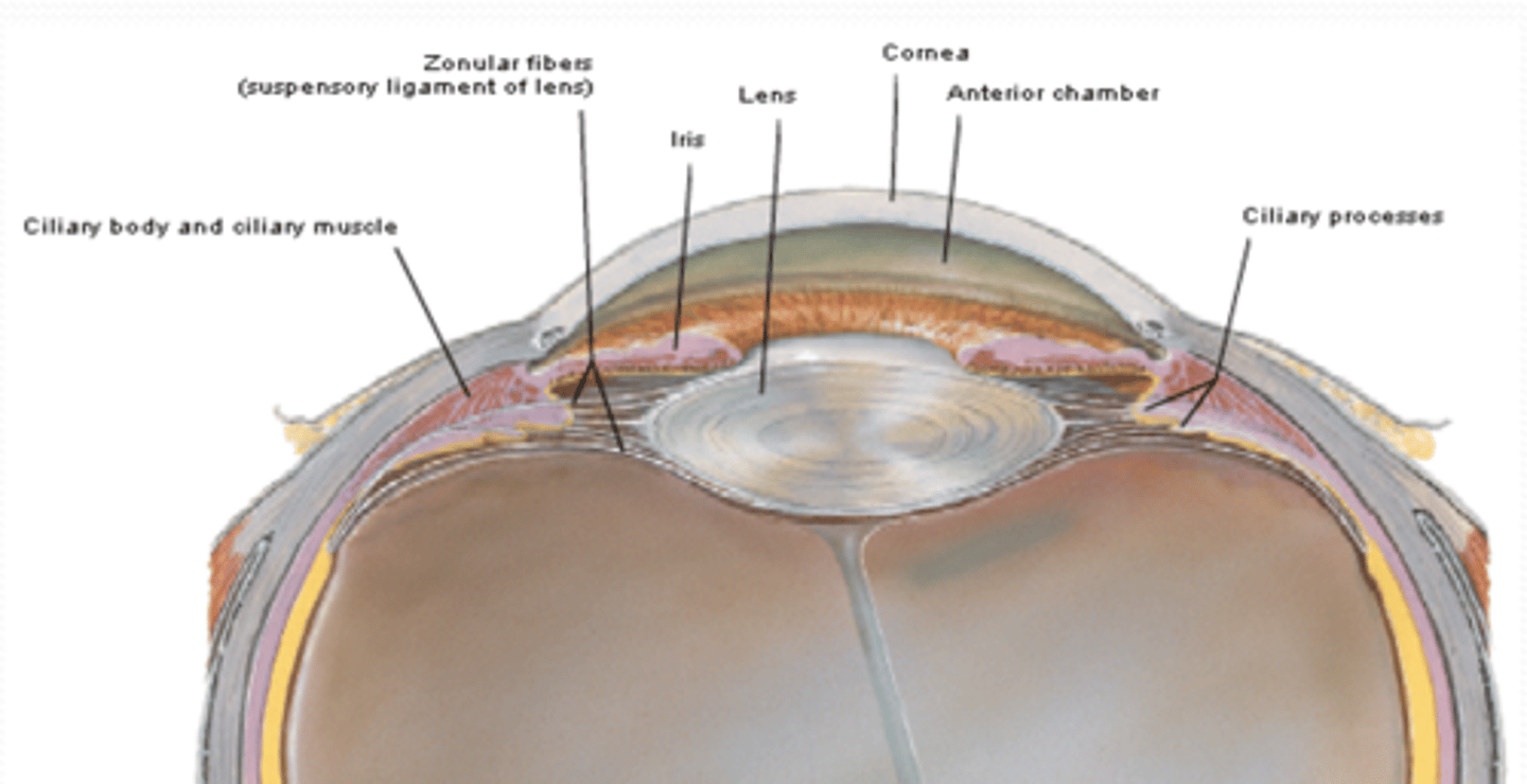
What is the function of the ciliary body (vascular layer) in the eye?
The ciliary body provides attachment for the lens and controls the accommodation of the lens by contracting its smooth muscles.
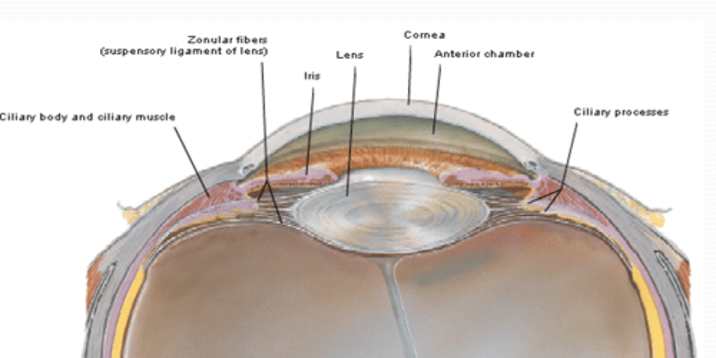
What are the ciliary processes (vascular layer) and what do they do?
The ciliary processes are folds on the internal surface of the ciliary body that secrete aqueous humor, which fills the anterior segment of the eyeball
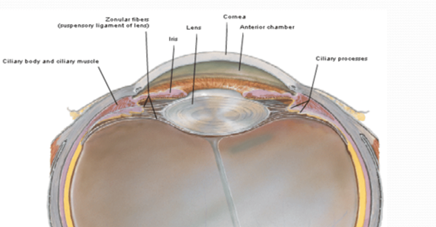
What are suspensory ligaments (vascular layer) and what is their function?
The suspensory ligaments connect the ciliary body to the lens, helping to control lens accommodation
What is the iris (vascular layer) and what is its function?
The iris is a thin contractile diaphragm with a central aperture (the pupil), which controls the amount of light that enters the eye.
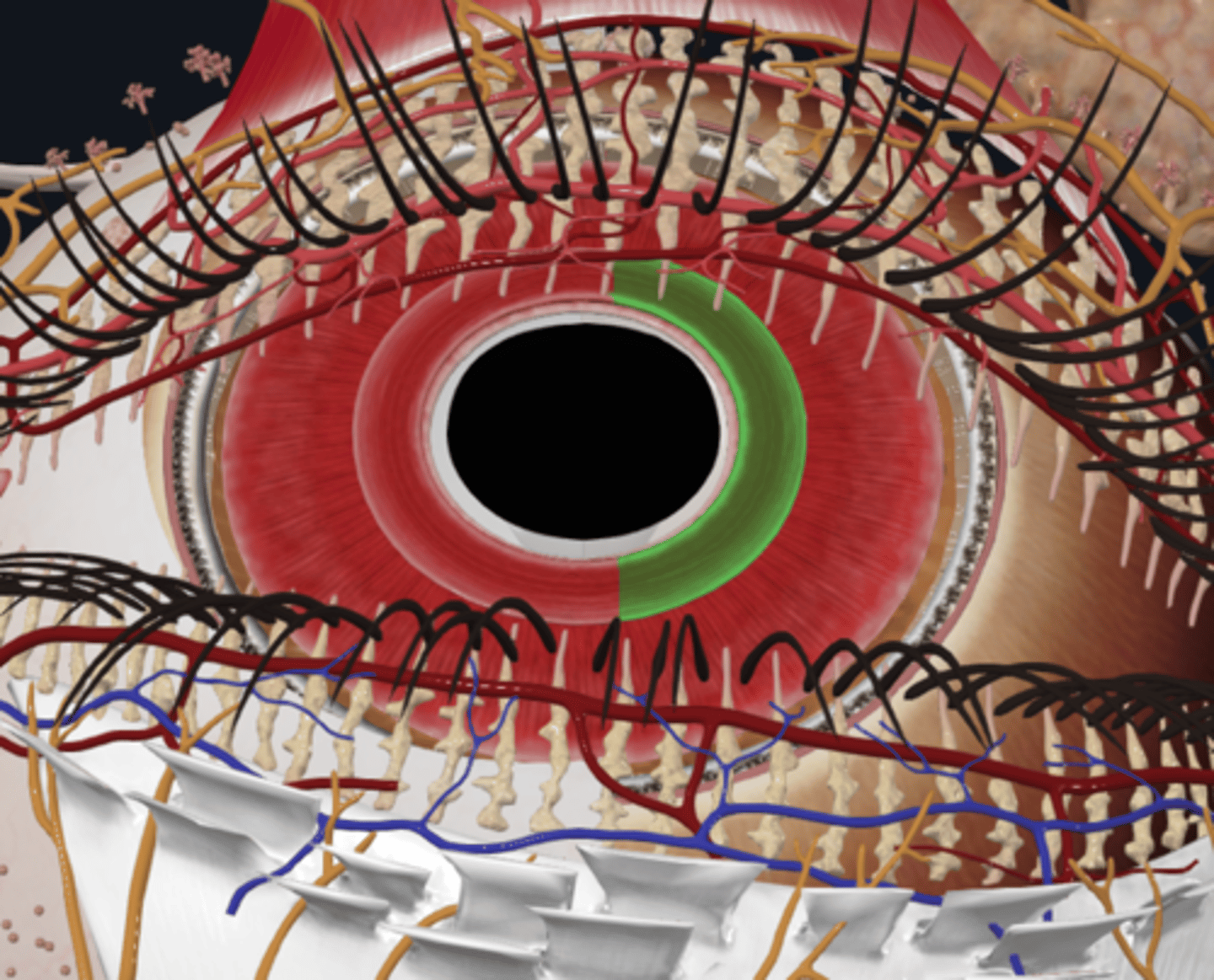
What muscle constricts the pupil and how is it stimulated?
The sphincter pupillae muscle constricts the pupil and is parasympathetically stimulated via the ciliary ganglion.
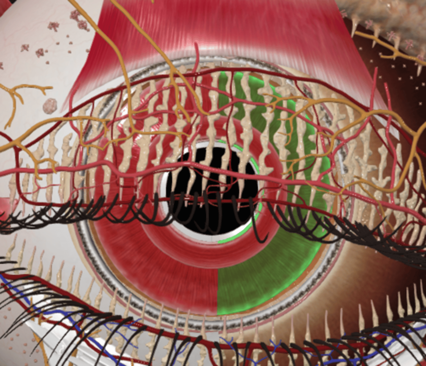
What muscle dilates the pupil and how is it stimulated?
The dilator pupillae muscle dilates the pupil and is sympathetically stimulated via the superior cervical ganglion.
What is the retina and what are its two layers?
1. Neural layer: Light receptive
2. Pigmented layer: Reduces light scattering by reinforcing the light-absorbing properties of the choroid
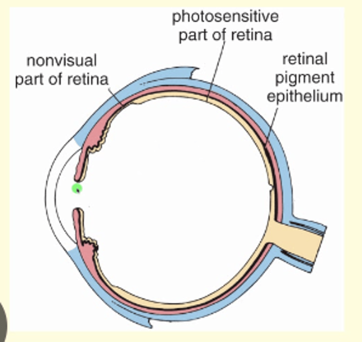
What is the non-visual retina?
The non-visual retina extends anteriorly over the ciliary body and the posterior surface of the iris.
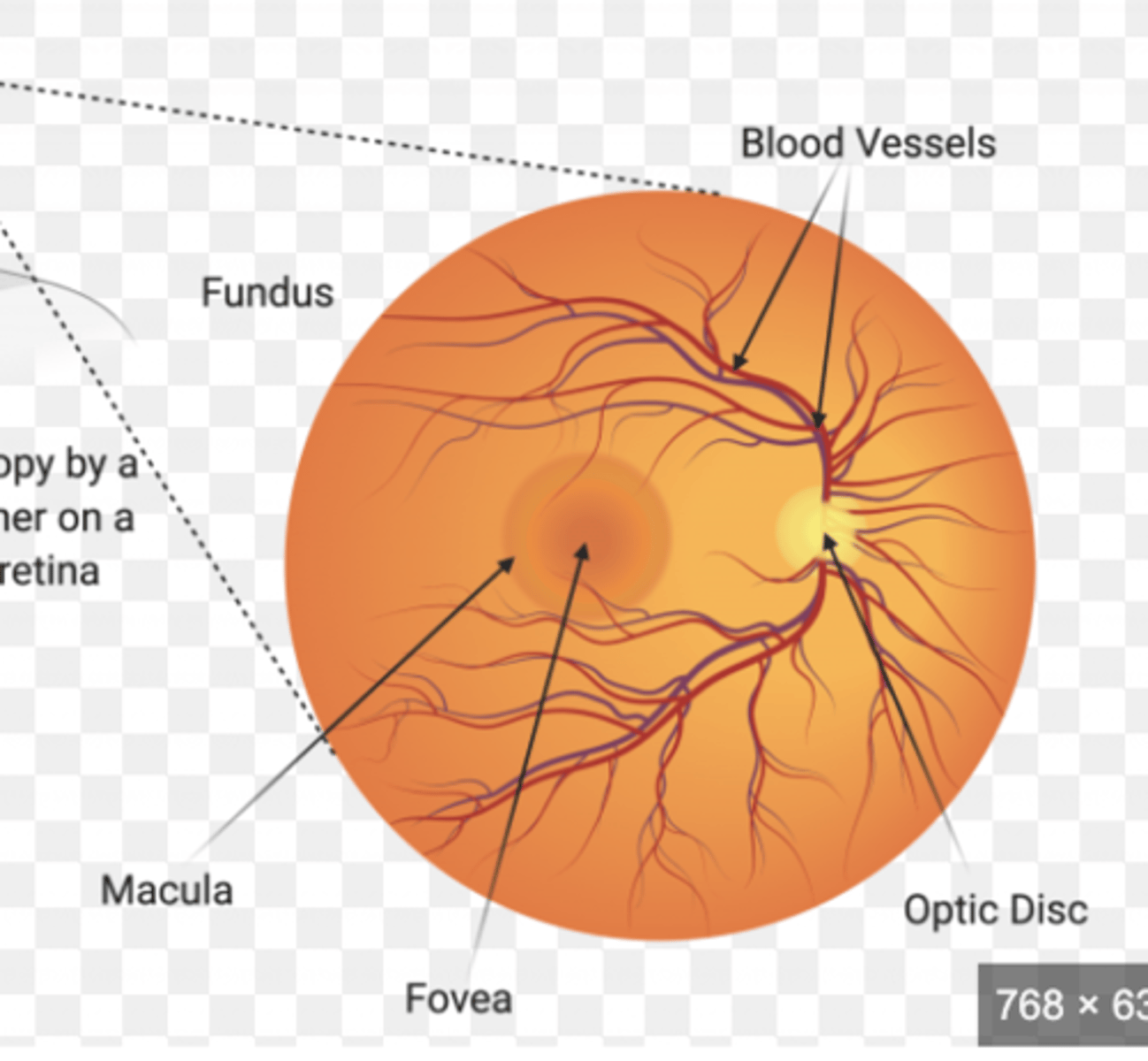
What is the fundus of the eyeball?
The fundus is the internal aspect of the posterior part of the eyeball where incoming light is focused
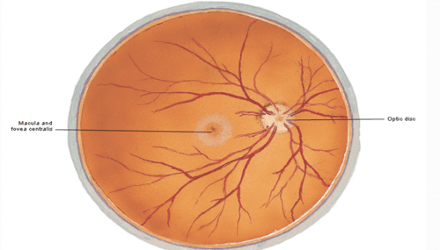
What is the optic disk and why is it insensitive to light?
The optic disk is the point where the optic nerve enters the eyeball, and it is insensitive to light because there are no photoreceptors there.
What is the macula of the retina specialized for?
The macula of the retina is specialized for visual acuity.
What is the fovea centralis and why is it important?
The fovea centralis is a small area (~1.5 mm) at the center of the macula and is the area of the most acute vision.
What is the function of the cornea in the refractive media of the eye?
The cornea refracts light entering the eye, focusing an inverted image on the light-sensitive retina of the optic fundus.
Where is the aqueous humor located and what is its function?
The aqueous humor occupies the anterior (between cornea and iris) and posterior (between iris and lens) chambers of the eye. It is produced in the posterior chamber by the ciliary processes and provides nutrients to the avascular cornea and lens.
What is the lens and how is its curvature modified?
The lens is a transparent, biconvex structure anchored by suspensory ligaments to the ciliary processes. Its curvature is modified by the ciliary muscle, which is parasympathetically stimulated via the ciliary ganglion.
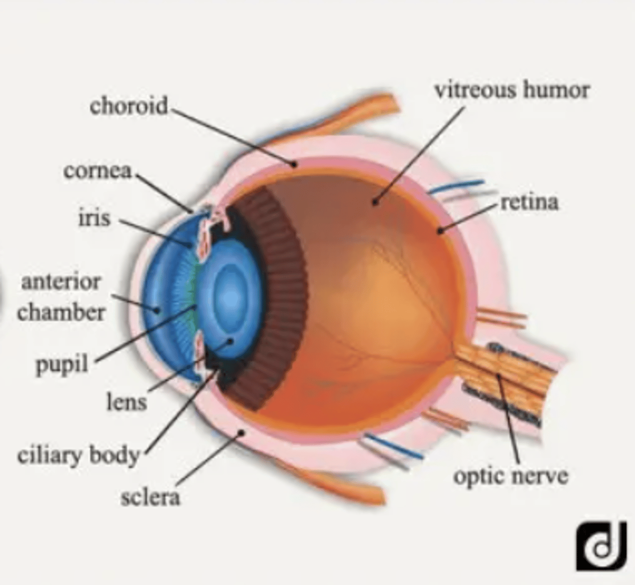
What is the vitreous humor and what is its function?
The vitreous humor is a watery fluid enclosed in the vitreous body, located posterior to the lens. It helps hold the retina in place and supports the lens.
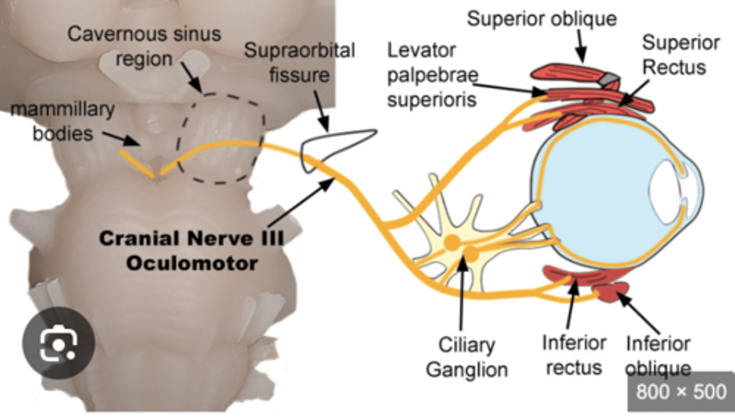
What is the course of the oculomotor nerve (CN III) and what does it innervate?
The oculomotor nerve travels in the lateral wall of the cavernous sinus and enters the orbit through the superior orbital fissure. It divides into superior and inferior divisions and innervates 5 out of 7 extraocular muscles. It also connects to the ciliary ganglion via short ciliary nerves to the ciliary body and iris.
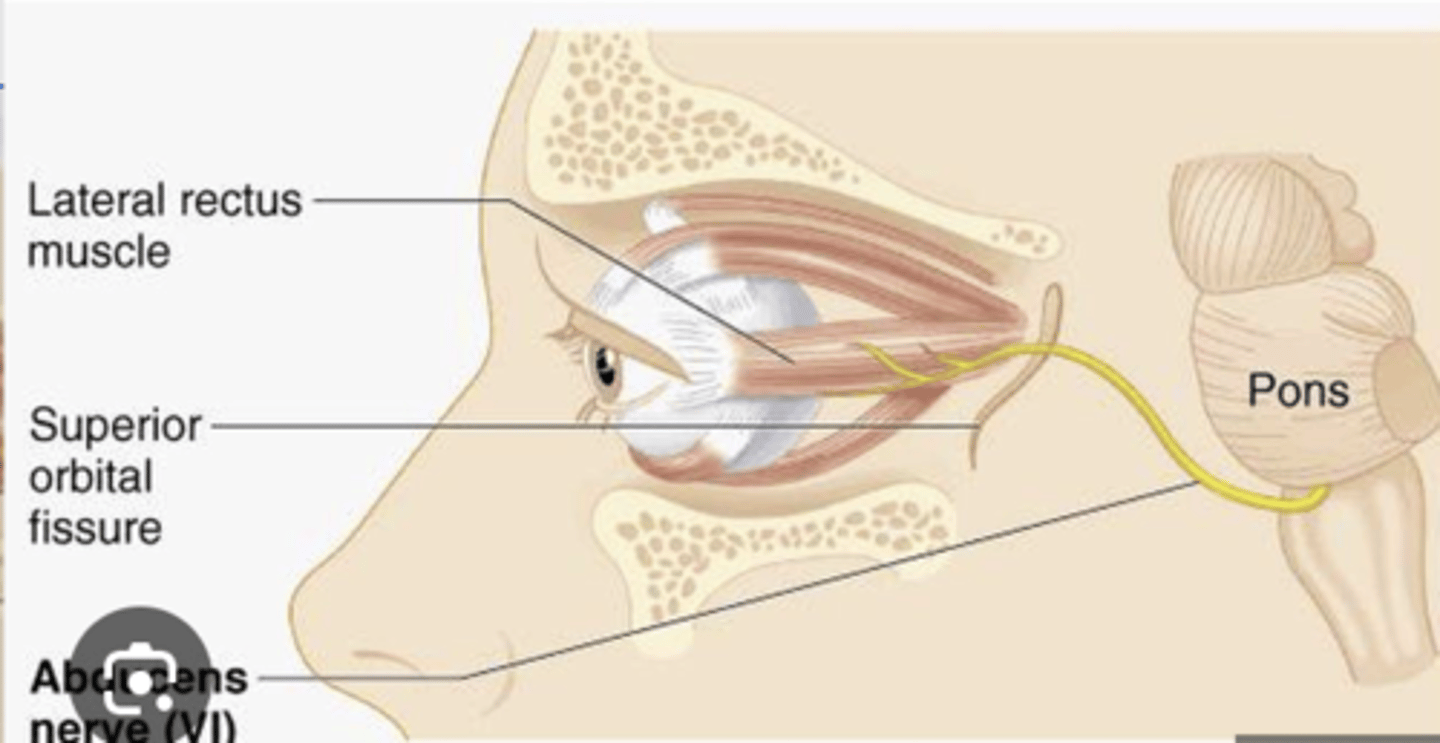
What is the course of the abducent nerve (CN VI) and what muscle does it innervate?
The abducent nerve travels in the cavernous sinus and enters the orbit through the superior orbital fissure. It innervates the lateral rectus muscle.
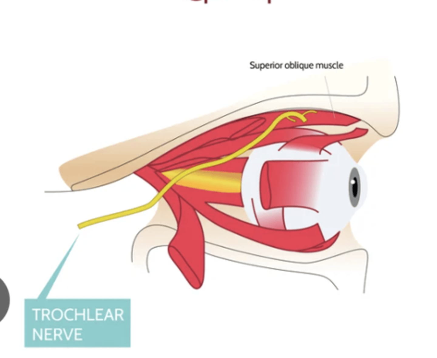
What is the course of the trochlear nerve (CN IV) and what muscle does it innervate?
The trochlear nerve travels in the lateral wall of the cavernous sinus and enters the orbit through the superior orbital fissure. It innervates the superior oblique muscle.
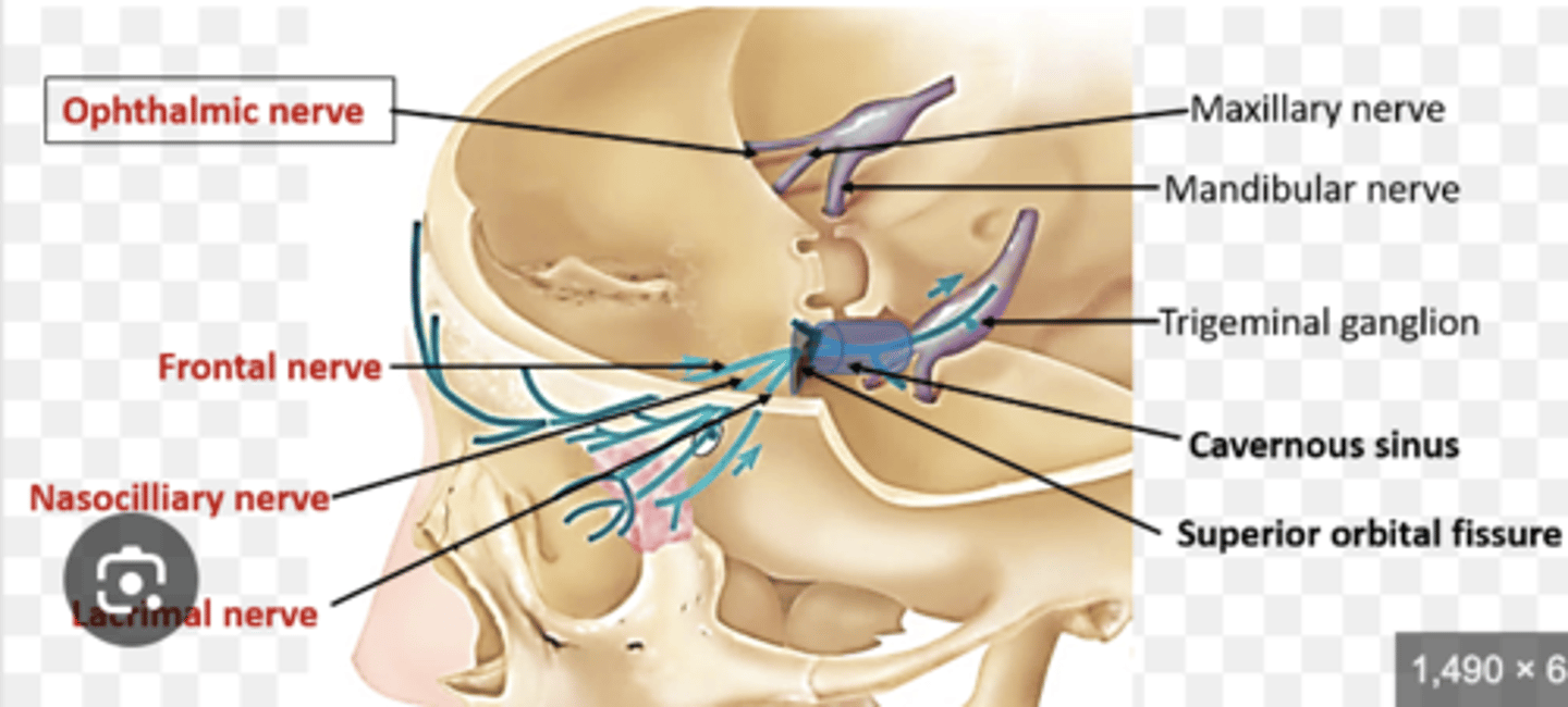
What branches does the ophthalmic division (CN V1) give rise to and what do they innervate?
The ophthalmic division gives rise to the frontal, lacrimal, and nasociliary branches.
The long ciliary nerve (a branch of nasociliary) innervates the iris and cornea
sympathetic fibers from the superior cervical ganglion innervate the dilator pupillae muscle.
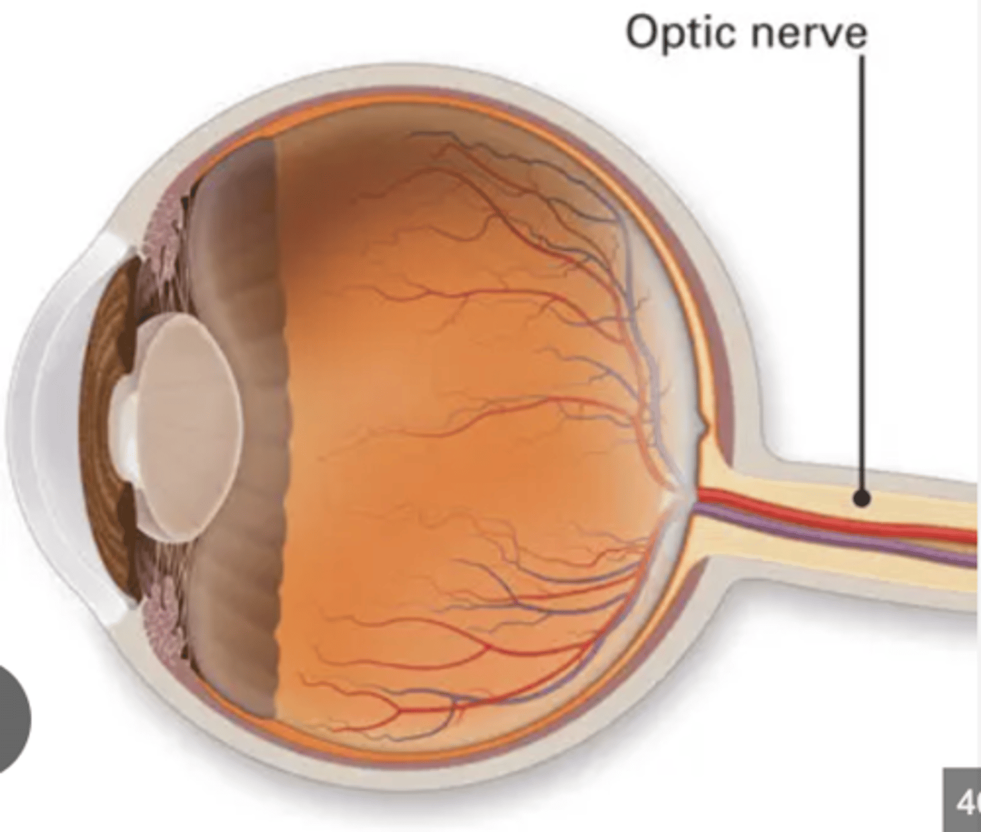
What is the function of the optic nerve (CN II)?
The optic nerve carries visual information from the retina to the brain for processing.
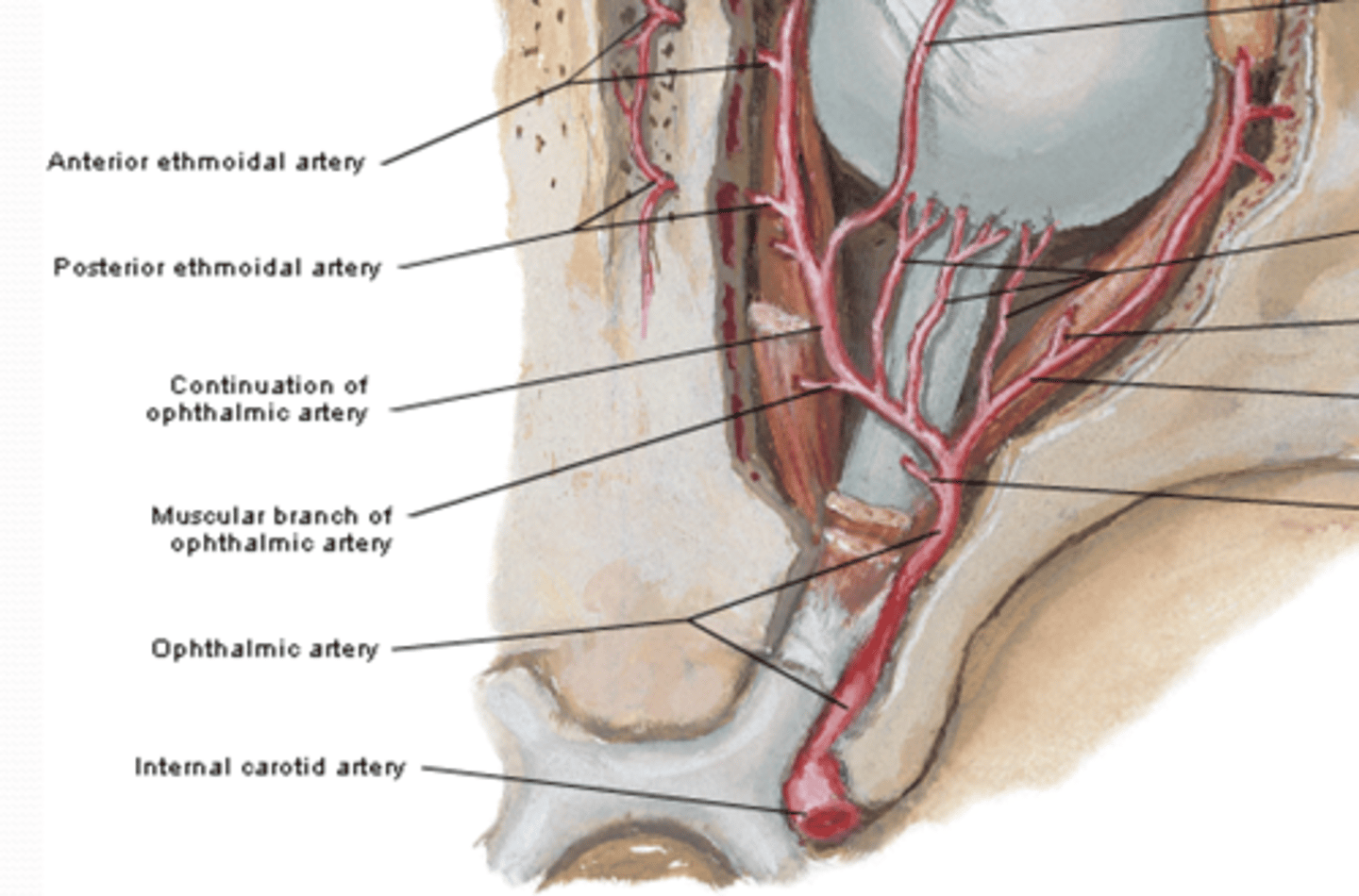
Where does the ophthalmic artery originate and what is its course?
The ophthalmic artery originates from the internal carotid artery after it emerges from the cavernous sinus. It runs parallel to the optic nerve through the optic canal
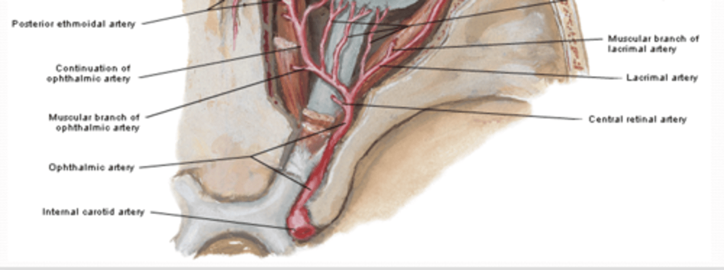
What is the function of the central artery, and what is its clinical relevance?
The central artery pierces the sheath of the optic nerve and runs with it to the eyeball. Its terminal branches are end arteries that supply the internal aspect of the retina.
***Occlusion of this artery results in blindness
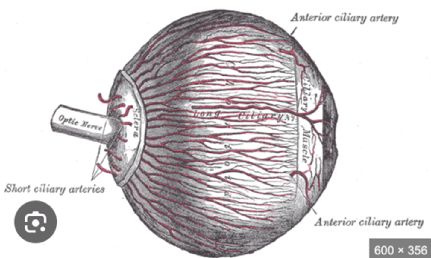
What structures does the ciliary artery supply?
The ciliary artery supplies
1. sclera
2. ciliary body
3. iris
4.choroid
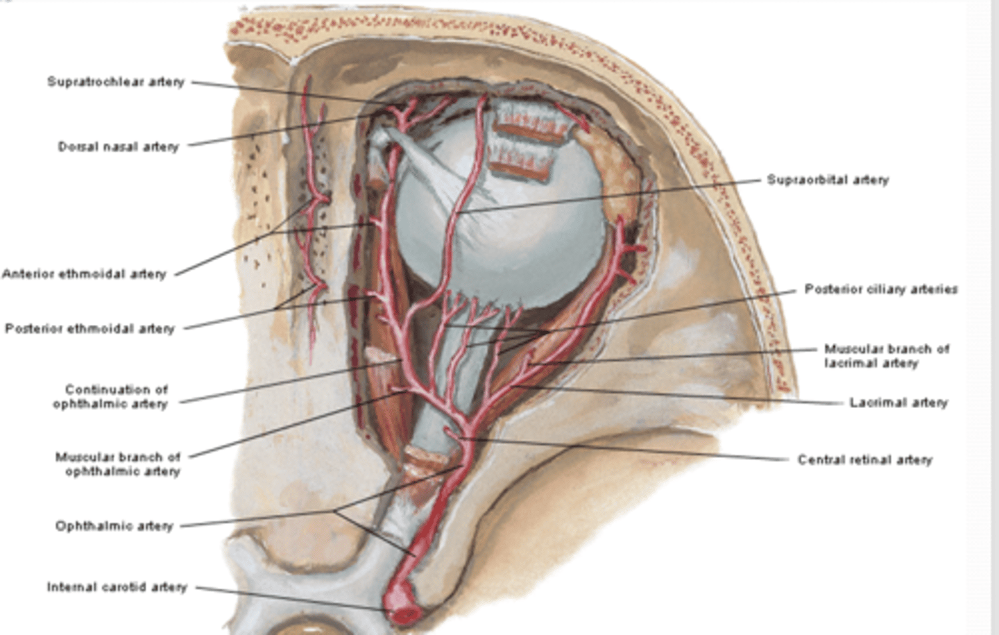
What structures are supplied by the lacrimal artery?
The lacrimal artery supplies
1. lacrimal gland
2. conjunctiva
3. eyelids.
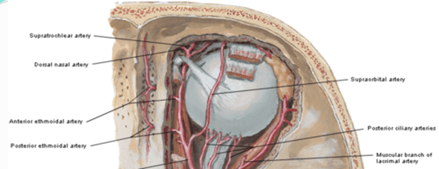
What areas do the anterior and posterior ethmoidal arteries supply?
The anterior and posterior ethmoidal arteries supply the sinuses.
Which structures do the muscular arteries supply?
The muscular arteries supply the orbital muscles.
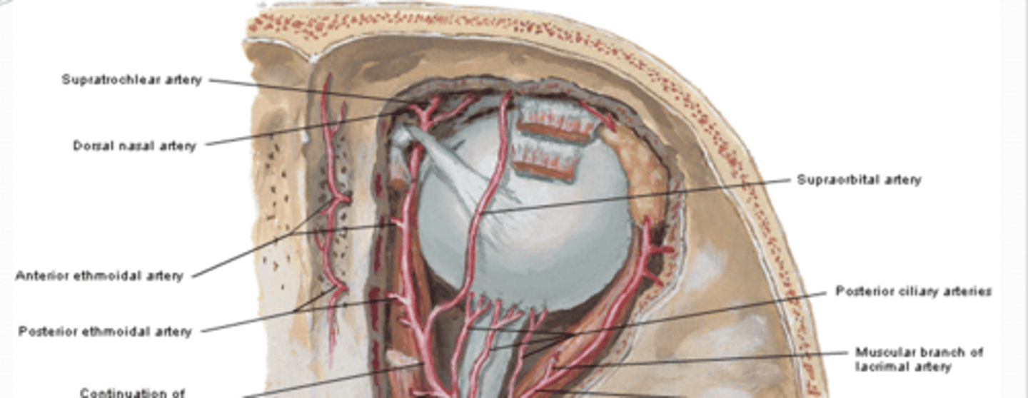
Where does the supraorbital artery emerge, and with which nerve does it travel?
The supraorbital artery emerges onto the face together with the supraorbital nerve.
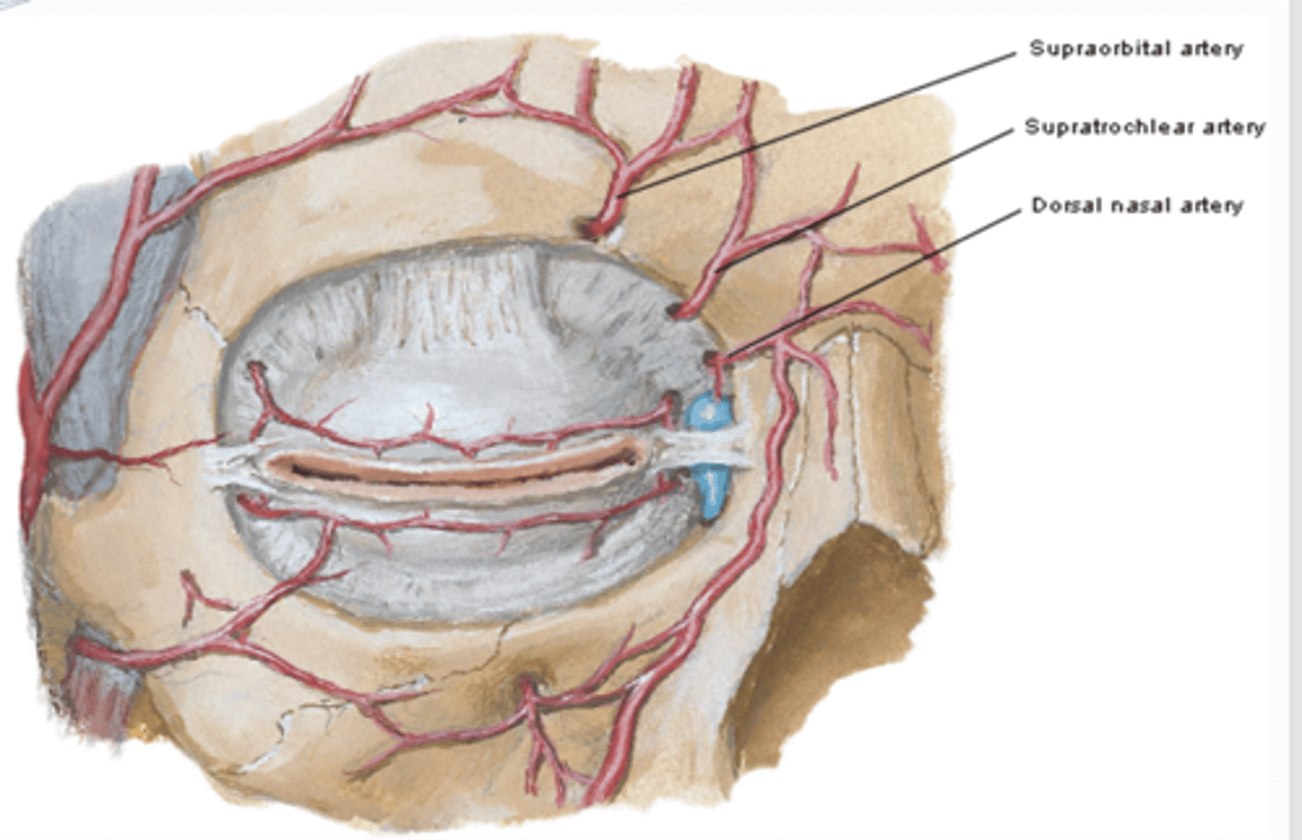
What is the supratrochlear artery and where does it emerge?
The supratrochlear artery emerges onto the face along with the supratrochlear nerve.

Where does the dorsal nasal artery travel?
The dorsal nasal artery travels on the medial side of the orbit.
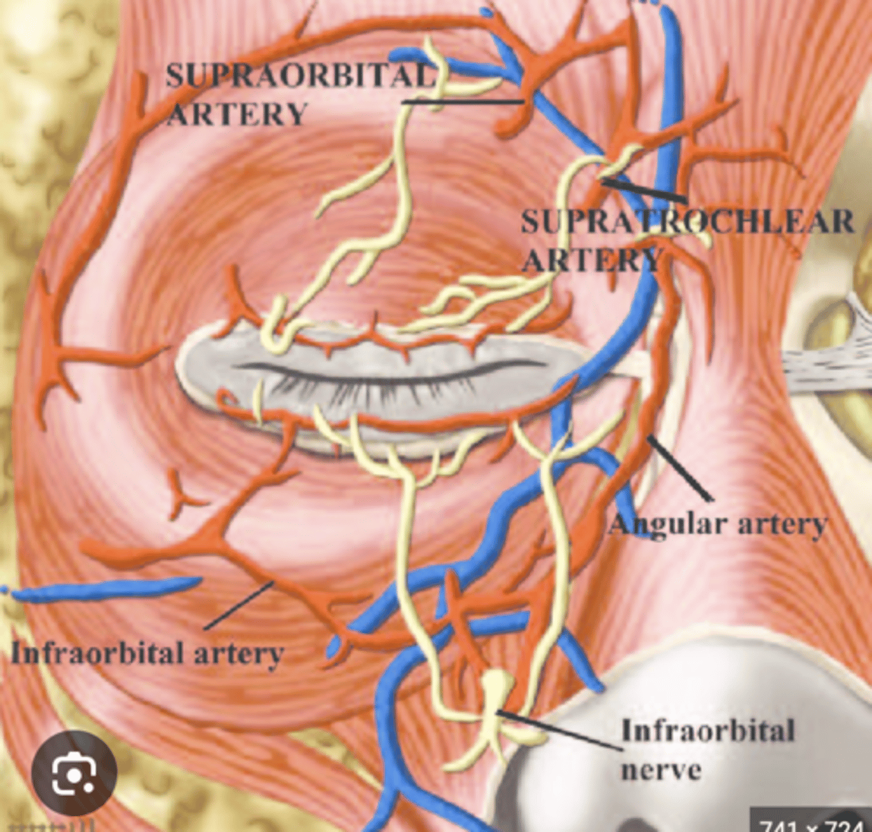
What does the infra-orbital artery supply and where does it originate?
The infra-orbital artery, a branch of the maxillary artery, supplies the orbital floor
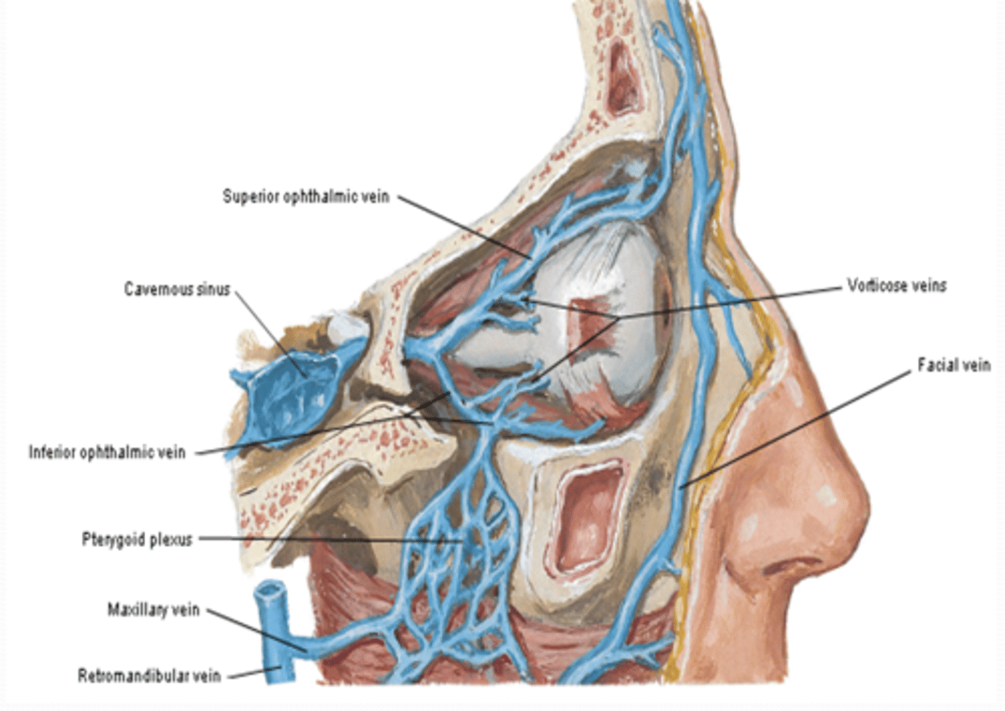
What is the course of the superior ophthalmic vein?
The superior ophthalmic vein is continuous with the facial vein anteriorly, crosses diagonally from medial to lateral above the optic nerve, exits through the superior orbital fissure, and terminates in the cavernous sinus. It drains structures within the orbit.
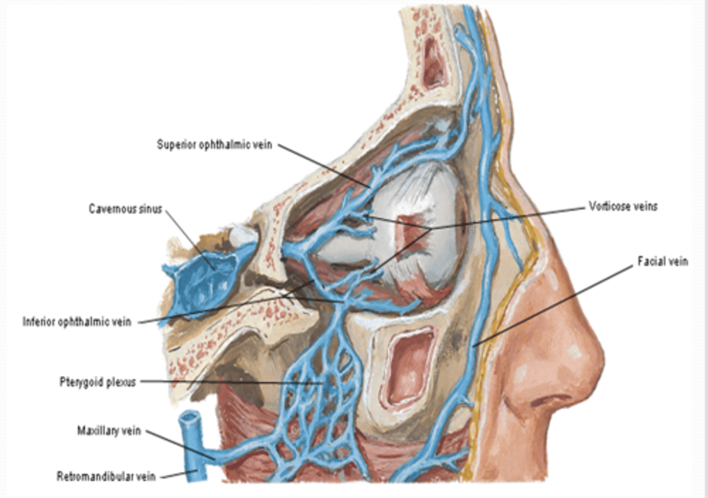
Where does the inferior ophthalmic vein run, and with which structures does it communicate?
The inferior ophthalmic vein runs on the floor of the orbit, begins at the plexus of veins on the floor, and communicates with the pterygoid venous plexus inferiorly and the superior ophthalmic vein before entering the cavernous sinus.
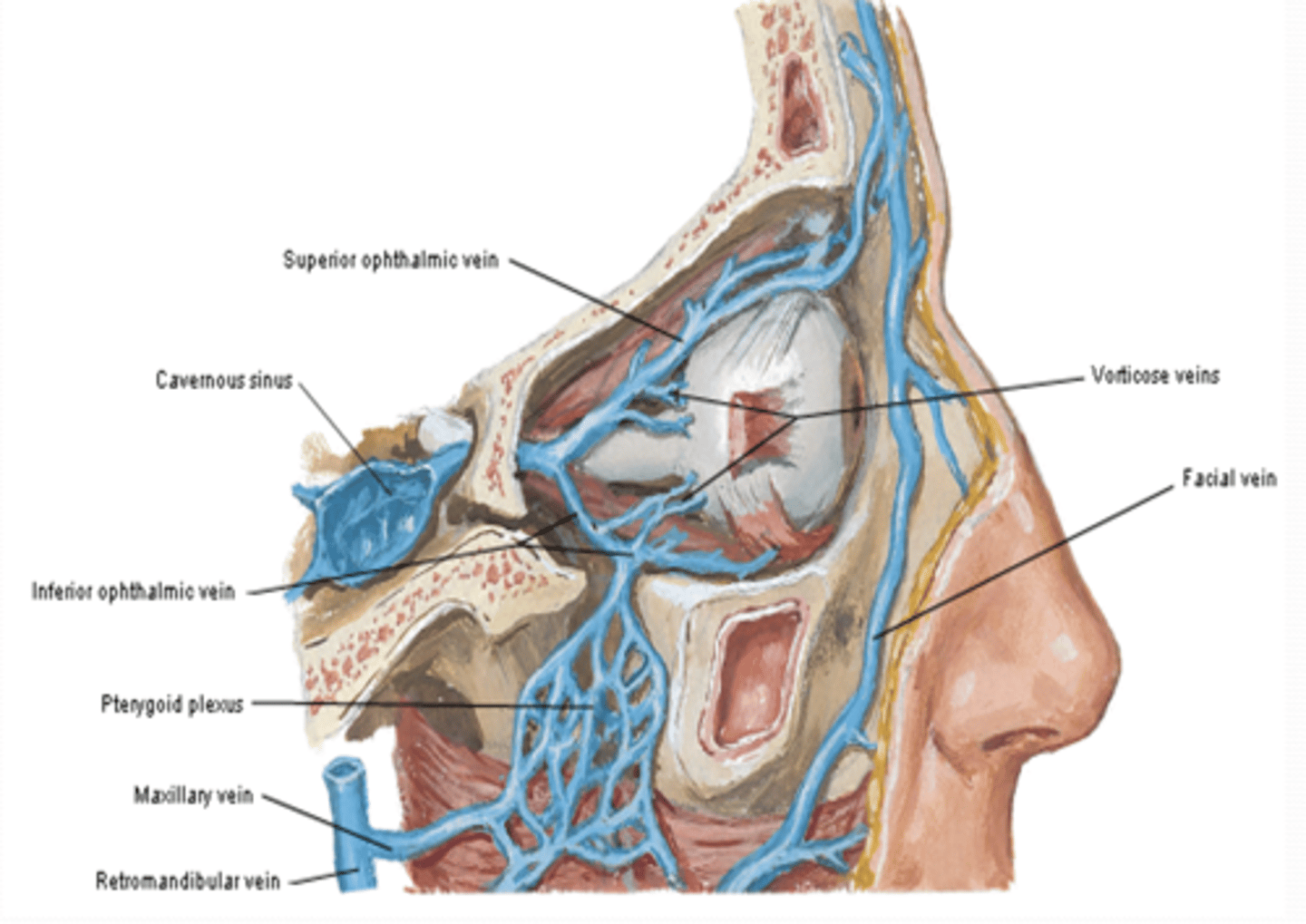
What do the vorticose veins drain, and where do they empty?
The vorticose veins drain the vascular layer of the eyeball and are tributaries of the ophthalmic veins.