Brain & Cranial Nerves
1/32
There's no tags or description
Looks like no tags are added yet.
Name | Mastery | Learn | Test | Matching | Spaced |
|---|
No study sessions yet.
33 Terms
CNS composition
brain and spinal cord
meninges- entire CNS surrounded by 3 membranes
layers of meninges that surround brain are the same as spinal cord
Dura mater- outer most layer; thickest and toughest
outer layer- (osteal, endosteal, or perosteal)
fused to perosteum
meningeal/ inner layer-
Venous sinus-
between outer and inner layer of dura, space with blood vessels and these
contains blood that has been to a tissue and is on its way back to the heart
Arachnoid layer- inside the dura mater
subarachnoid space-
inside arachnoid; cerebrospinal fluid filled space
forms a CUSHION between CNS and surrounding bone
SUPPORTS CNS
provides a CIRCULATORY function that would be accomplished by blood vessels elsewhere
Choroid plexuses-
small blood vessels or capillaries in ventricles that produce CSF
one in each of the four brain ventricles
CSF-
brain and spinal cord have interior cavities that are also filled with CSF
CSF produced in Ventricles (cavities) of the brain
flows through ventricles, central canal of spinal cord, and subarachnoid space
Pia mater- inner layer; lies on the surface of the CNS
Terms to describe regions of the brain at diff stages of development
Mature Brain
Cerebrum
diencephalon
Cerebellum (dorsal part)
mesencephalon (midbrain)
pons/pons varolii (ventral part)
medulla oblongata
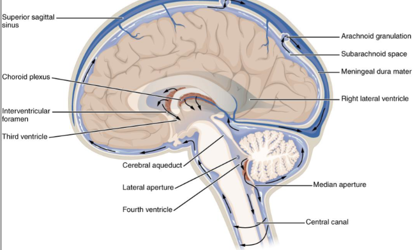
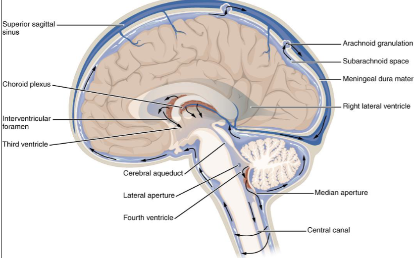
Brain Ventricles and CSF
lateral ventricles
2 most superior cavities
septum pellucidum/pellucidum septum
two cavities separated ny this thin wall/partition
septum- thin wall dividing two chambers
interventricular foramina/ foramina of Monro
CSF flows from lateral ventricles through this into 3rd ventricle
third ventricle/diencephaletic chamber
NO 1st or 2nd ventricle (lateral ventricles)
surrounded by diencephalon
aqueducts of Sylvius/ mesencephalic aqueduct/ cerebral aqueduct
from 3rd ventricle, CSF flows through this into the 4th ventricle
NOT IN CEREBRUM
fourth ventricle
4 Exits from this:
central canal of the spinal cord
CSF can go into
median foramen/ foramen of Magendie
single, midline opening
allows CSF to flow into subarachnoid space
lateral foramina/foramina of Luschka
2 lateral openings
allow CSF to flow into subarachnoid space
Blood flow
ventricles- cavities of brain that CSF is produced in
Choroid plexuses-CSF produced in ventricles of brain from the blood vessels
fluid must be reabsorbed into blood at same rate of production
b/c no room for swelling because CNS surrounded by bone
Superior sagittal sinus-
venous sinus in dura mater along the midline above the brain
indicated blood is going back to heart to be pumped somewhere else
Arachnoid villi/arachnoid granulations-
extensions of the arachnoid layer that extrude through the dura into the blood of the superior sagittal sinus
This is where CSF passes back into blood
Blood-brain Barrier (BBB)
not a lot of exchange between blood and tissues comparatively
Blood-brain barrier (BBB)
anatomic & physiologic factor that prevents passage of substances from blood into tissue of brain
endothelial cells that line blood vessels have tight junctions
prevent substances from getting between cells
Blood vessels, capillaries, in the brain surrounded by ASTROCUTES
they form physical barrier in addion to the wall of blood vessel
also secrete chemicals that affect permeability of vessels
Why some conditions cannot be treated in brain
chemicals in blood cant get into tissue
Parkinsons disease cant be given intravenous dopamine
dopamine cant get out of the blood vessels in the brain
Circumventricular organs
LACK the BBB
are in the wall surrounding the third ventricle
monitor concentrations and conditions in the blood
must respond to changes to maintain homeostasis
so there has to be more communication between the blood and these organs
Choroid plexuses have NO ASTROCYTES and are fairly permeable
although there is a “blood-CSF barrier” created by the tight junctions between the endothelial cells
Brain and blood supply
good blood supply CRITICAL to brain
has extremely high metabolic rate so it uses O2 rapidly and creates waste products rapidly
also must use carbs to get energy
cannot utilize fats or proteins as a source of energy
can increase blood flow by increasing heart rate
can increase respiratory rate to increase concentration of O2 in blood and decrease concentration of CO2
glucose + oxygen → carbon dioxide + water + energy
reaction shows how cells use O2 in the process of producing energy and produce CO2 as a waste product
carbon dioxide + water → carbonic acid ; carbonic acid → H+ + bicarbonate ion (HCO3-1)
when CO2 is produced, causes increase in number of H ions, or a drop in pH
in normal healthy adult, brain more sensitive to buildup of CO2 and H ions than it is to drop in O2 concentration
sensitive to all three concentrations
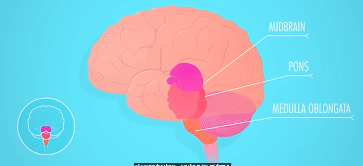
Brain Stem
brain stem
most inferior part, inside cranial cavity
similar appearance to spinal cord
composed of medulla oblongata, pons, and midbrain
medulla oblongata
most inferior part of brain stem
extends from foramen magnum to pons
composed primarily of ascending descending tracts
decussate- many of these nerve tracts cross midline
pyramids- large motor tracts that decussate
fibers cross midline; why right side= left and left= right
olives- lateral swellings of medulla where it has connections to the cerebellum through inferior cerebellar peduncles
some nuclei in medulla that maintain some basic life functions
include involuntary centers for heart rate, respiratory rate, and blood flow (vessel diameter)
pons (“bridge”)/ pons variolii-
part of the brain step superior to medulla
primarily ascending & descending tract
connected to cerebellum through middle cerebellar peduncles
nuclei in pons that affect respiratory center of the medulla to help control breathing cycles
pneumotaxic area and the apneustic area- centers of pons
midbrain/ mesencephalon
part of brain stem superior to pons
contains the aqueduct of Sylvius
Superior cerebellar peduncles connect midbrain to cerebellum
cerebral peduncles- ventral part of midbrain consists of a pair of fiber bundles
contain ascending and descending tracts that connect upper parts of the brain with lower parts of the brain and spinal cord
tectum- dorsal part of the midbrain
corpora quadrigemina- four small mounds
superior colliculi- two upper mounds involved in reflexive movement of the eyes due to a visual stimulus
inferior colliculi- lower mounds involved in reflexive movement of the head and trunk due to an auditory stimulus. “STARTLE” REFLEX
substantia nigra-
nucleus of cells near cerebral peduncles
involved with regulating SUBCONSCIOUS muscle movement
area degenerates in people with Parkinson’s disease
reticular formation
dispersed gray matter located in the spinal cord, medulla, pons, and midbrain
functions in the level of consciousness and arousal from sleep
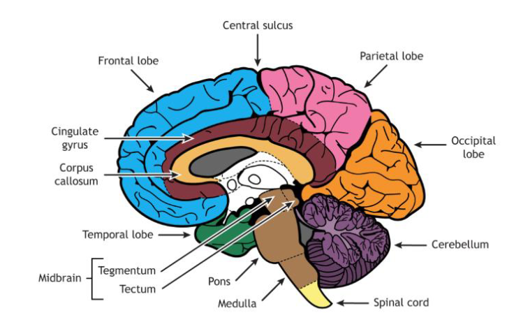
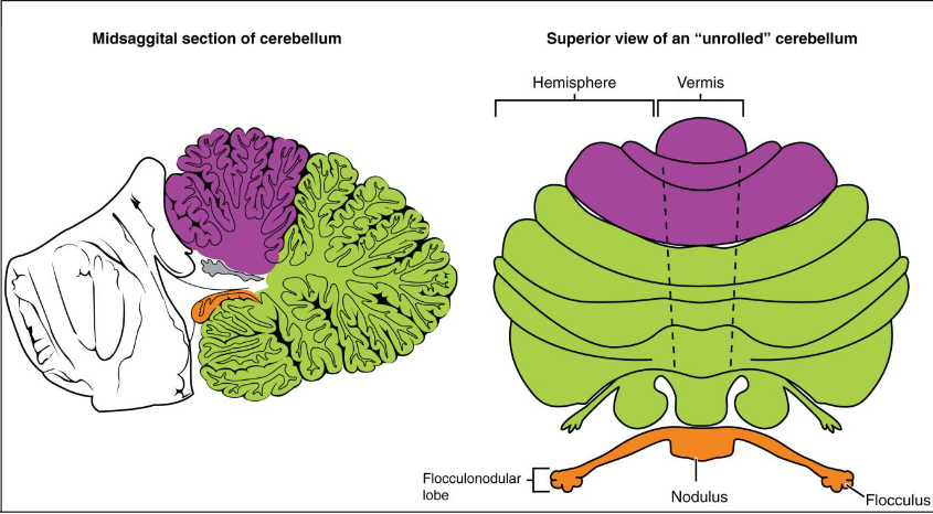
Cerebellum
dorsal to the brain stem
transverse fissure- deep groove between cerebrum and the cerebellum
contains the tetorium cerebelli
tentorium cerebelli- extension of the dura matter
has two hemispheres and a vermis
Falx cerebelli- extension of dura mater between two hemispheres
Arbor vitae- gray matter of cerebellum surrounds white matter and the internal white matter looks like a tree
Folia cerebelli- horizontal ridges of gray matter
involved in reflex control and coordination of skeletal muscle movement
affects muscle tone and muscles that maintain equilibrium or posture
Ataxia- disturbance of balance
caused by trauma & drugs
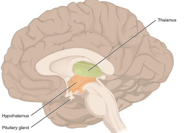
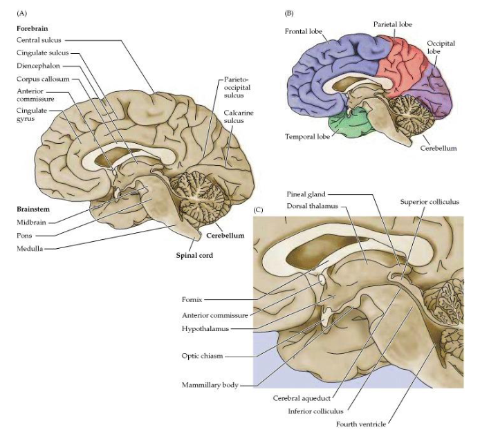
diencephalon
between midbrain and cerebrum
3rd ventricle in diencephalon
READ DESCRIPTIONS OF DIENCEPHALON CAREFULLY
integrates conscious & unconscious sensory information and motor commands
consists of epithalamus, thalamus, and hypothalamus
INTERMEDIATE MASS OF THE THALAMUS that extends through third ventricle
Epithalamus
above third ventricle and consists of pineal body and posterior commissure
pineal body/gland
endocrine gland that produces the hormone melatonin
posterior commissure connects cerebrum with midbrain
Habe nuclei of the epithalamus are involved in emotional responses to some odors
Thalamus
relay station for sensory and motor impulses to and from the cerebral cortex
involved in pain perception, temp perception memory, touch, ext.
part of the limbic system (emotional brain that affects memory) READ THE BOOK
Hypothalamus
structure responsible for maintaining homeostasis
affect body structures through autonomic nervous system &/or endocrine system
controls food intake, concentration of the blood, concentration of urine, etc. READ BOOK
Diencephalon in OPENSTAX pg 526-527
🧠 Brain Development Overview
Three primary brain vesicles become five secondary vesicles during development.
Prosencephalon splits into:
Telencephalon → becomes the cerebrum
Diencephalon → forms the thalamus, hypothalamus, and other structures.
The eye cup develops from the diencephalon → later becomes the retina, a rare case of CNS tissue becoming peripheral.
🔁 Why Diencephalon Matters
It remains the only brain region that keeps its embryonic name.
Located between the cerebrum and rest of the nervous system; acts as a passage for almost all brain signals.
Name translates to “through brain,” reflecting its role as a central connector.
👁 Key Developmental Connections
Retina originates from the diencephalon → connects to thalamus and hypothalamus via optic tract.
Optic tract also links to the midbrain (mesencephalon), which neighbors the diencephalon.
Cerebellum develops from the metencephalon → connects strongly to the pons; also links to the medulla and midbrain.
🧩 Adult Structures & CNS
Brain stem: Made of midbrain (mesencephalon), pons (metencephalon), and medulla (myelencephalon).
Cerebellum: Large, separate structure; no direct link to cerebrum.
Olfactory system: The only pathway that bypasses the diencephalon and connects straight to the cerebrum.
📚 Diencephalon Subregions
Lies deep beneath the cerebrum, forms walls of the third ventricle.
Includes any brain part with “thalamus” in its name:
Thalamus: Relay center between cerebrum and body.
Hypothalamus: Controls homeostasis via autonomic and endocrine functions.
Epithalamus: Includes pineal gland.
Subthalamus: Contains subthalamic nucleus (part of basal nuclei).
cerebrum
largest part of brain
part of brain involved with conscious thought and intellectual processes
2 cerebral hemispheres separated by a deep groove, the longitudinal fissure
falx cerebri- extension of the dura mater extends down into longitudinal fissure
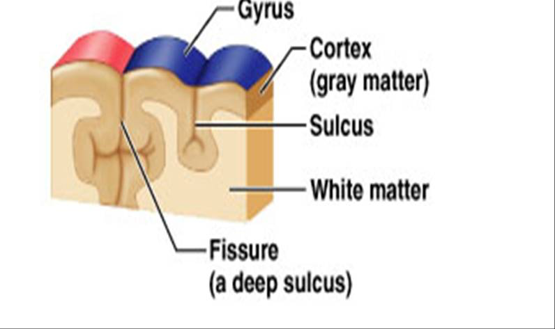
cortex- outer part of the cerebrum that is
composed of gray matter (contains cell bodies of neurons)
interior of cortex is white matter (contains myelinated fibers)
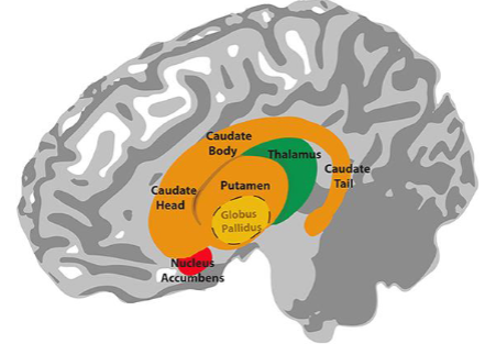
basal/cerebral nuclei/basal ganglia - deep areas of gray matter that are surrounded by white matter
corpus callosum
structure that contains fibers crossing the midline
main connection between right side of brain and left side
covered with gyri, sulci, and fissures
gyri- ridges
sulci- shallow grooves
fissures- deep grooves
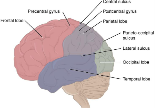
Cerebral hemisphere lobes
frontal lobe- separated from parietal lobe by central sulcus
precentral gyrus- most posterior gyrus of the frontal lobe (anterior to central sulcus)
post central gyrus- most anterior gyrus of the parietal lobe
Temporal lobe- lateral fissure separates frontal and temporal
Insula/island of Reil- within this fissure is a mass of gray matter
Parietal lobe- separated from the occipital lobe by the parietooccipital fissure
Functions of cerebral hemispheres
Each hemisphere receives sensory information from and sends motor commands to OPPOSITE SIDE OF THE BODY.
when giving function to a specific area, NOT PRECISE
some areas of brain are destroyed in some individuals, other parts can perform function of the missing tissue
Left
spoken and written language, numerical and scientific skills
Right
musical and artistic talent, insight, and imagination
White matter
white due to the presence of myelin
part of brain would contain fibers
Tracts- bundles of fibers in the CNS
Association fibers- connect gyri in the same hemisphere
commissural fibers- connect gyri in opposite hemispheres
CORPUS CALLOSUM- largest mass of commissural fibers
projection fibers- ascending and descending fibers
internal capsule- all of the fibers of the white matter together
Gray matter
in cerebrum consists of basal nuclei and the cerebral cortex
basal nuclei (basal ganglia)- paired masses of gray matter that are important in ordinary voluntary muscle movements
do not initiate the movement, but are involved in the coordination of the movement
claustrum, caudate nucleus, putamen, etc.
limbic system- basal nuclei, parts of cerebral hemisphere, and the diencephalon
functional grouping of structures, not anatomic
involved in EMoTIONS
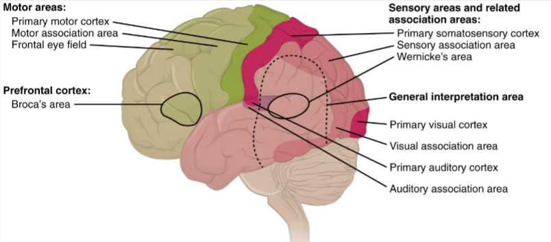
Functional areas associated with the cerebral cortex
READ CHAPTER 14 IN TEXTBOOK
motor area- area that controls muscular movement
sensory area- sensory information is conducted to here for interpretation
relatively small
association areas- surrounds sensory areas
larger areas that evaluate and interpret sensory information
primary visual area- receives information that produces a visual image
determines significance of an image
occipital lobe
general sensory area/ primary somatosensory- located on postcentral gyrus
sensory information from skin, muscles, and visceral receptors from various parts of the body come here
primary auditory area- superior part of the temporal lobe
primary olfaction area- medial surface of the temporal lobe
primary gustatory area- base of the postcentral gyrus
primary motor area- on the precentral gyrus
initiates impulses to muscles on the opposite side of the body
Premotor area/ somatic motor association area- anterior to the motor area
learned, complex movements loke writing
Speech motor area/ Broca’s area- on frontal lobe above the lateral fissure
speaking
also integrative centers that receive input from several association areas of the brain
Summary of ch 14
🧠 14.1 Sensory Perception
Special senses: Olfaction, gustation, audition, equilibrium, and vision (linked to specific organs).
General senses: Somatosensation (touch, pressure, vibration, temperature, pain) and visceral senses.
Sensory receptor types:
Structural: Free nerve endings, encapsulated endings, specialized cells.
Location-based: Interoceptors, exteroceptors, proprioceptors.
Function-based:
Chemoreceptors: For smell, taste, fluid balance, pain.
Mechanoreceptors: For touch, hearing, balance.
Thermoreceptors: Temperature detection.
Photoreceptors: Light sensitivity.
Sensory nerve pathways:
Spinal nerves: Mixed sensory/motor; sensory info enters via dorsal root.
Cranial nerves: Some purely sensory (olfactory, optic, vestibulocochlear), others mixed.
🧠 14.2 Central Processing
Sensory input routes:
Through spinal cord (body) or brainstem (head/organs) → thalamus in diencephalon.
Exception: Olfactory signals directly reach frontal/temporal lobes.
Major spinal tracts:
Dorsal column system: Carries touch & proprioception; decussates in medulla.
Spinothalamic tract: Carries pain & temperature; decussates in spinal cord.
Other sensory pathways:
Auditory: Processes frequency & localization via brainstem.
Vestibular: Influences cerebellum, spinal cord, and cortex.
Visual: Segregates field info; has dorsal (movement/action) and ventral (memory/form) streams.
🧠 14.3 Motor Responses
Motor system origin: Frontal lobe → premotor/supplemental areas → primary motor cortex.
Upper motor neuron tracts:
Corticobulbar & corticospinal tracts control voluntary movement.
Extrapyramidal system:
Maintains balance, posture, and tone via brainstem centers (superior colliculus, red nucleus, vestibular nuclei, reticular formation).
Lower motor neurons:
Synapse in spinal cord → skeletal muscle via neuromuscular junctions.
Motor unit size varies by precision (e.g., quadriceps vs. eye muscles).
Reflexes:
Simple circuits with sensory → motor neuron pathways.
Examples: Withdrawal reflex, corneal blink reflex, stretch reflex via muscle spindle.
Cranial Nerves
twelve pair of cranial nerves
motor, sensory, mixed
no completely motor nerves
Proprioception- sense of motion or body position
ability to know if your arm is straight or bent with eyes closed
primary motor nerves/motor nerves- some cranial nerves have almost all motor fibers but just a very small number of proprioceptive fibers that convey the sense of position
Oh Oh Oh To Touch And Feel Very Good Virgins Are Horny
Some Say Marry Money But My Brother Says Big Brains Matter More
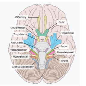
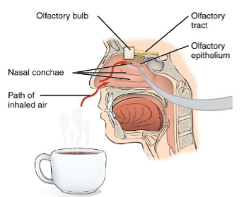
Cranial nerve I
olfactory nerve
sensory
convey the sense of smell or olfaction
nerve passes through the cribriform plate of the ethmoid bone
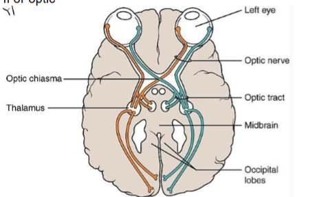
Cranial nerve II
optic
sensory
sense of vision
nerve passes from the back of the eye to the optic chiasm where some of the fibers cross the midline
optic tracts pass back from the optic chiasm
Cranial nerve III
oculomotor
primary motor
six muscles attached tothe outside of the wall of the eye
extrinsic ocular muscle point the eye in different directions
four of these six muscles are innervated by this nerve
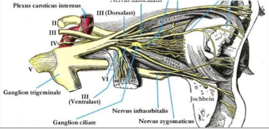
Cranial nerve IV
trochlear nerve
primary motor
nerve innervates one of the two extrinsic ocular muscles that is not innervated by cranial nerve III
muscle called the superior oblique
smallest of the cranial nerves

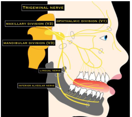
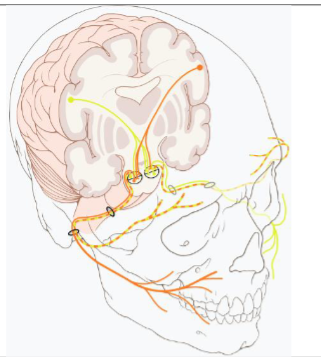
cranial nerve V
trigeminal
mixed
largest of the cranial nerves
motor- this nerve innervates the muscles of mastication
relatively strong muscles of the face that are used in chewing
sensory- nerve conveys sensory information from the anterior part of the tongue but not taste
conveys sensory information about the cheeks, skin, and teeth
Tic Douloureax/ trigeminal neuralgia- extremely painful condition with this nerve. MOST SEVERE PAIN in humans
Cranial nerve VI
abducens
primary motor
lateral rectus muscle- nerve innervates the sixth extrinsic ocular muscle

Cranial nerve VII
facial
mixed
motor- nerve innervates the relatively weak muscles of facial expression
Bell’s Palsy- paralysis of this nerve causes one side of the face to sag
Sensory- nerve conveys sense of taste from the anterior part of the tongue
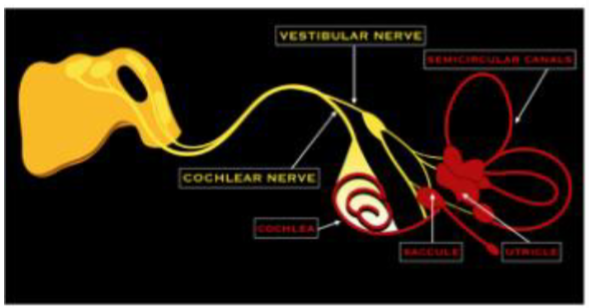
Cranial nerve VIII
vestibulocochlear. auditory, acoustic, statoacoustic
sensory
sensory- nerve conveys sense of hearing and equilibrium
Cranial nerve IX
glossopharyngeal
mixed
motor- nerve innervates the muscles involved in swallowing and gagging
sensory- nerve conveys sensory information from the throat and posterior part of the tongue including taste

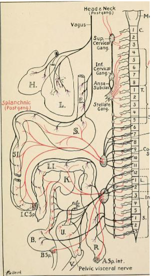
Cranial nerve X
vagus
mixed
“wanderer”- READ TEXT FOR DETAILS
fibers in the neck, head, thorax, and abdomen
sensory and motor fibers run to and from the pharynx, larynx, lungs, heart, and digestive tract
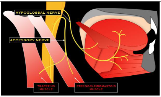
Cranial Nerve XI
Accessory Spinal
spinal accessory
primarily motor
innervates muscles of the upper back and neck

Cranial nerve XII
hypoglossal
primarily motor
innervates muscles of the tongue
Sayings
Some Say Marry Money But My Brother Says Big Brains Matter More
Oh Oh Oh To Touch And Feel Very Good Virgins are Horny