BIO_V 111 UBC - Key Terms and Facts
1/88
There's no tags or description
Looks like no tags are added yet.
Name | Mastery | Learn | Test | Matching | Spaced |
|---|
No study sessions yet.
89 Terms
Cells
Small individual units of life
4 structural features of a cell
Cell membrane
Cytoplasm
Ribosomes
RNA
Cell membrane (Structure of a cell)
A semipermeable membrane which encloses the cell and acts as a barrier between the cell’s internal and external enviornment
Cytoplasm (Structure of a cell)
The liquid component of a cell plus the floaty bits, e.g. organelles. (Analogy; Bubble Tea)
Ribosomes (Structure of a cell)
Tiny structures which carry out protein synthesis, essential for cell division and repair.
Proteins
A large molecule made of chains of amino acids, carries out most work in cells (e.g. structure, transport, defense, communication, speeding up chemical reactions).
Prokaryotic Cells (Types of Cells)
Simple, single-celled organisms without a nucleus (e.g. bacteria, archaea), the earliest form of life.
Eukaryotic Cells (Types of Cells)
More complex cells with a nucleus and organelles, evolved from prokaryotes and make up plants, animals, fungi, and protists.
Prokaryotic vs Eukaryotic Cells
Prokaryotic Cells
No nucleus
Single, circular chromosomes
No membrane-bound organelles
Eukaryotic Cells
Nucleus that contains DNA
Linear chromosomes
Membrane-bound organelles, e.g. mitochondria
Both
+6 characteristics of life
4 structural features of a cell
Organelles
Sub-cellular structures performing specific functions/features inside a eukaryotic cell.
Mitochondria
Break down food molecules (like glucose) to release energy, produces ATP (cell’s energy currency)
Chloroplasts
Found in plants/algae, site of photosynthesis (light energy is converted into sugar)
Ribosomes
Tiny structure in cells responsible for protein synthesis (linking amino acids together through instructions in DNA, via RNA).
How did eukaryotic cells arise (What is the theory of endosymbiosis?)
Likely from prokaryotes through endosymbiosis, where one cell lived inside another and became organelles (e.g. mitochondria, chloroplasts). Eukaryotic cells arose from prokaryotic cell, where once cell engulfed the other. Rather them digesting it, they formed a mutually beneficial relationship.
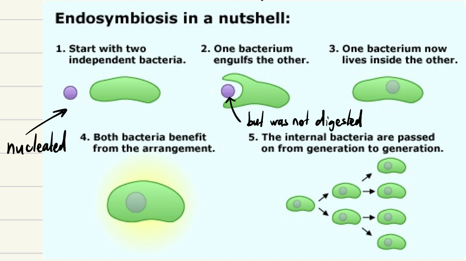
Theory of Endosymbiosis
Start with two independent bacteria
One bacterium engulfs the other
One bacterium now lives inside the other (nucleated, but not digested)
Both bacteria benefit from the arrangement
Internal bacterial are passed on from one generation to another.
Evidence supporting Eukaryotes are descendants of Ancient Prokaryotes
Circular DNA - both mitochondria/chloroplasts have their own circular DNA
Double Membranes - mitochondria and chloroplasts are surrounded by 2 (separate) membrane
Reproduction - Mitochondria and chloroplasts reproduce
Genes - Mitochondria/chloroplasts have their own DNA. The genes are more similar to genes found in prokaryotes than to the genes of eukaryotes.
Genetics
Study of genes, genetic variations, how genetic traits/conditions are passed down by parents/grandparents across generations.
DNA (Deoxyribonucleic acid)
Double stranded molecule, composed of 2 strands of polynucleotides that coil around each other
Stores genetic recipes (genes) for all the proteins that make up an organism.
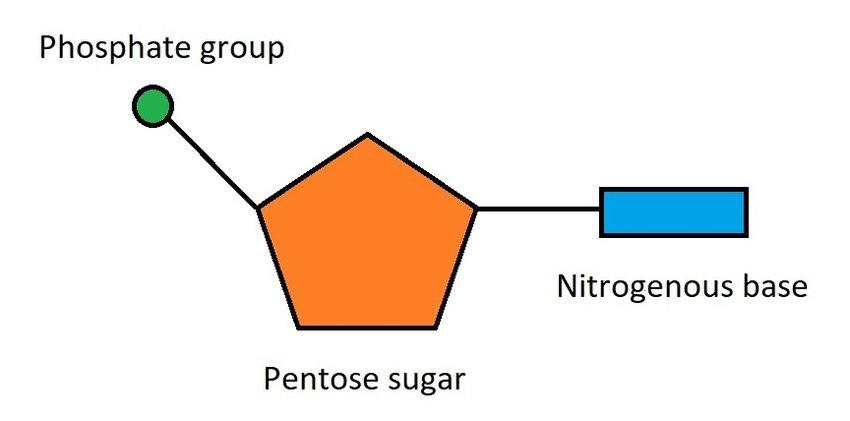
Nucleotide
Composed of:
A sugar group
A phosphate group
One of 4 types of nitrogenous base - (A) Adenine, (T) Thymine, (C) Cytosine, (G) Guanine
Nitrogenous Bases
Adenine (A) always binds with Thymine (T)
Guanine (G) always binds with Cytosine (C)
The sequence of steps is important because it contains the instructions to code for a specific protein.
Genome
The entire set of DNA found in a cell/organism, often measured as the number of base-pairs in the DNA.
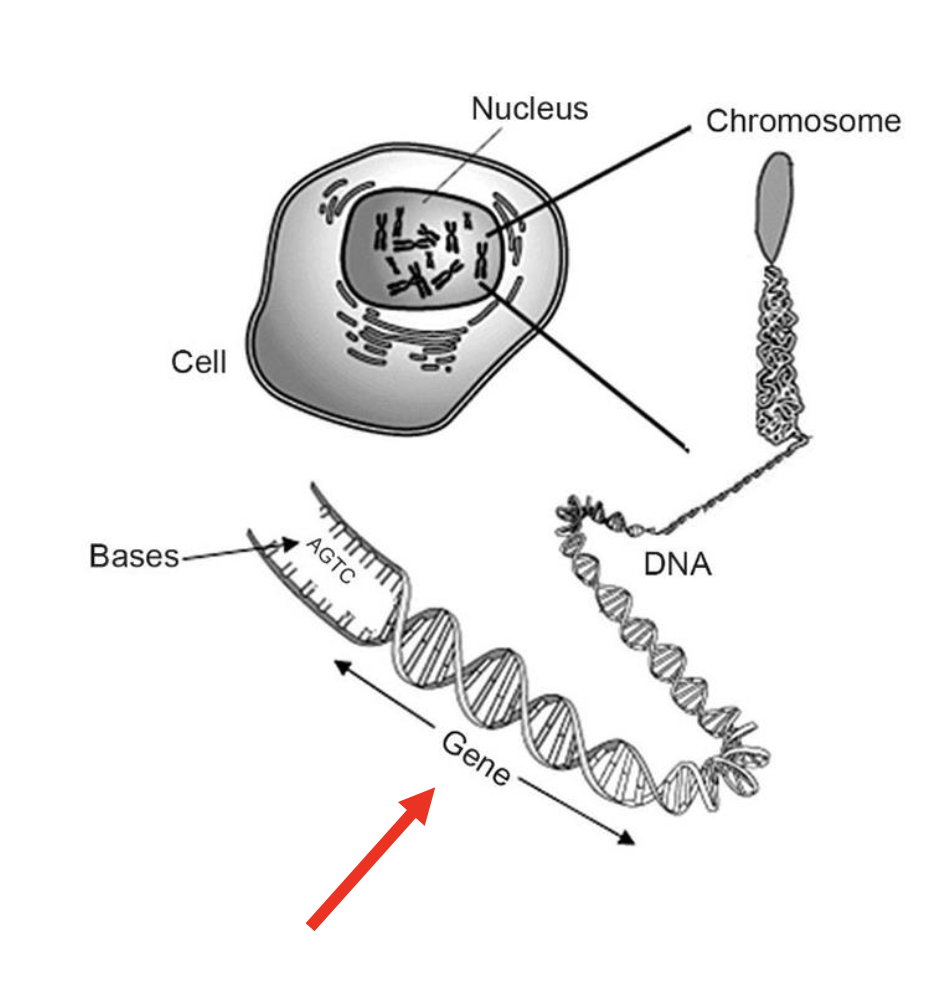
Chromosome
A single, tightly packaged DNA molecule with proteins that carries many genes.
It’s DNA + packaging proteins (so it fits inside the nucleus).
It contains many genes (not just one).
Humans have 46 chromosomes in most cells (23 pairs).
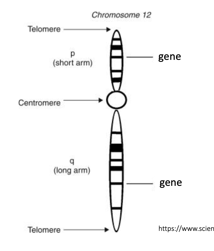
Parts of a chromosome
Centromere – Region where sister chromatids connect and spindle fibers attach during cell division.
Arm – Sections of the chromosome extending from the centromere (short p and long q).
Telomere – Protective DNA-protein caps at chromosome ends, preventing deterioration or fusion.
Genes
Small sections of DNA that code for a specific protein.
Phenotype
The observable traits or characteristics of an organism (physical, physiological, or behavioral), shaped by both its genes and environment.
Alleles
Different versions of the same genes, responsible for variation in inherited traits (e.g. fur color).
Arise due to mutations (or changes in the sequence of nucleotide) in the DNA
Mutation
Changes in sequence of nucleotide in the DNA.
Causes: Mistakes when DNA is replicating, environmental factors damaging DNA (e.g. UV Light)
Mutations can change: amino sequence and function of the protein.
Gene to Protein
Transcription (of DNA to RNA) → DNA is copied into mRNA using RNA polymerase.
Translation (of RNA to Protein) → mRNA is read in 3 letter blocks (codons), each one producing a specific amino acid, amino acids join together to produce a protein.
3 terms to describe the chromosomes in a nucleus
Total number of chromosomes found within a cell
Ploidy of cells (number of sets of chromosomes in a cell)
Haploid number (n - sets of chromosomes number of different types of chromosomes in a compete set)
Ploidy
Number of sets of chromosomes in a cell
Haploid (n or 1n) = 1 of each chromosome type or one complete set of chromosomes set,
Diploid (2n) = 2 of each chromosome, or 2 complete sets of chromosomes
Polyploid (3n, 4n, etc.) = 3 or more of each chromosome, or 3 or more complete sets of
chromosomes
Haploid Number (n)
Number of chromosomes in a single complete set of chromosomes
Number of chromosomes in a gamete of a diploid organism
Haploid number for humans is 23.
Diploid (2n)
A cell with two sets of chromosomes, one from each parent.
Human Gametes
Human gametes (sperm or eggs) are haploid.
Haploid means they carry one set of chromosomes (n = 23).
That includes:
1 of each autosome (chromosomes 1–22)
1 sex chromosome (X or Y).
Differences in size (length) (chromosomes)
Longer chromosomes = more nucleotides
Human autosomes are labelled 1-22 based on length (#1 being the
longest chromosome)
Differences in centromere location (chromosomes)
Centromeres are the constriction point on a chromosome.
Each chromosome has one centromere, which divides chromosomes into two arms
(may be of unequal length)
Codons
A sequence of three nucleotides in DNA or RNA that serves as a unit of genetic code, specifying a particular amino acid or signaling the start or stop of protein synthesis.
Homologous Chromosomes
A matched pair of chromosomes (one inherited from each parent (same size, have the same CENTROMERE location, same sequence of genes)
Unreplicated vs Replicated chromosomes
Unreplicated Chromosome: 1 DNA molecule with 1 centromere
Replicated Chromosome: 2 DNA molecules joined at 1 centromere
Sister chromatids
2 identical chromatids (on replicated DNA)
Each chromatid is 1 DNA molecule
Two sister chromatids that are attached at the centromere are one chromosome
Genotype
Set of alleles or genes that are carried by an individual or a cell - relating to specific trait (or traits) we’re interested in.
Mitosis
Parent cell (cell to undergo cell division) produce two progeny/daughter cells that are genetically identical to the parent cell and each other
Occurs in somatic cells (cells of the body other than sperm or eggs)
Purposes of Mitosis
essential for growth – i.e. you started as one fertilized cell; now your body is composed of trillion cells;
replacing damaged/worn-out cells
asexual reproduction in some organisms
mitotic errors have been linked to cancer
Cell Cycle
G1 (or Gap 1 phase)
S (or DNA synthesis phase)
G2 (or Gap 2 phase)
Mitosis (or M phase)
Cytokinesis
Interphase (Cell Cycle)
G1, S and G2 make up interphase, the longest part of a cell’s life
G1 (Interphase - Cell Cycle)
The cell is performing its functions, e.g. being a liver cell or skin cell. (DNA is NOT replicated)
If cell is not dividing, enters Go phase: Performs main function indefinitely
• e.g., neurons, muscles cells in your heart are in G_0 phase.
If cell recieves the signal to start dividing, the cell begins to prepare in G1 through: growing, duplicating organelles, accumulating nucleotide, obtains energy reserves.
G1 Checkpoint - Cell is checked before it can proceed to S phase
Sufficient number of organelles?
Cell adequate size?
Is the DNA damaged?
S-Phase (Interphase - Cell Cycle)
Synthesis, when the DNA replicates (and sister chromatids are formed)
Two strands of a DNA molecule separate from each other. Each strand serves as a template to synthesize a new complement strand
Result – two DNA molecules, each containing one original “old strand” (dark blue) and one new “daughter strand (light blue).This process is called semiconservative DNA replication (not
testable).
G2 Phase (Interphase - Cell Cycle)
Final preparations are made before cell divides
May be additional growth, more organelles may be duplicated
G2 Checkpoint - All below criterias must be met:
All DNA is replicated
DNA is undamaged
Mitosis Steps (M Phase - Interphase - Cell Cycle)
Mitosis is divided into 4 or 5 phases:
Prophase
Prometaphase
Metaphase
Anaphase
Telophase
Prophase (Mitosis)
Chromosomes condense (thickening and visible), nucleus disappears.
Previously replicated DNAs coil tightly into sister chromatids.
Spindle apparatus begin to form (spindle fibers - "Tiny cell ropes that move chromosomes during division.")
Nuclear membrane disappears
Prometaphase (Mitosis)
Nuclear envelope breaks down
Microtubles (tiny tubes in cells for transport) contact chromosomes at kinetochores.
Metaphase (Mitosis)
Chromosomes line up in the middle of the cell
Anaphase (Mitosis)
Sister chromatids separate, move to opposite sides of the cell by spindle fibers
Telophase (Mitosis)
Chromosomes are on complete opposite ends
New nuclei form on each side to make these two new cells
Cells start to decondense
Cytokinesis
Splits the cytoplasm following mitosis
plasma membrane starts to pinch in to create two progeny/daughter cells
cell divison complete - two new daughter cells formed.
Meiosis
Type of cell division found in sexually reproducing organisms.
Goal: diploid parent cell to produce 4 genetically distinct haploid daughter cells or gametes (eggs or sperm).
Process: Meiosis I and Meiosis II
Mitosis vs Meiosis
Mitosis
Somatic Cell
5 phases
Diploid (parent) to Diploid (daughter cells)
Produces cells used for growth, repair and replacement of damaged/worn-out cells.
Daughter cells are genetically identical to each other and the parent cell
Meiosis
Germline cells in gonads
Genetic Variation (inheritance, further mutations)
8 phases
Diploid to Gamete
Produces Gametes for Sexual Reproduction
Gametes/daughter cells are genetically different from each other (ideally) and genetically different from the parent cell.
Similarities
Both are types of nuclear division
DNA replicated in Interphase
Starts with a single parent cell
Same basic steps occur
Early Prophrase I (Meiosis I)
Chromosomes condenses, nuclear membranes disassemble (so spindle fibers can reach chromosome).
AND Synapsis - each chromosome pairs up and bonds with a corresponding homologous chromosome, forming a tetrad.
Tetrad
Group of four sister chromatids in paired homologous chromosomes.
Chromsomes include genetic information called genes, inherited from each parent. Different version of the same gene on each chromosome are alleles.
Late Prophase I (Meiosis I)
Crossing-over and recombination.
While homologous chromosomes are tightly paired, non-sister chromatids exchange bits of DNA/segments of alleles:
in a process called crossing-over.
outcome is called recombination
Creates genetic variety in off springs.
Metaphase I (Meiosis I)
Homologous chromosomes line up at equator, attach to spindle fibers from opposite poles.
Outcome: Gametes receive a random combination of maternal and paternal chromosomes.
Anaphase I (Meiosis I)
Spindle fibers separate homologous chromosomes in each tetrad and pull them to opposite poles of the cells.
Telophase I & Cytokinesis I (Meiosis I)
Nuclear envelope may reform.
Cytokinesis – creates two HAPLOID cells
Sister chromatids still attached at centromere
Meiosis II
Sister chromatids separate. Very similar to mitosis; but now the cell is haploid (if original parent cell was diploid).
Homeostasis
The ability of an organism to maintain a stable internal environment despite changes in the external environment.
Examples:
Humans maintain a stable body temperature (~37 °C) even if it’s cold or hot outside.
Cells regulate water balance, pH, and ion concentrations.
Plants regulate gas exchange and water through stomata.
Homozygous genes
Both copies of a gene are the same allele.
e.g. GG and gg
Heterozygous genes
The two copies of a gene are different alleles.
Two different alleles of the same gene on your homologous chromosomes.
Example: Gg.
Trait
Any observable characteristics of an organism, at any level (molecular, developmental, physiological, morphological, behavioral.
Phenotype
The state of any measurable characteristic or trait of a species or type of animal being,
(e.g. eye color, skin roughness, body length, adaptiveness to temperature)
Determined by: Genotype and Enviornment
Genotype
The alleles of a gene that are carried by an individual.
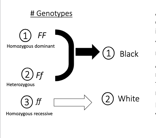
Dominant alleles (Relationships between alleles)
Produces the (dominant) phenotype in individuals who have at least one copy of this allele (FF or F_). The dominant allele can come from either parent.
Typically a dominant allele codes for a functional protein
Recessive alleles (Relationships between alleles)
Produces a (recessive) phenotype only if an individual has two copies of the recessive allele (one inherited from each parent).
Typically a recessive allele codes for proteins that are non-functional or have reduced function.
Notation - Autosomal alleles
Dominant allele → written as capital letter (e.g. F), recessive allele is written in lower case.
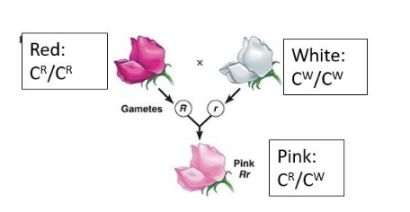
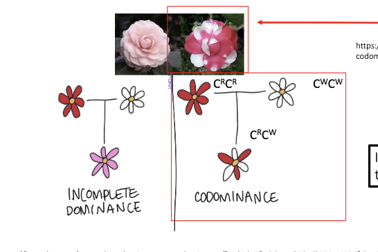
Incomplete dominance (Non-dominance - Alelles)
An allelic relationship where one allele is not dominant over the other allele. The phenotype of the heterozygotes tends to be intermediate between the phenotype of the homozygotes.
Don’t use upper and lowercase lettering when alleles have a non-dominant relationship - use numbering instead.
True-breeding parents
Homozygous for the alleles of interest
Co-dominance (Non-dominance - Alelles)
Allelic relationship in which the heterozygote exhibits the phenotype of both alleles at the same time.
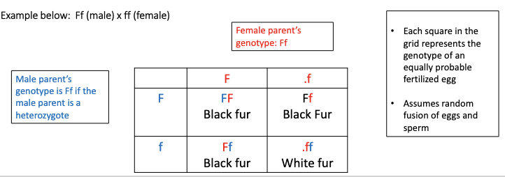
Punnett Square
Square grid for predicting possible genotypes/phenotype of offspring from a particular cross.
All possible gamete genotypes for one parent are placed on the top axis.
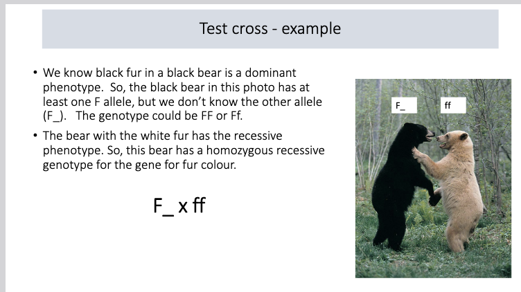
Test cross
Crossing an individual with the dominant phenotype but potentially an unknown genotype (eg. B_, BB or Bb) with an individual(s) that is homozygous recessive (bb, tester) for the genes under consideration.
The phenotypes of the offspring are examined. If any offspring have the recessive phenotype, the parent with the dominant phenotype must be a heterozygote.
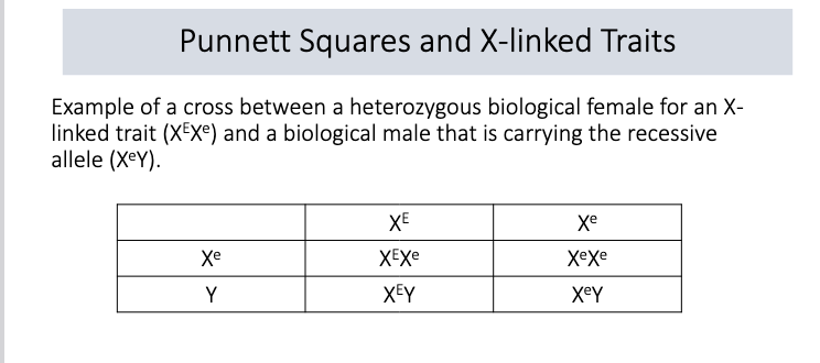
X-linked genes
Biological females have the genotype XX. So a female carries two alleles for an X-linked gene. (XE XE, XE Xe, Xe Xe)
Biological males have the genotype XY. So a male carries one allele for an X-linked gene (XE Y, Xe Y)
This means that males can exhibit the recessive phenotype if they carry just one recessive allele.
Calculating probability of producing an offspring with two or more phenotypes
“And” - white fur and female - MULTIPLY
“Or” - black fur or white fur, ADD
Autosomal
Any chromosome that is not a sex chromosome.
e.g. Humans = 46 chromosomes (23 pairs)
22 pairs are autosomes (numbered 1 through 22).
1 pair is the sex chromosomes (XX in females, XY in males).
Steps for Mode of Inheritance Qs
Carefully read the scenario and scan the data
Define your genes/alleles based on this hypothesis
Use a Punnett Square to make prediction about the expected genotype/phenotype frequencies for the F1 and F2 generation based on your hypothesis
Compare the expected frequencies with the observed frequencies.
Autosomal dominant
Need only ONE copy of the allele to show the trait
Example: Huntington's disease
H = Huntington's (dominant)
h = normal (recessive)
Genotypes:
HH = affected
Hh = affected (heterozygous but still shows trait)
hh = normal
Classic cross: Hh × Hh → 3 affected : 1 normal ratio
Autosomal recessive
Need TWO copies of the allele to show the trait
Example: Cystic fibrosis
C = normal (dominant)
c = cystic fibrosis (recessive)
Genotypes:
CC = normal
Cc = normal (carrier)
cc = affected
Classic cross: Cc × Cc → 3 normal : 1 affected ratio
X-linked trait inheritance: Female
e.g. A female (XX) inherits an X-chromosome from both parents.
If the male parent (XY) carries the dominant allele on his one X-chromosome, the female offspring must also have the dominant phenotype because she will inherit this dominant allele.
A female can only have the recessive phenotype if she inherits the recessive allele from both parents. (X^c X^c)
X-linked trait inheritance: Male
The male parent gives their Y chromosome to male offspring and their X chromosome to their female offspring (so a male cannot pass an X-linked gene to male offspring).
X-linked dominant
X-linked recessive
Somatic Cells
All the body’s cells except the reproductive cells (sperm and egg).
Number of possible gamete genotypes that could be produced (if genes aren't linked or they're linked w/cross-over) + state what n is
2n, where n = number of heterozygous genes