2: Development of limbs
 Call Kai
 Learn
Learn Practice Test
 Spaced Repetition
 Match
Match Flashcards
 Knowt Play
1/47

There's no tags or description
Looks like no tags are added yet.
Name | Mastery | Learn | Test | Matching | Spaced | Call with Kai | Chat |
|---|
No analytics yet
Send a link to your students to track their progress
48 Terms
syndactyly
skin webbing between fingers/toes. most frequently: 3rd and fourth fingers; 2 and 3rd toes . more common in foot
synostosis
fusion of phalanges
embryological basis of syndactyly
separate digital rays fail to form in 5th week or webbing of fingers fails to break down between 6th and 8th weeks. lack of differentiation between 2 or more digits. normally mesenchyme in periphery of hand and foot plate condenses to form the primordial of fingers and toes and thinner tissue between breaks down.
characteristic syndrome produced by thalidomide
amelia and meromelia. hands and feet attached to trunk by small, irregularly shaped bones, intestinal atresia and cardiac defects
Meromelia
gross defect of limb
amelia
complete absence of a limb
atresia
absence of a normal body opening; occlusion; closure
Polydactyly
supernumery of fingers and toes. often completely formed and lacks proper muscle fixation. . ulnar or radial side and foot on fibular side
cutaneous syndactyly
webbing of skin between fingers and toes due to failure of tissue to break down
osseous syndactyly
fusion of bones
structural defect underlying congenital dislocation of hip
socket doesnt fully cover ball
CHD is associated with breech presentation (i.e. buttocks rather than head delivered first). Speculate on why this might be so?
Breech presentation may place undue pressure on the developing hip joint: fails to complete normal development
mesenchyme
primitive undifferentiated connective tissue. derived from mesoderm. proliferation allows lengthening of limbs
talipes equinovarus
most common type of clubfoot. soles of feet turned medially and feet are sharply planter flexed. feet are fixed in tiptoe position
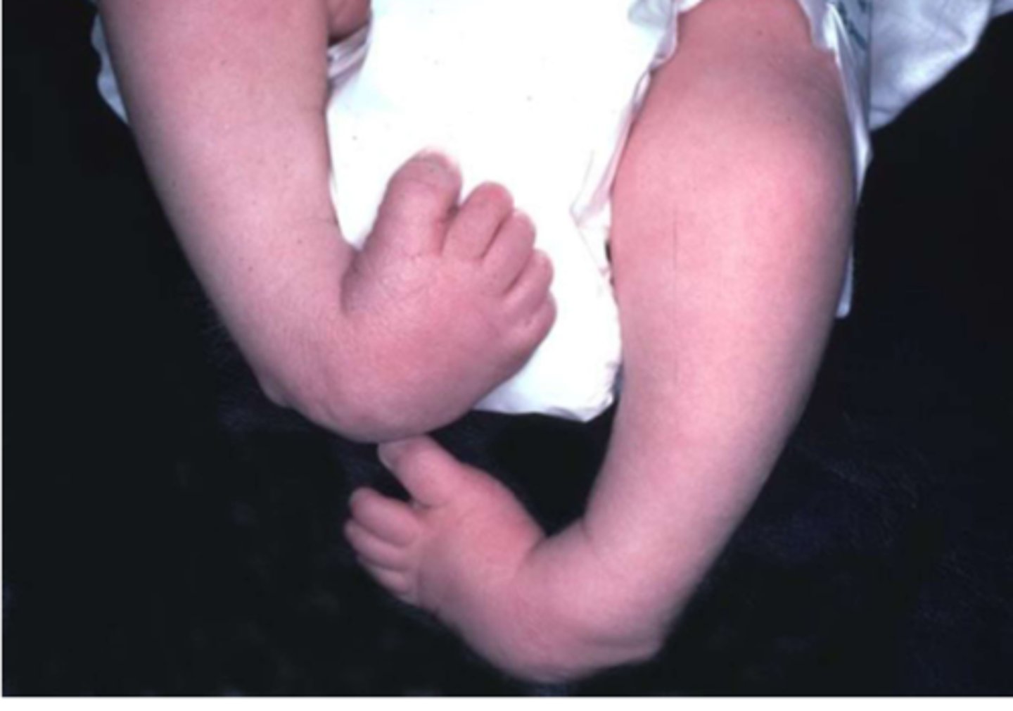
treatment for clubfoot
ponseti method. baby's foot being slowly manipulated in a better position and then being put in a cast. repeated weekly for 8 weeks. achilles tendon cut to release foot. special boots worn to keep in position until 4/5
The AER marks the boundary between the dorsal and ventral ectoderm. This forms an axial line. What does this mean in terms of sensory nerve supply to the limb?
s considerable overlap between adjacent dermatomes in terms of nerve supply so damage to one dorsal spinal root does not lead to anaesthesia to the entire dermatome as that area is innervated by other adjacent nerves. The line of junction between 2 dermatomes supplied from discontinuous spinal levels is known as the axial line. There is no overlap in nerve supply at the axial line as this marks the embryonic boundary between flexor and extensor compartments (C5 not near T1)
AER
ridge of thickened ectoderm at apex of developing limb bud. stimulates outgrowth of limb buds. maintains undifferentiated state in mesenchyme immediately underlying it. enables proliferation and lengthening of limb bud. limb growth is proximal to distal. induces development of digits and disappears when no longer needed
notochord clinical relevance
nucleus proposus in middle of IV. herniates and presses on spinal nerve (slipped disc) and commonly at l4-5 with l5 root nerve most affected causing radiating pain down the leg to big toe and weakness of extensor pollicis longus
c8 dermatome test
touch distal ventral aspect of little finger. ulnar nerve.
ulnar nerve roots
c8-t1
ulnar nerve type
mixed
t10 dermatome testing
touch umbillicus
Axillary nerve (C5-C6) test
regimental badge area
t4 surface anatomy marker
nipples
t6 surface anatomy marker
xiphistermun
t12 anatomy marker
pubis
Cervical nerves C5, C6 and C7 emerge above the corresponding numbered vertebra, while cervical nerve C8 emerges below vertebra C7. Explain why this is so
extra spinal nerve between 1st cervical vertebrae and skull so 7 cervical vertebrae and 8 cervical nerves. from t1 down, spinal nerves exit below corresponding vertebra
mytome
muscle/group of muscles recieve nerve supply from a single cord segment
dermatome map above shoulder
c2: above neck
c3: neck
c4: clavicle
c5: below clavicle
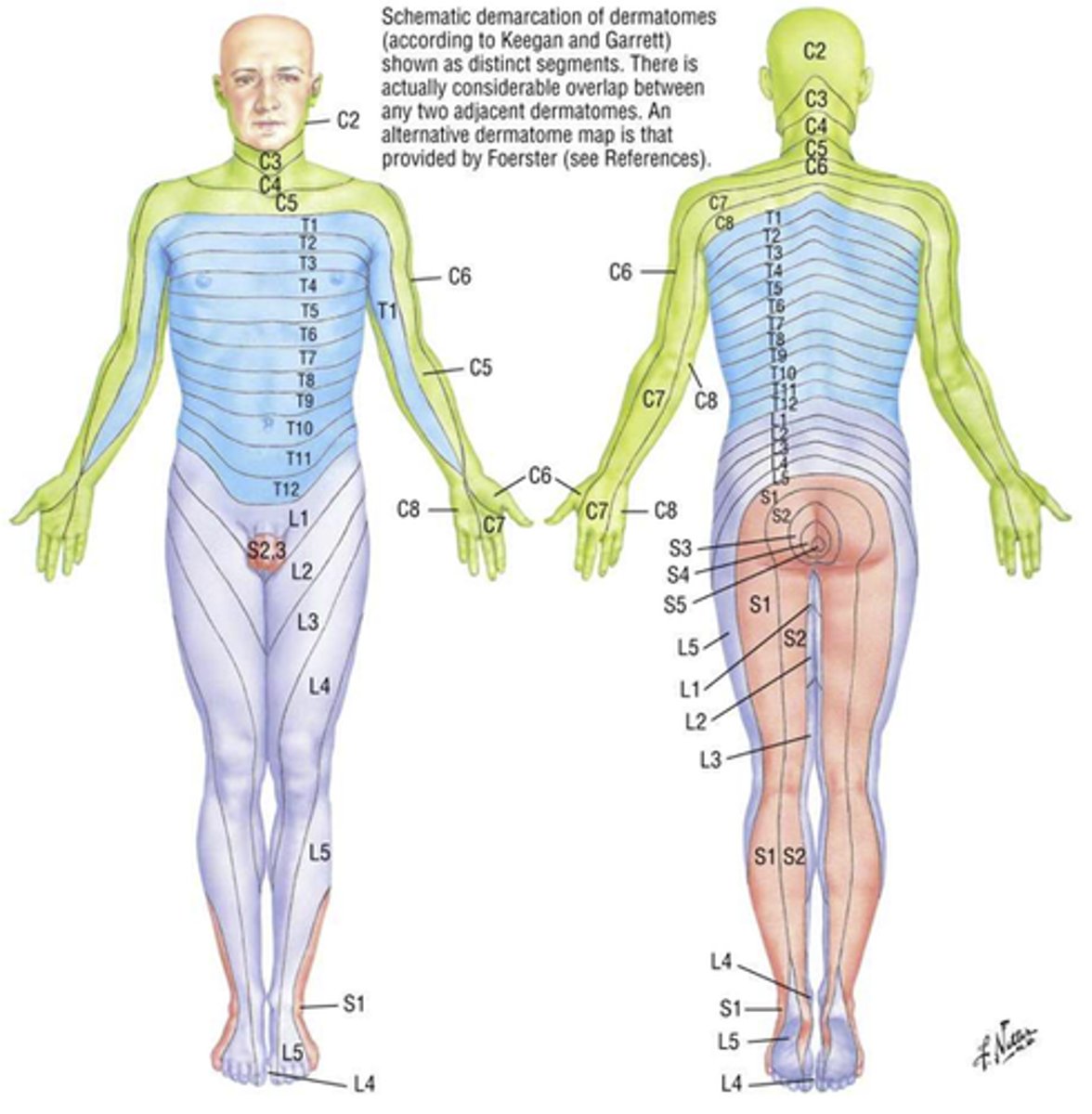
dermatome map trunk
t1: middle arm
-t12 pubis
dermatome map sub trunk
l1: below pubis but not genitals
brachial plexus
C5-T1
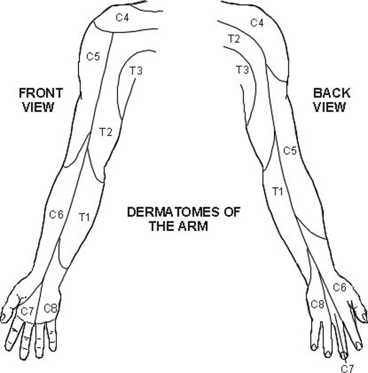
dermatome vs cutaneous distribution of peripheral nerve. clinical significance
dermatome strip of skin supplied by single spinal nerve whereas cutaneous distribution of peripheral nerve area of skin supplied by peripheral nerve. mostly arise from plexus which receives imput from many spinal root nerves. locate where damage has occured
pattern and outgrowth of limb bud starts to appear when
end of fourth week
epimysium
connective sheath covering entire muscle bundle
deep muscles of back supplied by which branch of spinal nerve
dorsal ramus
spinal cord
receives afferent fibres carry sensory nerve fibres through dorsal roots and sends efferent fibres which carry motor and autonomic nerve fibres through ventral roots
ectoderm tissues
mouth, nostril and nervous tissue, teeth, sweat glands. neurofibromatosis, sturge webber syndrome, tuberosis sclerosis
ventral side of thumb. dermatome and cutaneous nerve area
c6 and median nerve
describe epemere and hypomere
musculature into two regions. epemere goes dorsally to form erector spinae muscles and innervated by posterior ramus of spinal nerve roots. hypomere ventrally to form rest of musculature and innervated by anterior ramus of spinal nerve root
gross anatomy of spinal cord
has one enlargements from rostral to causal in cervical and lumbar regions. enlargements of spinal cord result from enlargement of grey matter that contains neural machinery to operate limbs
Hoxa genes
week 3 embryo development
3 germ layers. ecto, meso, endo
somites
rounded elevations of mesoderm which appear either side of neural tube on dorsal aspect of embryo from base of skull to tail region
ectoderm
forms epidermis and nervous system
fourth week of development
limb buds appear as small projections on lateral body wall
what core is limb bud
mesenchymal
how are digits formed
apoptosis of tissue between them