Mod 11 - Apoptosis
1/22
There's no tags or description
Looks like no tags are added yet.
Name | Mastery | Learn | Test | Matching | Spaced | Call with Kai |
|---|
No analytics yet
Send a link to your students to track their progress
23 Terms
Apoptosis =
= programmed cell death.
In this lecture, we will identify the proteins involved in apoptosis of mammalian cells and worm cells (nematode C.elegans).
…
T/F: Apoptosis is a normal part of life [cycle of a cell].
true!
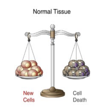
It's estimated that in the average adult human, more than a million cells are dying per second. Over the course of a year, this is the equivalent to the mass of an entire adult human.
It’s part of homeostasis. Cells are always being added to our bodies, by the process of cell division, but cells are also always being taken away by the process of programmed cell death.
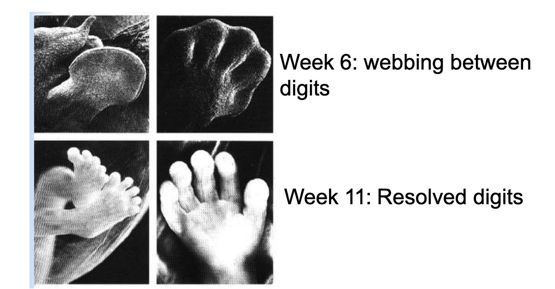
The different stages of development in the hands and feet of the human embryo:
notice the webbing disappears by week 11. this is due to apoptosis. w/o it, we would still have webbed hands.
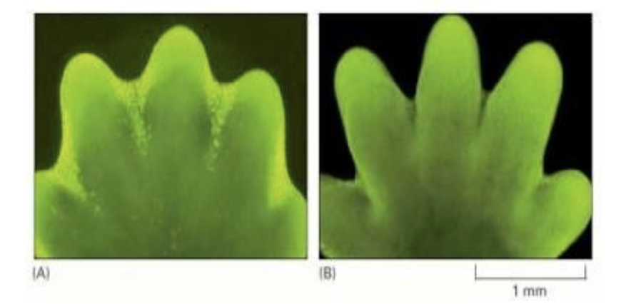
How can a TUNEL assay help us SEE apoptosis.
The image is of a developing mouse paw.
The mouse embryo has been stained w/ a dye that specifically labels cells undergoing apoptosis.
The apoptotic cells appear as bright green dots between the developing digits in the image on the left. On the right, we see that one day later, the inter‐digital cell death has eliminated the tissue between the developing digits, and few, if any, apoptotic cells can be seen.
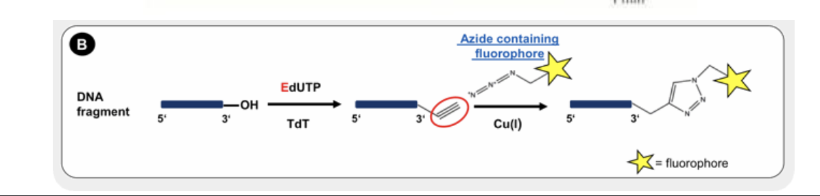
So how do we specifically label apoptotic cells?
The TUNEL assay takes advantage of the fact that DNA in apoptotic cells is nicked (contains DNA breaks). Because of this, fluorescent dUTP can be incorporated into the nicks via enzymes called terminal deoxynucleotidyl transferase. So the fluorescence highlights apoptotic cells.
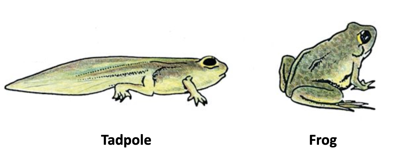
Both frogs and humans lose their tails from when they were tadpoles and embryos, respectively lol.
Thanks to …
… apoptosis!
For the induction of apoptosis in the tadpole tail, it’s specifically due to an increase in thyroid hormone.
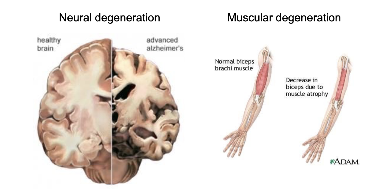
While cell death is part of the normal turnover of cells in our body, inappropriate cell death can also lead to disease. e.g…
In Alzheimer's disease, neurons in the hippocampus and certain regions of the cerebral cortex die. In Huntington's disease, neurons in the striatum die.
In Parkinson's disease, dopamine neurons in the substantia nigra undergo apoptosis.
Duchenne muscular dystrophy has inappropriate cell death.
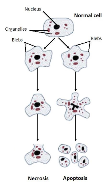
What are the two ways in which cells can DIE?
thru damage! like a cell that undergoes necrosis due to external damage. so the cell will swell —> release contents into surrounding tissue —> can lead to infection.
thru apoptosis! cells commit suicide in response to stress or damage. normal regulation so —> the debris is contained and recycled —> no infection.
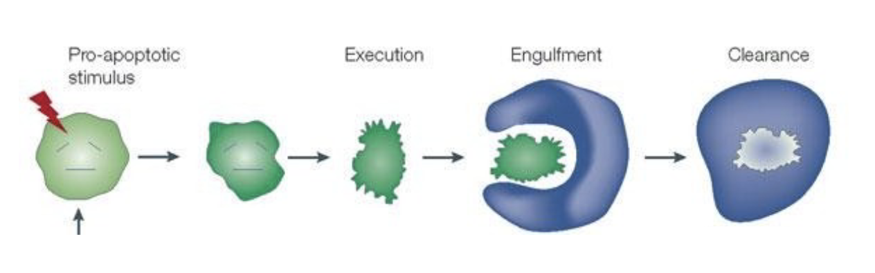
What are the biological analogies for 1. killing the cell. 2. ridding the body. 3. destroying evidence.
killing the cell = cell execution
ridding the body = engulfment
destroying evidence = clearance
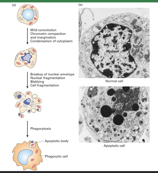
Outline the process of apoptosis:
The chromatin is compacting and becoming condensed. The nuclear envelope breaks down, the contents of the nucleus are fragmented, the DNA is broken down, and the proteins are degraded. The cytoplasm also undergoes a process of condensation as cellular components aggregate. The mitochondria become permeabilized and mitochondrial proteins are released into the cytosol. The cell membrane begins to move and change shape, creating the blebs or protrusions. The cell fragments, creating compartments of the cell that contain the debris of the dead cell. Each of these little cell fragments will be phagocytized and the components will be recycled and reused.
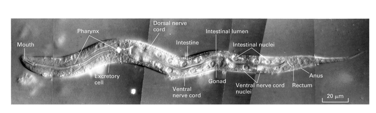
Now we will talk about the worm: nematode C. elegans…
How many somatic cells EXACTLY does it have? How many cells underwent cell death?
947 somatic cells.
The lineage of every cell has been traced back to the single fertilized zygotic cell. This single cell undergoes rounds of mitotic divisions leading to the 947 distinct somatic cells, and the function and structure of each of these cells has been identified.
131 cells underwent apoptosis.
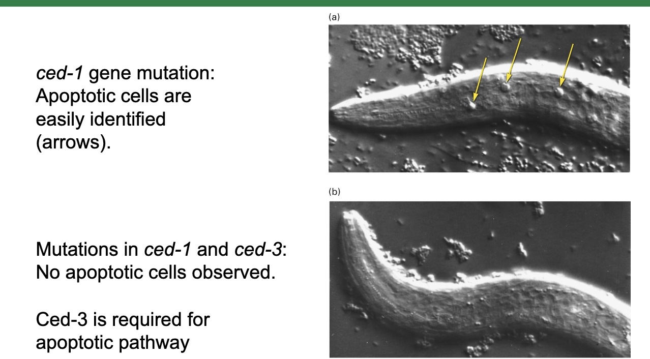
Mutations in genes are required to PREVENT apoptosis. What are those genes called?
cell death genes (ceds) hahaahhaha.
What genes in the worm will we discuss?
ced-1: a loss of function mutation in this gene still allows cells to undergo apoptosis. so it’s not necessary for apoptosis to occur.
ced-3, ced-4, ced-9, egl-1: necessary for inducing apoptosis. if there’s mutations in these, apoptosis will not occur.
What 4 worm genes are necessary for inducing apoptosis?
ced-3, -4, -9, egl-1
Loss-of-function mutations in ced-3 or ced-4 do what?
Loss-of-function mutation in ced-9 does what?
egl-1 does what?
Loss‐of‐function mutations in ced‐3 or ced‐4 prevent apoptosis.
In contrast, in a loss‐of‐function mutation in ced‐9, all of the cells die. This is because CED‐9 is an inhibitor of apoptosis that acts by inhibiting the activation of this caspase holoenzyme.
EGL‐1 is the signal for apoptosis that activates the pathway by inhibiting the inhibitor (CED‐9)
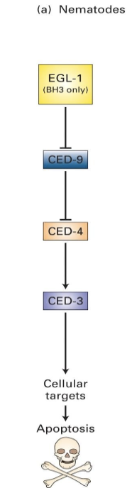
What is the caspase holoenzyme?
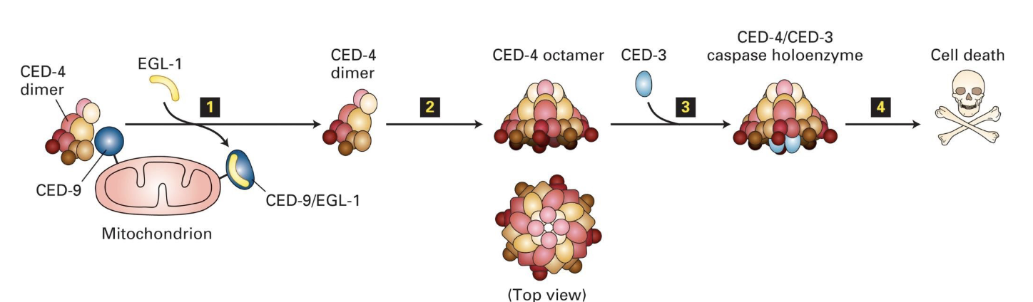
caspase holoenzyme = a protease that will target many diff. proteins for degradation! it’s formed as a complex of ced-4 + ced-3 coming tg.
What are the mammilian homologues of those genes and the caspase holoenzyme?
egl-1 == Bid & Bim
ced-9 == Bcl-2
caspase holoenzyme composed of ced-3 & ced-4 == apoptosome composed of caspase-9 and Apaf-1
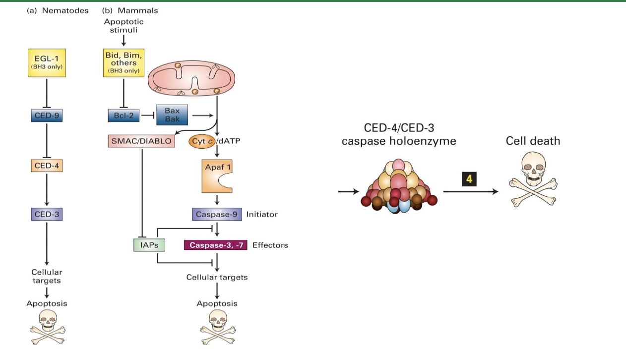
How does the caspase holoenzyme activate?
CED-9 normally binds to CED-4 dimers, keeping them inactive.
When EGL-1 binds to CED-9, it releases CED-4, which then joins with CED-3 to form the caspase holoenzyme.
This activated caspase degrades cytosolic and nuclear proteins, leading to apoptosis.
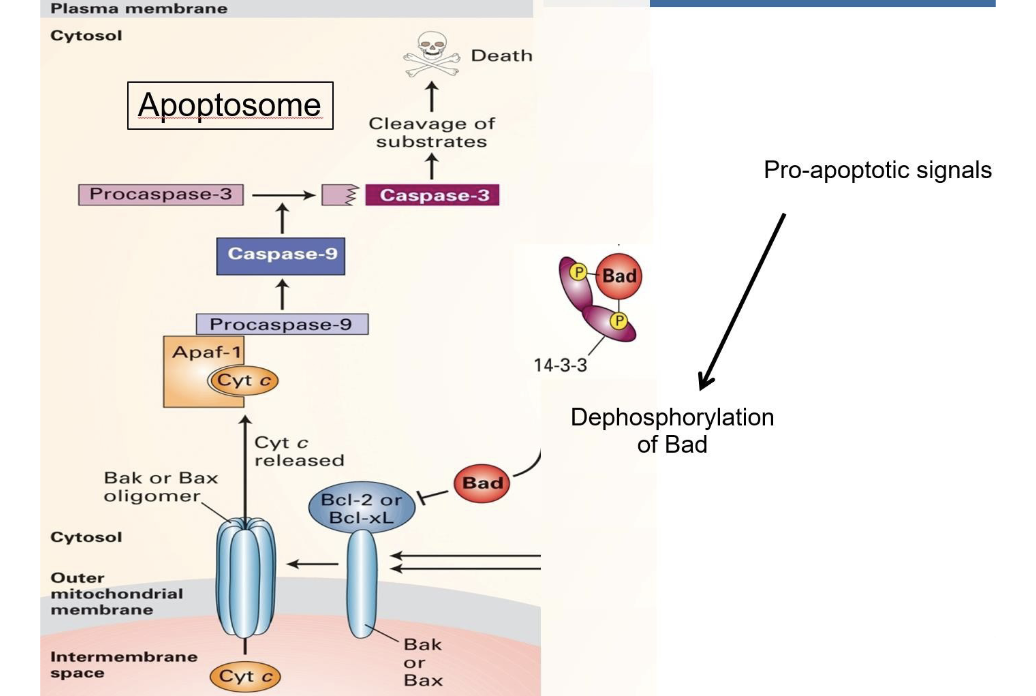
How does Bcl-2 work (mammalian homolog of ced-9)?
Bcl-2 is anchored to outer membr. of mitochondria.
Normally Bcl-2 functions to maintain low permeability of membr.
But inactivation of Bcl-2 leads to pores forming in the membr., which is exactly what causes apoptosis.
Go into detail about how exactly Bcl-2 ends up making pores form:
In mammalian cells, Bad (the apoptotic signal) is inactive when phosphorylated and bound to 14-3-3.
When dephosphorylated and unbound from 14-3-3, Bad binds to Bcl-2, which then activates Bax.
Bax reorganizes in the outer mitochondrial membrane and clusters to form pores.
These pores allow cytochrome C and other proteins (which were originally in the intermembr. space) to escape into the cytosol, triggering apoptosis.
Cytochrome C is essential to forming the apoptosome.
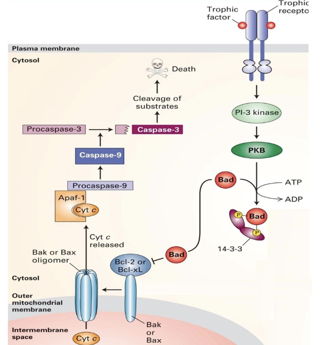
While apoptotic signals promote Bad’s dephosphorylation and apoptosis, what prevents apoptosis and keeps the cell alive?
trophic factors prevent apoptosis and keep the cell alive.
How do trophic factors work?
Trophic factors initiate a kinase cascade that leads to phosphorylation of the Bad protein.
However, once those trophic factors are removed, the Bad protein can be …
…dephosphorylated and apoptosis can occur!