Lines and Tubes (Chest, Cardiovascular Devices, Biliary, and Shunt)
 Call Kai
 Learn
Learn Practice Test
 Spaced Repetition
 Match
Match Flashcards
 Knowt Play
1/30

There's no tags or description
Looks like no tags are added yet.
Name | Mastery | Learn | Test | Matching | Spaced | Call with Kai | Chat |
|---|
No analytics yet
Send a link to your students to track their progress
31 Terms
Chest Tubes
Used when lung has collapsed away from chest wall - air/fluid is in the pleural space
Emergency procedure - large diameter tube inserted into the pleural space to allow air or fluid to escape, allows lungs to re-inflate
Chest Tube Placement
Upper thorax if for collapsed lung,
Lower thorax if for drainage of fluid,
Keep external end of chest tube below the level of the insertion site
Chest Tube Precautions
KEEP drainage collector lower than chest level,
DO NOT TIP the drainage protector,
AVOID tension on the tubing,
COVER if the seal is broken immediately cover the punction site with an airtight seal (Vaseline covered gauze works well) and call for help
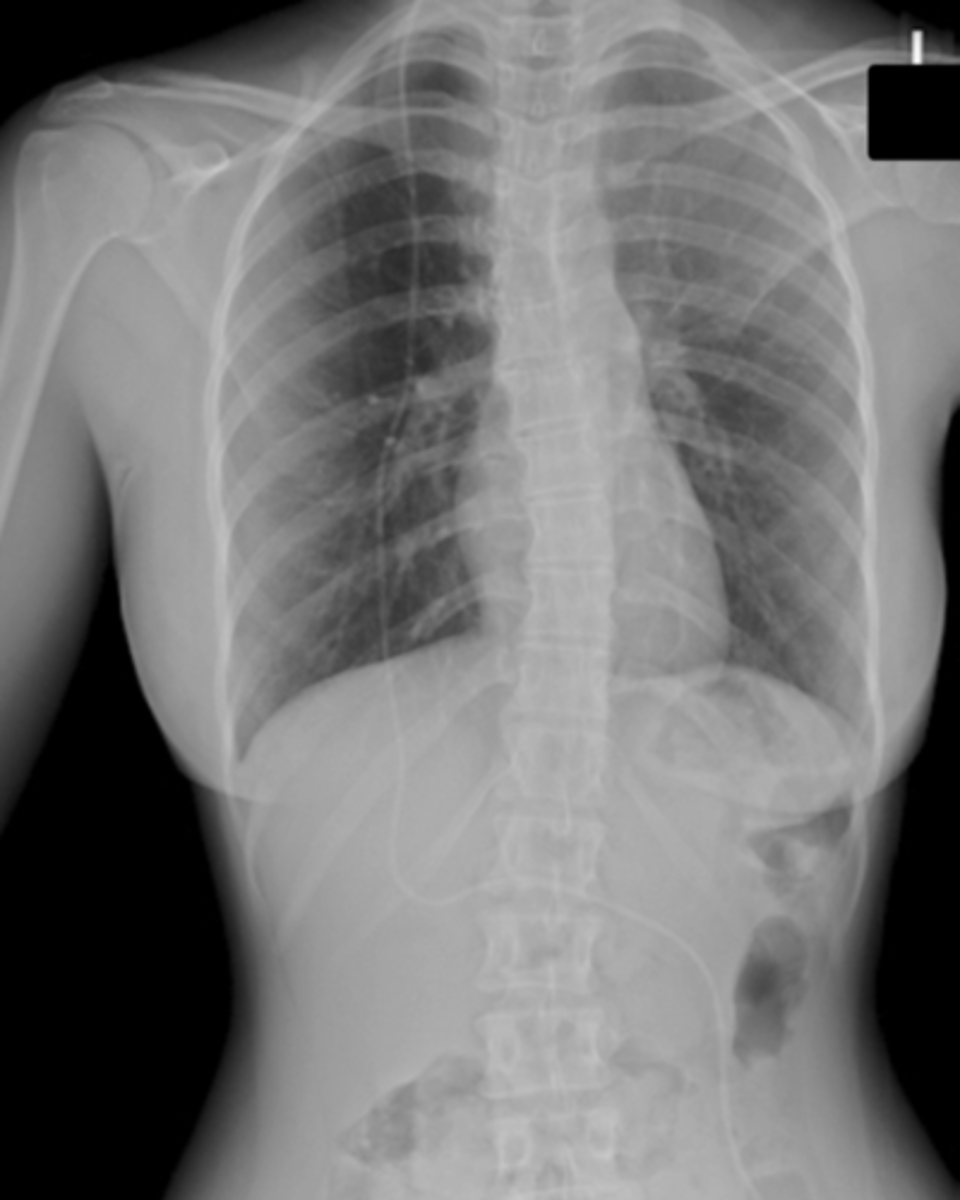
Chest Tube X-Ray (image)
PICC Line
Type of central venous line, placed near heart (SVC)
PICC Line Function
Administration of medication long-term IV therapy, chemotherapy
PICC Line Pathway
starts (normally in arm) and ends at SVC just superior to the right atrium
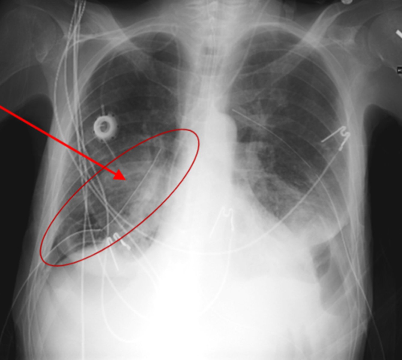
PICC Line X-Ray (image)
Port-A-Cath
Long term IC therapy (chemotherapy),
Preserves vessel puncture site,
surgically placed,
special diaphragm disk under skin,
special right angle needle is used to puncture the diaphragm,
may be used in CT for contrast injections
Port-A-Cath placement
ends just superior to right atrium
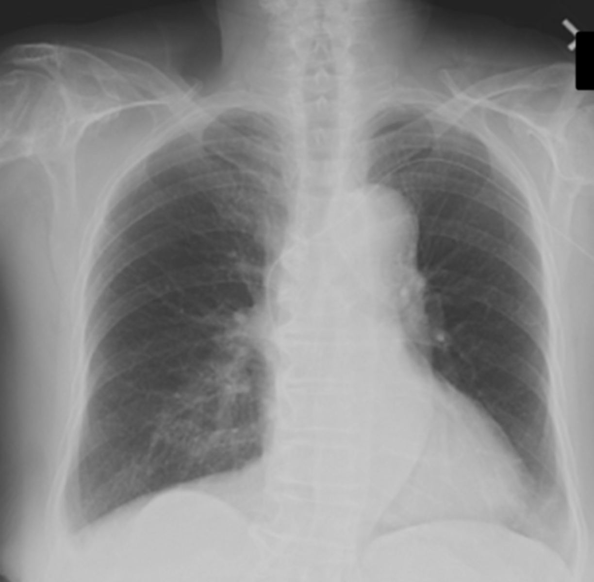
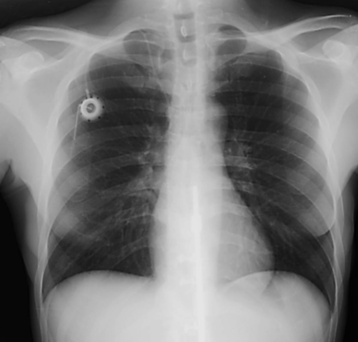
Port-A-Cath X-Ray Image
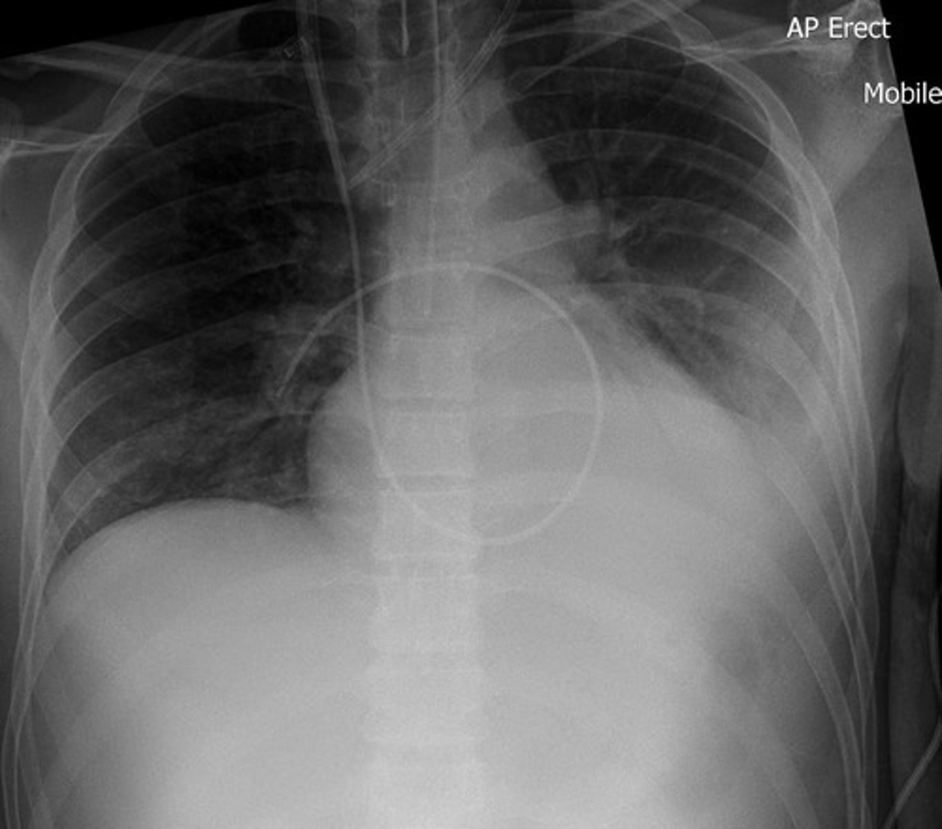
Swan Ganz Line
Multiple lumen line, Blood gas samples, pressure readings, administering meds
Swan Ganz Line Pathway
Subclavian vein - SVC - RA - RV - Pulmonary Artery
Swan Ganz Line X-Ray (image)
Pulled Central Venous Line or PICC
Direct pressure on puncture site,
call for help,
patient in steep left latera Trendelenburg position (keeps air on the right side of the heart)
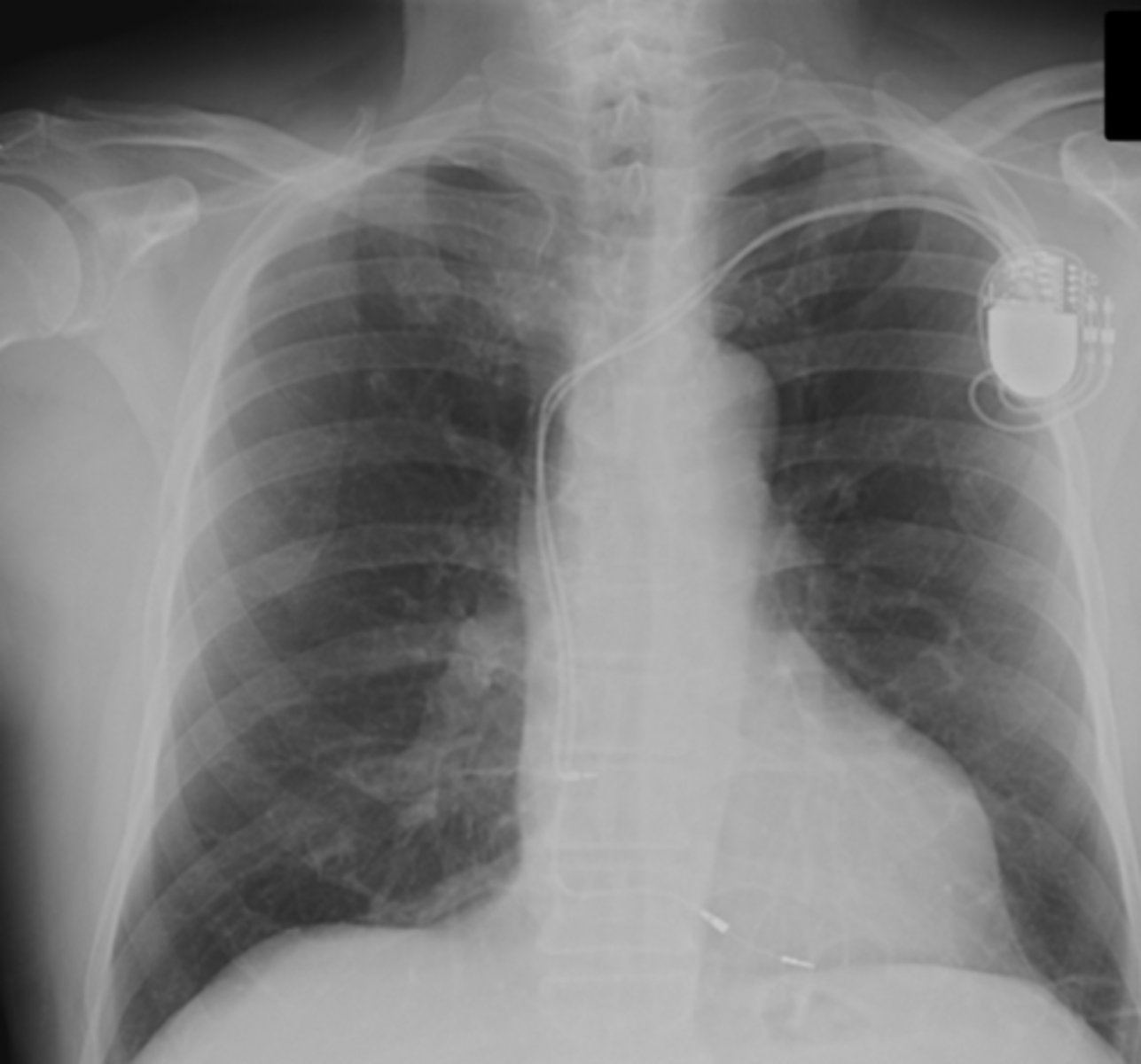
Pacemakers
temporary vs. permanent,
often two electrodes (RA and RV)
Pacemaker Pathway
Subclavian V - SVC - RA - RV, electrode wedged against RV wall
Pacemaker X-Ray (image)
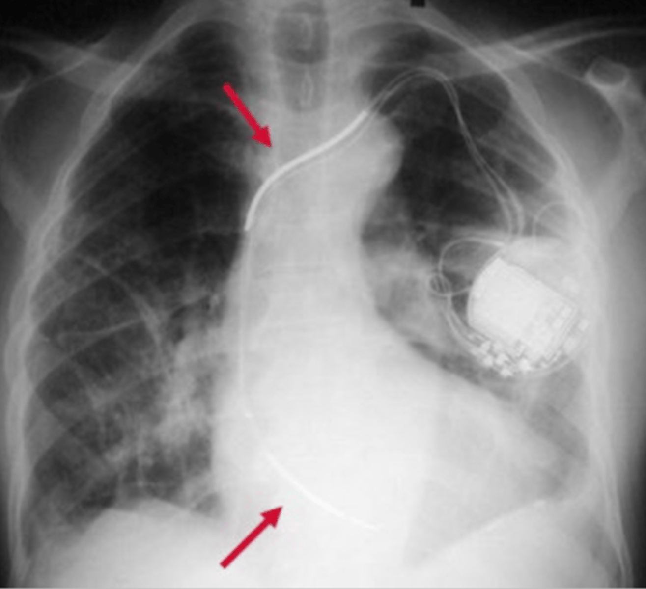
Automatic Implantable Cardioverter Defibrillator (AICD)
Corrects arrhythmias and helps the heart beat more regularly
AICD positioning
One electrode in the SVC or brachiocephalic vein and the second electrode (if used) placed in the apex of the right ventricle
AICD X-Ray (image)
Intra-Aortic Balloon Pump (IABP)
Temporary treatment,
helps heart pump more blood with less energy,
Inflates during diastole which helps blood enter the coronary arteries,
Deflates during systole so that blood can reach the body
IABP X-Ray (image)
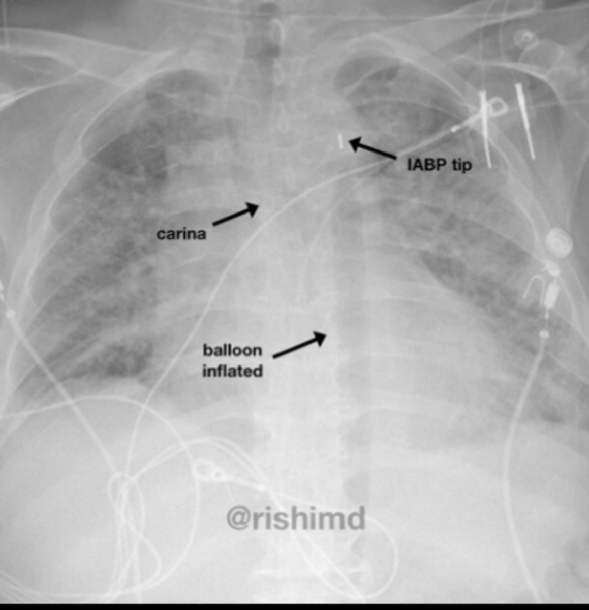
IABP marker
balloon can be seen and as a shadow and has a SMALL METALIC MARKER that is meant to be distal to the left subclavian artery
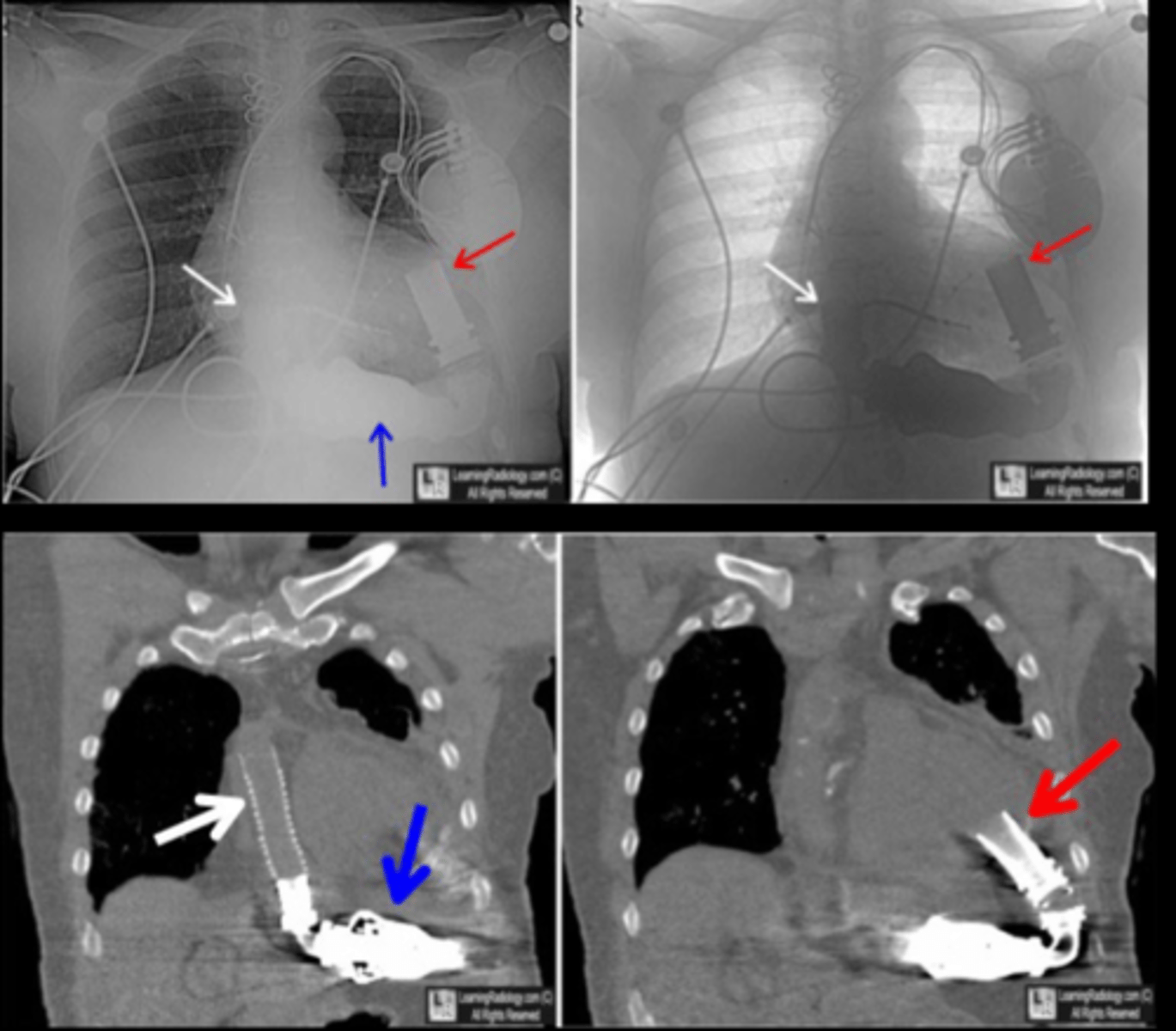
Left Ventricular Assist Device (LVAD)
Pump used when patient is in end-stage heart failure,
Helps the heart pump blood to the rest of the body,
allows patient to return home,
can be used as a bridge to heart transplant or as a permanent alternative to heart transplant
LVAD Images
Cannula for ascending aorta (white arrows); pump (blue arrows); LV cannula (red arrows)
Electrocardiogram Leads (ECG/EKG)
Can interfere with visualization of central lines and other implanted devices on image,
If able, attempt to redirect, depending on the reason for exam
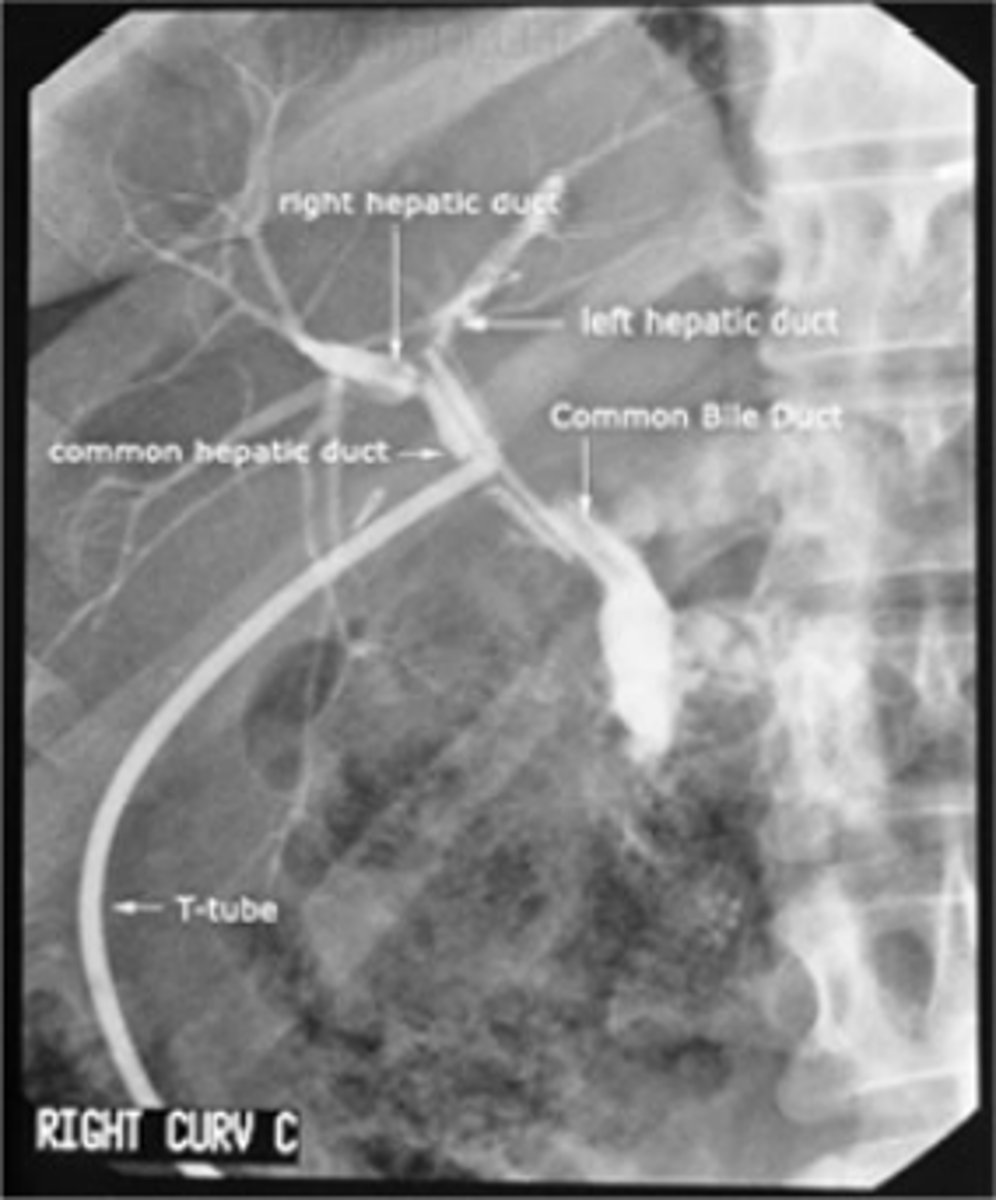
T-Tube
Inserted during/post op cholecystectomy,
documents patency of common bile duct, and presence of residual gallstones
T-Tube Pathway
common hepatic duct - common bile duct - outside of body
T-Tube (image)
Ventriculoperitoneal (CSF) Shunt X-ray (image)