Looks like no one added any tags here yet for you.
Review and write SOAP notes
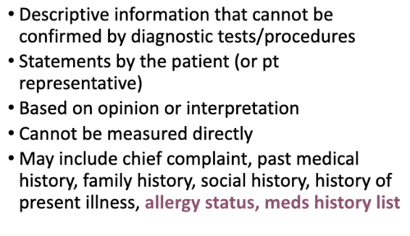
S: subjective
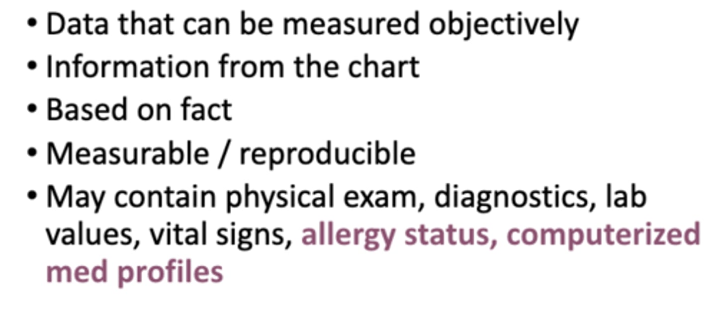
O: objective
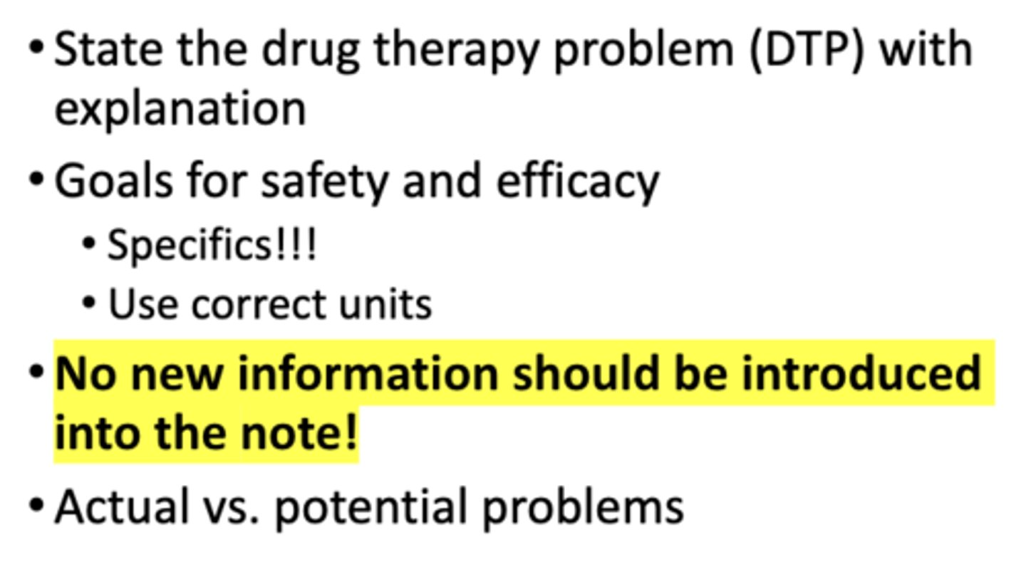
A: assessment
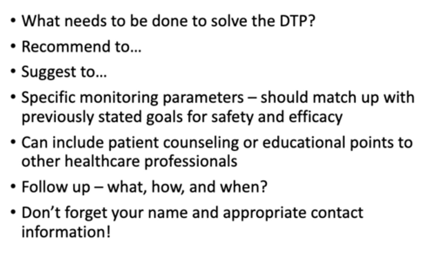
P: plan
What is subjective info?
What is objective info?
What is assessment info?
What is plan info?
What are the basic components of a SOAP note?
Describe and write SBAR notes
S: situation
B: background
A: assessment
R: recommendation
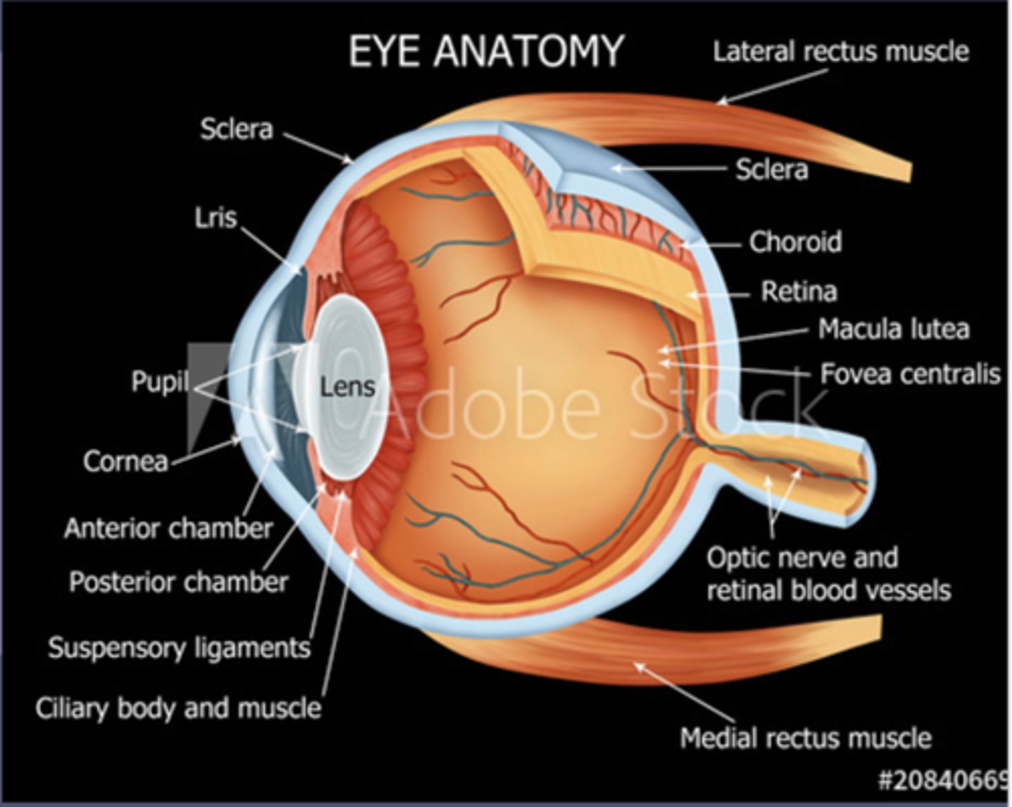
Identify parts of the eye
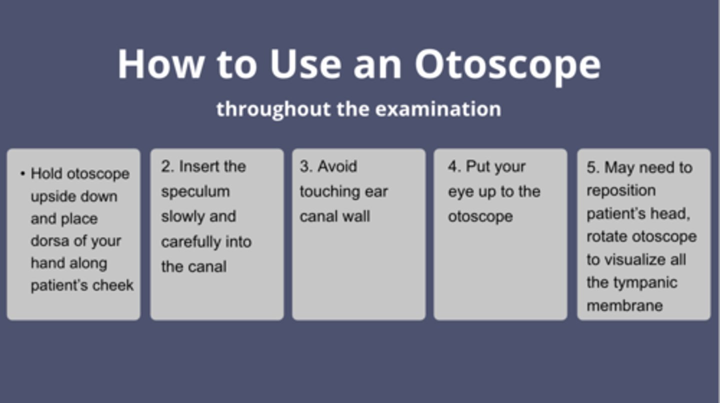
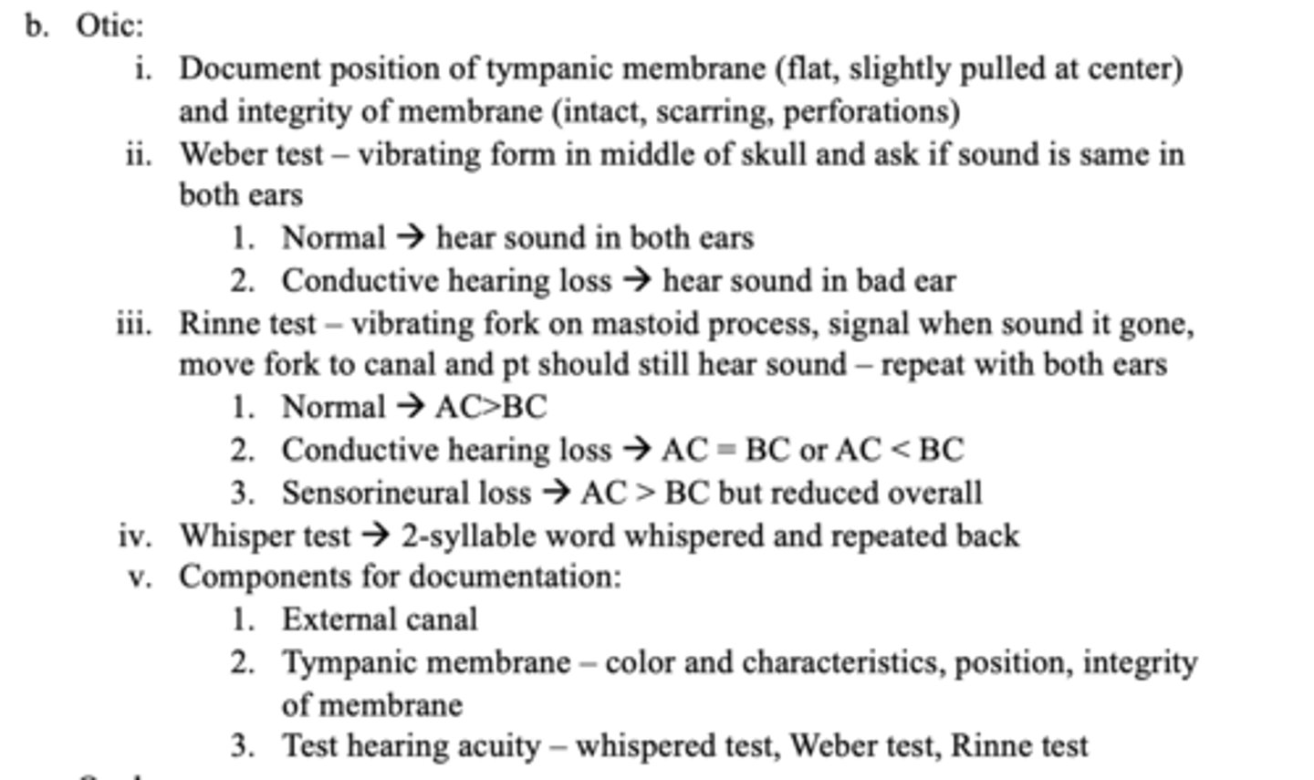
List the steps you would use in order to inspect the ear using an otoscope
How to use: largest speculum, tilt pt head away from you, pull up and back with adults and kids older than 3
- Hold upside down and side of your hand along pt cheek
- Insert slowly and carefully into canal
- Avoid touching ear canal wall
- Put your eye up to otoscope
- May need to move pt head or otoscope to see all of tympanic membrane
What are we looking for? Inspecting external canal AND Inspect tympanic membrane
Perform an eye examination using a pen light or ophthalmoscope light
a. Lens and pupil exam -> looking for foreign bodies, color differences, and symmetry
b. Pupil reflex -> slightly nasal but symmetrical on both sides
c. Pupillary response -> equally round and reactive to light, direct and consensual light reaction
Describe a normal eye
Define medical terminology in relation to eye and ear assessment
Design an OTIC treatment plan for a patient based on HPI triaging of a chief complain and patient interview using SCHOLAR-MACs
Design an OCULAR treatment plan for a patient based on HPI triaging of a chief complain and patient interview using SCHOLAR-MACs
inspect eyelids and eyebrows, sclera, and inspect conjunctival sac
True or False: the tympanic membrane separates the external and the middle ear
True
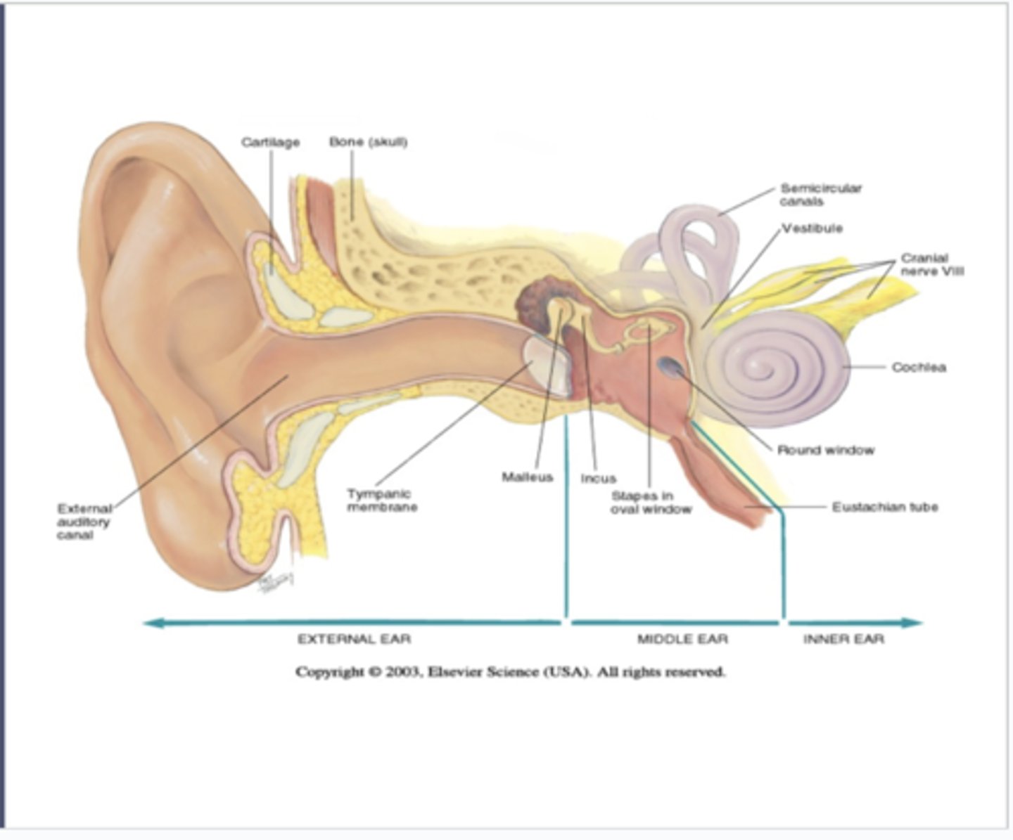
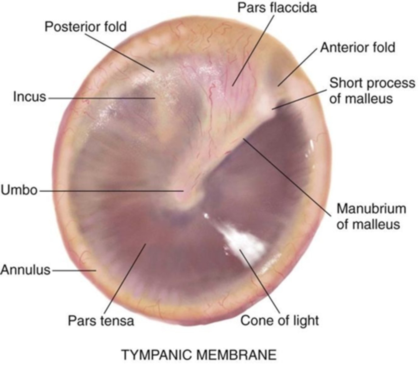
What color is the tympanic membrane?
translucent membrane with pearly gray color
What does the external canal look like?
clear
no redness, swelling, or lesion
What does the tympanic membrane look like?
Color and Characteristics: pearly gray, landmarks intact, cone of light is visible
Position: flat and slightly fulled in at the center
What are we looking for in a lens and pupil exam?
foreign bodies, color differences, and symmetry
What are we looking for in pupil reflex?
slightly nasal, but symmetrical on both sides
What are we looking for in pupillary response?
equally round and reactive to light (direct and consensual)
True or False: the external canal is 2.5-3 cm, stops at tympanic membrane, and has cerumen glands that lubricate and protect the ear
True
True or False: the external canal separates external and middle ear, translucent membrane with pearly gray color
False
This is the tympanic membrane
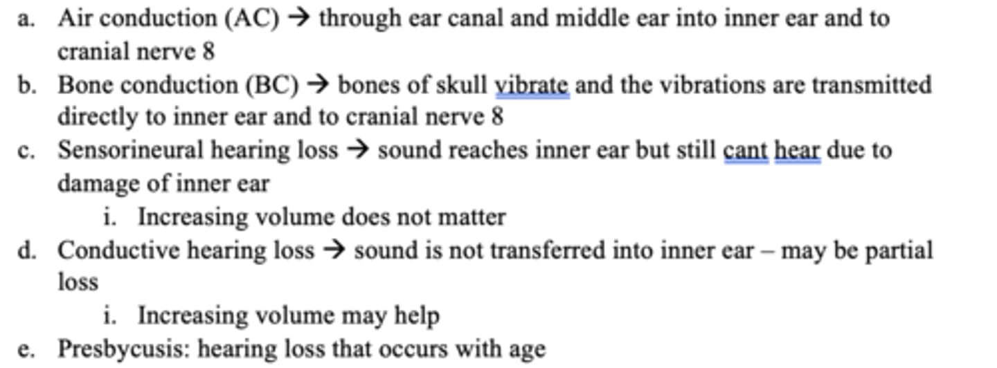
What is air conduction (AC)?
Air conduction is the primary pathways vibrations are sensed here (through the ear canal and middle ear into the inner eat to cranial nerve 8)
What is bone conduction (BC)?
bones of the skull vibrate (those vibrations are transmitted directly to the inner ear and to the cranial nerve 8)
What is sensorineural hearing loss?
sound reaches the inner ear but still cannot hear because of damage (of the inner ear)
True or False: increasing sound volume can help in sensorineural hearing loss
False
Volume doesn't matter
What is conductive hearing loss?
sound is not transferred from the outer/middle ear to the inner ear and there may be partial loss
True or False: increasing volume in conductive hearing loss is beneficial
True
What is presbycusis?
hearing loss that occurs with age
What causes sensorineural hearing loss?
ototoxic meds, loud noises, infections, trauma, birth defect, age, hereditary
What causes conductive hearing loss?
ruptured eardrum, impacted cerumen, head trauma, birth defect, hereditary
What is the eyelid?
multilayer tissue that protects the anterior part of the eye,
keeps eyes lubricated via tears (directed to lacrimal ducts),
has muscles for movement
What is the sclera?
white, noninnervated layer that provides rigidity and protects internal structures
What is the cornea?
dome-shaped, innervated and response for the principal refractive element
Where are eyedrops absorbed?
cornea
What is aqueous humor?
maintains intraocular pressure
What does it mean if high intraocular pressure (IOP)?
risk factor for glaucoma
What is the iris?
visible colored portion that regulates light coming in
What is the pupil?
central opening of the iris
What is the retina?
processes light signals, optic disc sends info to the brain, and macula is responsible for visual activity
What is retinal detachment?
separation of the retinal layer from the pigment layer (large floaters, blackout curtain, and flickering lights)
Counseling points for eye drops:
1. Wash hands
2. Remove contact lenses, if applicable
3. Inspect expiration date
4. If the eye drops are a suspension, shake well.
5. Remove cap.
6. Tilt head back
7. Pull down on the lower lid to create a well or pouch
8. Apply 1 drop of solution. Do NOT touch the tip of the dropper to the eye
9. Gently close the eye
10. Gently apply pressure to the tear duct with your finger
11. Repeat steps 6-10 if more than 1 drop of solution needs to be used
12. Blot excess solution from around the eyes with a tissue
13. Wait 5 minutes to administer other drops. Administer drops 10 minutes prior to eye ointments/gels
Counseling points for ear drops
1. Wash your hands with soap and water
2. Gently clean your ear with a damp cloth
3. Shake bottle (if suspension) for 10 seconds
4. Tilt the affected ear or lie on your side
5. To open the ear canal:
*Adult: Pull ear down and back
*Child younger than 3: Pull up and back
6. Open bottle: make sure dropper is not chipped or cracked.
7. Place the correct number of drops in the ear. Gently press on the small skin flap over the ear to help drops stay in ear canal.
8. Keep ear tilted for a few minutes
9. Replace and tighten the cap
10. Wash hands