CSCS Chapter One Key Terms
 Call Kai
 Learn
Learn Practice Test
 Spaced Repetition
 Match
Match Flashcards
 Knowt Play
1/87

There's no tags or description
Looks like no tags are added yet.
Name | Mastery | Learn | Test | Matching | Spaced | Call with Kai | Chat |
|---|
No analytics yet
Send a link to your students to track their progress
88 Terms
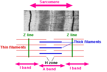
a-band
The region of a sarcomere that contains thick myofilaments (myosin) and appears darker under a microscope. It is involved in muscle contraction.
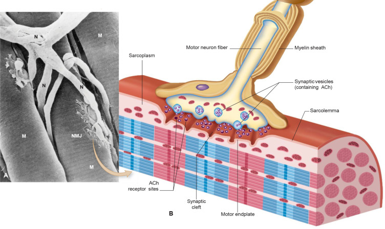
acetylcholine
A neurotransmitter that transmits signals across the neuromuscular junction, facilitating muscle contraction.
actin
A protein that forms thin myofilaments in muscle fibers and plays a crucial role in muscle contraction by interacting with myosin.

action potential
a rapid, temporary change in electrical voltage across a cell membrane that neurons use to send signals

all-or-none principle
a neuron or muscle fiber will fire a complete action potential at full strength if a stimulus reaches the required threshold, and will not fire at all if the stimulus stays below that threshold

alveolar pressure
Alveolar pressure is is the pressure of the air inside the alveoli (tiny air sacs) inside the lungs
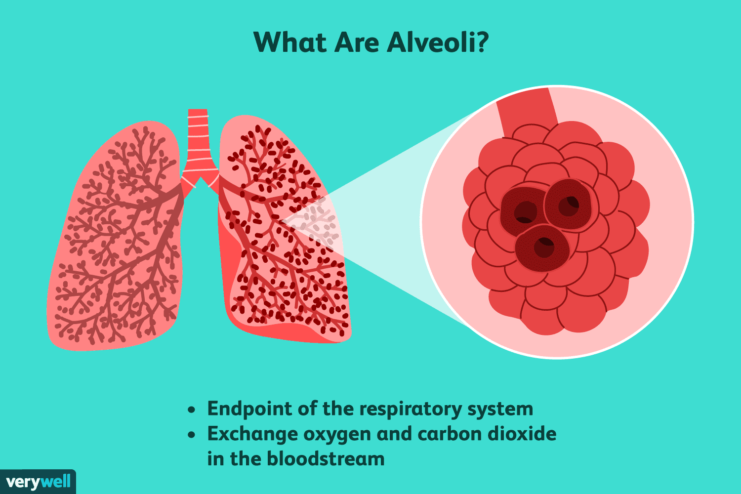
alveoli
tiny, balloon-like air sacs at the very end of your lung's breathing tubes where your body trades fresh oxygen for waste carbon dioxide
anatomical dead space
the volume of air in the conducting airways that does not take part in gas exchange, around 150ml in an adult

aortic valve
a one-way gate in the heart located between the left ventricle and the main artery, called the aorta

appendicular skeleton
consists of bones that form upper and lower limbs, the shoulders and pelvic girdles.

arterial system
a branching network of strong muscular tubes that carry oxygen rich blood from the heart to every organ and tissue in the body
arteriole
a very small blood vessel that branches out from an artery and leads directly into smaller capillaries
artery
a major blood vessel that carries blood away from the heart to tissues and or organs throughout the body
arteriovenous oxygen difference
the difference in oxygen content between arterial and venous blood. (a-vO2 difference) indicates cell oxygen extraction and metabolic demands
atrioventricular (av) bundle
Known as bundle of his, group of specialized heart muscle fibers. carry electrical signals from av node to lower chambers of the heart
atriventricular (av) node
a group of specialized cells in the heart that acts as electrical connector between upper (atria) and lower (ventricle chambers.
atrioventricular (av) valves
one way flaps of tissue that sit between the upper and lower chambers of the heart. Consists of mitral valve on left, tricuspid valve on the right
atrium
the two upper chambers of the heart that collect blood returning from the body and lungs
axial skeleton
formed of 80 bones that include the skull, vertebral column, and thorax.
biaxial joints
a freely movable synovial joint that allows movements around two distinct axes and two anatomical planes. example is knuckle joints
bone periostium
a dense fibrous membrane that covers the outer surface of almost all bones. Attachment point for tendons and ligaments
bradychardia
condition where resting heart rate drops below 60 bpm. Due to aging, medications, heart problems or high levels of cardiovascular health
bronchi
large air passageways that branch off from the windpipe (trachea) and lead into the lungs
bronchiole
tiny air passageways in the lungs that branch off from bronchi to deliver air to alveoli
capillary
smallest blood vessel, enables transfusion of oxygen, nutrients and waste to pass between blood and tissues
cardiac output
the total volume of blood your heart pumps through your body every minute. Cardiac output = stroke volume x heart rate (bpm)
cartilaginous joints
a connection between bones where the surfaces are entirely joined by cartilage, a tough yet flexible connective tissue
cross bridge
when the globular head of a thick filament (myosin) binds with active site on thin filament (actin)
cross bridge cycling
the repetitive molecular process where myosin head groups bind to, pull on, and detach from actin fillaments to generate muscle force and shortening
depolorization
rapid change in muscle or nerve cell membrane potetnial from a negative to a positive state. triggering an action potential that leads to muscle contraction
diastole
relaxation and filling phase of the cardiac cycle when the heart ventricles refill with blood between contractions
diastolic blood pressure
the lowest arterial pressure during the relaxation phase of the cardiac cycle between heart beats.
diffusion
passive movement of gas molecules (ie oxygen/co2) from areas of varying pressure. occurs passively.
distal
located further from the center of the body/trunk.
double product
also called rate pressure product (rpp), a direct measure of the workload and energy demand placed on heart. formula: rrp = HR x Systolic Blood Pressure
ejection fraction
the percentage of blood pumped out of the hearts main pumping chamber (left ventricle) with each heart beat. EF = (SV/EDV) x 100 edv=end-diastolic volume
electrocardiogram
a diagnostic procedure that records the hearts electrical activity via electrodes while an individual performs activity on a treadmill or cycle
end-diastolic volume
the maximum amount of blood in the ventricles right before the heart contracts. Marks the end of diastole (filling phase)
endomysium
the innermost layer of loose connective tissue surrounding and enclosing each individual muscle fiber
epimysium
dense layer of fibrous connectiove tissue that wraps around an entire skeletal muscle
extrafusal fibers
the standard skeletal msucle fibers that generate the force and tension needed for body movements and posture
fasciculi
bundles of individual skeletal muscle fibers. contained by the perimysium
fast twitch fiber
skeletal muscle cells optimized for rapid, high force, and explosive contraction using anaerobic metabolism
fibrous joints
fixed connections between bones that are united by dense collagen fibers, lack joint cavity (skull sutures)
fick equation
vo2 - Q x (a-v)O2 difference. Math rule that shows how much oxygen your body uses when you exercise by multiplying how much blood your heart pups by how much oxygen your muscles use.
frank starling mechanism
the hearts built in ability to pump harder when more blood fills it (like a rubber band)
fused tetanic contraction
smooth, continuous, maximal muscle contraction caused by a very high frequency of nerve signals
golgi tendon organ
a sensory receptor located where the muscle meets its tendon. it measures muscle tension and triggers relaxation to prevent injury
heart rate
the number of times your heart beats per minute. direct measure of cardiovascular work
hemoglobin
iron-rich protein in red blood cells, carrying oxygen from the lungs to working muscles during exercise
hyaline cartilage
specialized tissue covering ends of bones where they articulate, act as shock absorber and smooths joint movements
h-zone
central region of muscle fiber subunit (sarcomere). Contains only thick filaments (myosin)
i-band
light colored zone inside a msucle fiber that shortens when your muscle contracts
inferior
situated toward the feet relative to another part of the body
intrafusal fibers
specialized muscle fibers found inside a muscle spindle, detects muscle stretch and rate of stretch
left bundle branch
electrical pathway in the heart that carries signals to the lower-left chamber (left ventricle), causes ventricles to contract.
maximal heart rate
the maximum amount of bpm your heart can beat during extreme effort
maximal oxygen uptake
Vo2 max, the highest amount of oxygen your body can absorb, transport and use during extreme exercise
mean arterial pressure
(the average blood pressure in a persons arteries during a single heartbeat. MAP = diastolic pressure + 1/3 (pulse pressure).
metabolic equivalent (MET)
1 MET: the amount of energy (and oxygen) your body uses at rest. Measurement of work during physical activity. 3 = light, 3-6 moderate, 6> vigorous
minute ventilation
the total volume of air you breath in and out of your lungs in one minute. Tidal volume x Respiratory Rate
mitral valve
one way valve between left atrium and ventrical. preents blood flow back into atrium
motor neuron
specialized nerve cell (and the muscle fibers it innervates) which carries electrical impulses from the brain and nervous system to skeletal muscles.
motor unit
a single nerve cell (motor neuron) and all the muscle fibers it controls
multiaxial joints
a freely movable synovial joint that allows movement in three different directions or planes of motion
muscle fiber
a single cylindrical muscle cell that contracts to create movement.
muscle spindle
a tiny sensory receptor inside a muscle belly that detects muscle length and speed of stretch. composed of intrafusal fibers
myocardium
the msucular middle layer of the heart that contracts and pumps blood
myofiber
a single complete muscle cell
myofibril
cylindircal fiber running the length of a muscle cell, made up of sarcomeres
Myofilament
microscopic thread-like protein strands of actin or myosin. building blocks of sarcomeres
myosin
microscopic protein, thick filament, tiny motor myosin heads latch to actin binding sites
Myosin adenosine triphosphate (ATP)
the chemical energy source and reaction that powers muscle contraction. the energy currency of muscle cells.
neuromuscular junctions
the chemical connection point where a motor neuron meets skeletal muscle fiber to signal movement
oxygen uptake
the amount of oxygen the body takes in, delivers and uses to create energy for muscles during exercise
parasympathetic nervous system
bodys rest and digest braking system: lowers HR, reduces blood pressure, restores metabolic balance after physical activity
perimysium
layer of connective tissue that groups individual muscle fibers into bundles called fascicles
physiological dead space
the total volume of inhaled air that does not take part in O2/CO2 exchange with blood
pleura
two-layer fluid filled membrane that surrounds the lungs and lines the chest cavity
pleural pressure
the pressure in the thin, fluid filled space between the lungs and the chest wall, prevents lungs from collapsing
power stroke
moment when muscle cell generats force to cause muscle contraction. myosin heads latch and pull on actin filaments
proprioceptor
specialized sensory receptor in your muscles, tendons, and joints that aid in body awareness, balance and control
proximal
indicates a body part that is close to the center of the body or the point where a limb attaches
pulmonary valve
a one way valve that enables blood flow from the right ventricle to the pulmonary arteries
purkinje fibers
specialized electrical conducting cells located in the inner walls of the hearts ventricles. maximizes cardiac output, increases hr, prevents backflow
P-wave
initial upward bump on an ecg/ekg, shows the electrical activation and contraction of the atria
qrs complex
spike on ecg, indicates depolorization, contraction and repolarization of the hearts ventricles during contraction
rate pressure product
simple n