3. Cell Injury: Cell Death, Necrosis, & Apoptosis
1/91
There's no tags or description
Looks like no tags are added yet.
Name | Mastery | Learn | Test | Matching | Spaced |
|---|
No study sessions yet.
92 Terms
normal to have some degree of cell turnover and is essential to maintaining homeostasis and is a normal part of development
programmed cell death
response to severe cell injury
pathologic cell death
What are the two major types of cell death?
necrosis and apoptosis
third form of cell death that is the process of cell self-digestion which does not always result in cell death and can be an adaptive behavior
autophagy
True or false: A cell can have more than 1 type of cell death occurring within a tissue at the same time.
true
implies death of a cell due to swelling of the cell (oncosis), which results in eventual rupture of the cell membrane
necrosis
When the process necrosis is regulated, what is it often referred to as?
necroptosis
What does necrosis typically illicit? Why?
inflammation; the release of the cell contents into the extracellular matrix (interstitium)
programmed cell death which typically affects individual cells through the process of condensation, shrinking, and breakdown into membrane bound fragments which can be phagocytosed by phagocytic cells, most commonly macrophages
apoptosis
Through what pathways can apoptosis occur?
intrinsic or extrinsic
What happens to cell size with necrosis?
enlarged (swelling)
What happens to cell size with apoptosis?
reduced (shrinkage)
What happens with the nucleus during necrosis?
pyknosis → karyorrhexis → karyolysis
What happens with the nucleus during apoptosis?
fragmentation into nucleosome size fragments
What happens to the plasma membrane during necrosis?
disrupted
What happens to the plasma membrane during apoptosis?
intact with altered structure, especially the orientation of lipids
What happens to the cellular contents during necrosis?
enzymatic digestion and may leak out of the cell
What happens to the cellular contents during apoptosis?
intact or may be released in apoptotic bodies
Is there adjacent inflammation with necrosis?
yes, frequently
Is there adjacent inflammation with apoptosis?
no
Is necrosis physiologic or pathologic?
invariably pathologic (culmination of irreversible cell injury)
Is apoptosis physiologic or pathologic role?
often physiologic as a means of eliminating unwanted cells or may be pathologic after some forms of cell injury, especially DNA damage
What is known as the death receptor pathway?
the extrinsic apoptotic pathway
What begins the process of apoptosis in the extrinsic pathway?
activation of plasma membrane receptors called death ligand receptors
Once the death ligand receptor is activated, what happens? What does this do?
activated secondary messengers; lead to the activation of caspases
What is known as the mitochondrial pathway of apoptosis?
intrinsic apoptotic pathway
How is the intrinsic pathway triggered?
by many different stressors (either damage to the cytocavitary network or through irreversible DNA damage), which signals mitochondria to release caspases which then trigger the apoptosome, which induce apoptosis
Which caspses execute apoptosis?
C
C
C
C
caspase 3
caspase 6
caspase 7
caspase 12
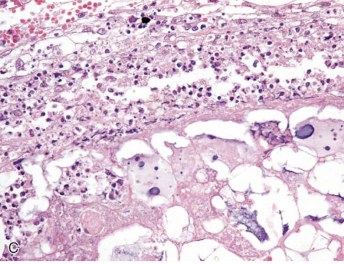
How does apoptosis appear histologically?
looking for cellular and nuclear fragmentation
Morphologically, apoptosis is a process of chromatin condensation referred to as ________, nuclear fragmentation known as ________, and ________ of the plasma membrane to form membrane bound ________ ________, which contain ________ ________, ________, and condense ________.
pyknosis; karyorrhexis; blebbing; apoptotic bodies; nuclear fragments; organelles; cytosol
Is there usually inflammation associated with apoptosis? Why or why not? What are typically recruited for clean up?
no; the fragments are membrane bound; phagocytic cells (macrophages)
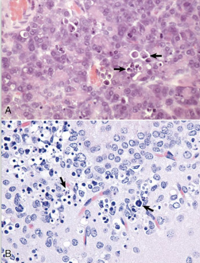
What is this an image of?
apoptosis
What is the most common form of cell injury? What does this cause? Can this be reversible?
hypoxia; depletion of ATP and the inability of certain Na+/K+ ATPase pumps to regulate the electrochemical gradient within the cell; yes
When does cell injury commonly become irreversible and often lead to necrosis?
when calcium influxes into the cell from the extracellular compartment
There is about 4X as much calcium in the ________ compartment than ________.
extracellular; intracellularly
Where is calcium highly sequestered?
inside the cell into the ER and mitochondria
What results in the release of intracellular sequestered calcium from these key places? What will this do?
influx of extracellular calcium; sets off a cascade of enzymes which ultimately results in further depletion of ATP, nuclear damage, and membrane damage
What does necrosis typically result in?
inflammation
What are the hallmarks of necrosis?
P
K
K
pyknosis
karyorrhexis
karyolysis
condensation of the nucleus
pyknosis
fragmentation of the nucleus
karyorrhexis
swelling of the nucleus
karyolysis
During necrosis, cells tend to have intense ________ ________ due to the denatured proteins and loss of ribosomes. Later, cells can have ________ ________ and appear swollen, rounded, and frequently detach from the neighboring cells and basement membrane.
cytoplasmic eosinophilia; cytoplasmic pallor
What is this an image of?
necrosis
Necrosis presents in different ways depending on what?
T
N
T
tissue involved
nature of the injurious agent
time elapsed after injury
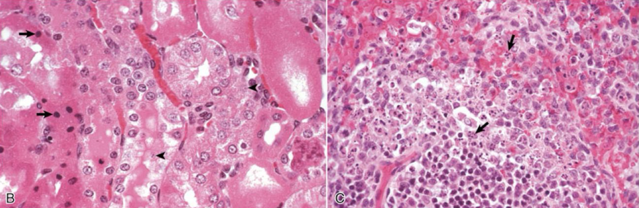
denaturation of cytoplasmic proteins and the retention of the cell membrane
coagulative necrosis
What is coagulative necrosis almost always due to?
H
I
T
hypoxic change
ischemia
toxic injury
With enough time, what will happen to coagulative necrosis?
will break down and become liquefactive
What is not hindered in coagulative necrosis?
degradation of nucleic acids
Cells with coagulative necrosis will have what type of nuclei?
pyknotic, absent, or karyorrhetic nuclei
True or false: In coagulative necrosis, the initial injury and subsequent cellular acidosis results in the denaturing of lysosomes which delays proteolysis and the breakdown of cell membranes.
true
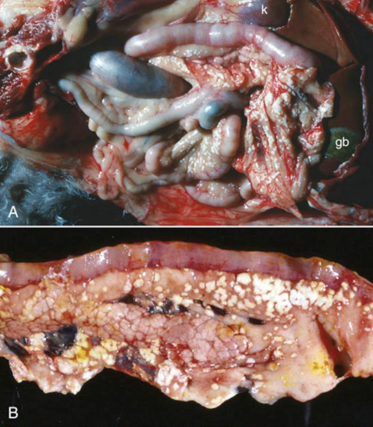
What is this an image of?
coagulative necrosis
What is an example of coagulative necrosis?
renal infarction
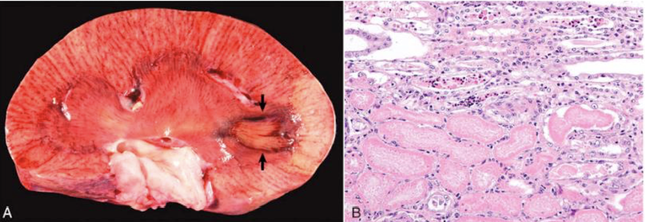
What is this an example of?
coagulative necrosis
older/more advanced lesion when compared to coagulative necrosis with a loss of architecture with possible calcification and a cheese-like appearance
caseous necrosis
Grossly, how can caseous necrosis present?
C
G
L
Y
crumbled
granular
laminated
yellow-white color
True or false: What causes coagulative necrosis is the same that causes caseous necrosis, but are caused by specific agents so not every coagulative necrosis becomes this.
true
What is this an image of?
caseous necrosis

What is this an image of?
caseous necrosis
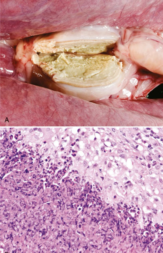
What is this an image of?
caseous necrosis
What can older lesions of caseous necrosis do?
organize and form laminations
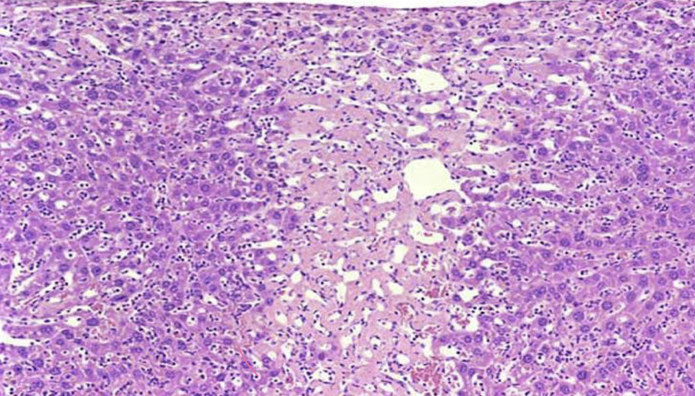
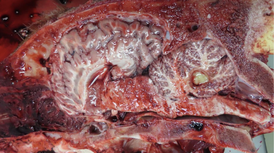
lysed cells converted to a fluid phase
liquefactive necrosis
What is liquefactive necrosis initially?
coagulative necrosis
In the brain, what type of necrosis will it always become?
liquefactive
term for liquefactive necrosis in the brain
malacia (softening)
True or false: Liquefactive necrosis may first manifest as a translucency of the tissue, but later on it will become yellow, soft, and swollen.
true
Liquefactive necrosis is the ________ stage of necrosis in the ________ due to the lack of ________ interstitium which often upholds the tissue structure. It is rich in ________ and ________ ________, which results in further breakdown.
final; CNS; fibrous; lipids; lytic enzymes

What is this an image of?
liquefactive necrosis
What is this an image of?
liquefactive necrosis

What is this an image of?
liquefactive necrosis
type of coagulative necrosis which most often occurs on the distal extremities such as the limbs, tail, pinnae, or dependent portions of organs such as the mammary gland and lungs
gangrenous necrosis
What are the types of gangrenous necrosis?
W
D
G
wet gangrene
dry gangrene
gas gangrene
grossly, tissues will appear red-black and wet in appearance and saprophytic (clostridium sp) bacteria are involved
wet gangrene
grossly, appears red-black and dry with no proliferating bacteria and no fluid
dry gangrene
form of wet gangrene in which the saprophytic bacteria produce gas in the tissues, which can often be palpated as crepitus where the anaerobic bacteria are proliferating and producing toxins
gas gangrene
True or false: The differentials will change based on the appearance of the gangrene.
true
When will wet gangrene form?
when necrotic tissue is infected by saprophytic bacteria (clostridial bacteria)
What is dry gangrene a result of?
decreased vascular perfusion which can be from loss of blood supply or intense vasoconstriction and commonly occurs on the extremities
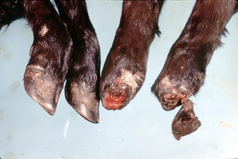
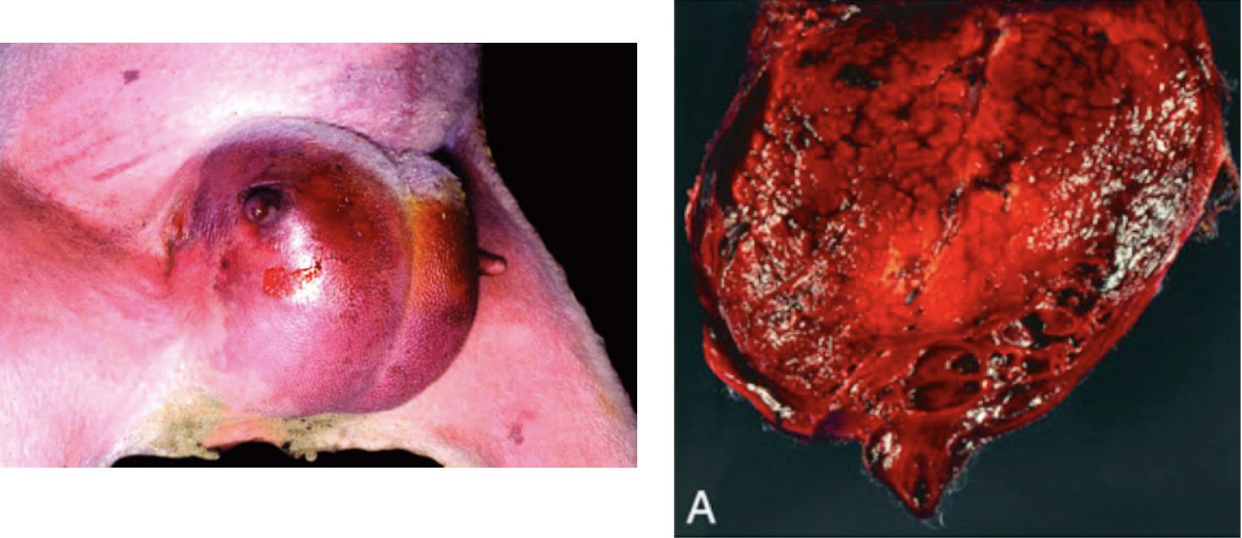
What is this an image of?
dry gangrene (ischemic necrosis)
What is this an image of?
wet gangrene (from gangrenous mastitis)
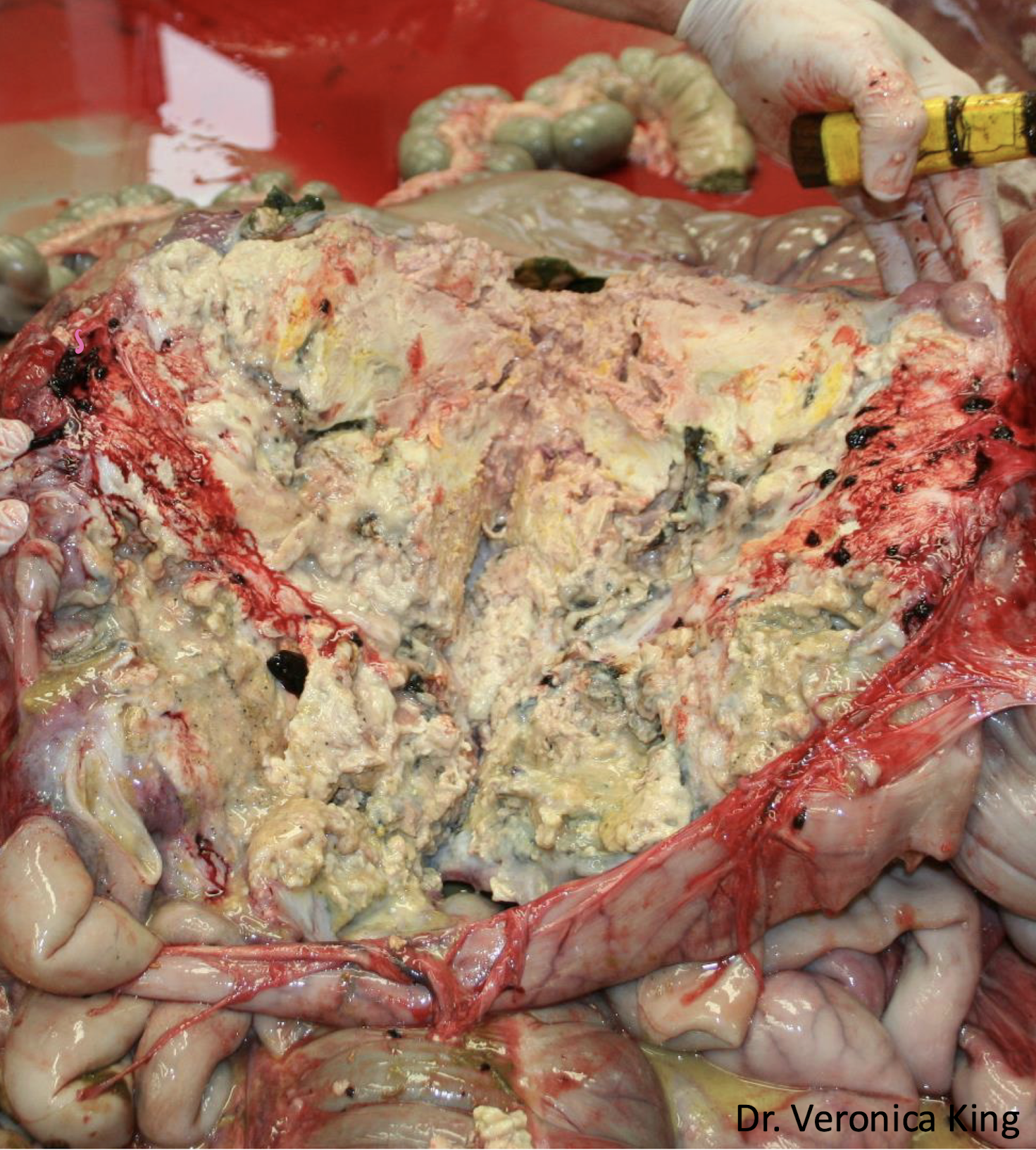
grossly, this type of necrosis appears like irregularly shaped areas of white to yellow discoloration within the adipose tissue, which if you palpated this, would feel firm and chalky (soaponification)
fat necrosis
True or false: Fat necrosis is not always fatal.
true
Histologically, fat necrosis elicits an ________ process, which often recruits ________ and ________ to the sites. Necrotic adipocytes have pale ________ and ________ cytoplasm, rather than colorless, punctate vacuoles.
inflammatory; macrophages; neutrophils; eosinophilic; basophilic
What happens to fat cells when they go necrotic? What is this called?
they will not look white, they will appear pink to blue; soaponification
What are the types of fat necrosis?
E
T
N
I
enzymatic necrosis
traumatic necrosis
nutritional
idiopathic
typically the most prevalent type of fat necrosis that most commonly occurs in carnivores as a consequence of pancreatitis where the pancreatic enzumes (lipases, amylases) are released into the surrounding pancreatic fat
enzymatic necrosis
type of fat necrosis that is pressure induced which may be due to direct injury or ischemia
traumatic necrosis
What is an example of traumatic necrosis?
down cow
What is nutritional fat necrosis commonly caused by?
low antioxidant diet and typically low in vitamin E
Where is idiopathic fat necrosis the most commonly found?
in abdominal fat of cattle in overconditioned cows and the falciform fat of horses and ponies
What is this an image of?
fat necrosis
What is this an image of?
fat necrosis