unit 2: transfer of genetic information
1/70
There's no tags or description
Looks like no tags are added yet.
Name | Mastery | Learn | Test | Matching | Spaced |
|---|
No study sessions yet.
71 Terms
polygenic trait
trait where the phenotype depends on the interaction of many genes
mendel's laws of inheritance
• principle of dominance
• law of segregation
• law of independent assortment
principle of dominance
recessive alleles will be masked by dominant alleles
⇒ not true for every gene
law of segregation
when gametes form, alleles are separated so that each gamete carries one and only one allele for each gene
⇒ not true for every gene
law of independent assortment
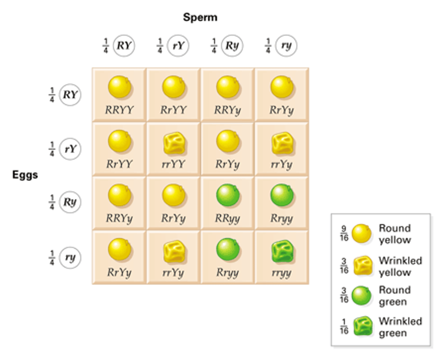
segregation of alleles for one gene occurs independently to that of any other gene (not true for every pair of genes)
⇒ all four gametes (of eggs and sperm) are equally likely to be made during meiosis
epistasis
the impact of one gene masks the expression of another ⇒ usually because the genes impact different steps in the same biochemical pathway
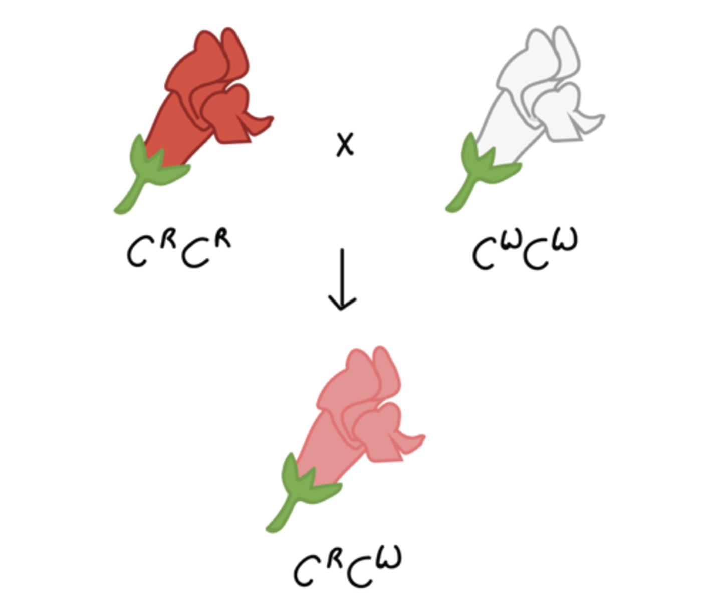
incomplete dominance
neither allele is completely dominant so heterozygote offspring would express a blend of both traits (red flower x white flower ⇒ flower with PINK petals)
co-dominance
both alleles are expressed fully and equally in heterozygote (red flower x white flower ⇒ flower with BOTH red and white petals)

sex-linked genes
• X-linked genes
• Y-linked genes
X-linked genes
genes on the X chromosome
⇒ sons will receive only one copy of these genes and are never heterozygous
⇒ fathers can never pass their alleles onto their son
Y-linked genes
genes on the Y chromosome
⇒ sons will always receive their father's allele, and are never heterozygous
⇒ daughters never have these genes at all
somatic mutation
genetic mutations in non-reproductive cells meaning the mutation is not passed on to offspring
⇒ can happen anytime due to error in replication/exposure to mutagens
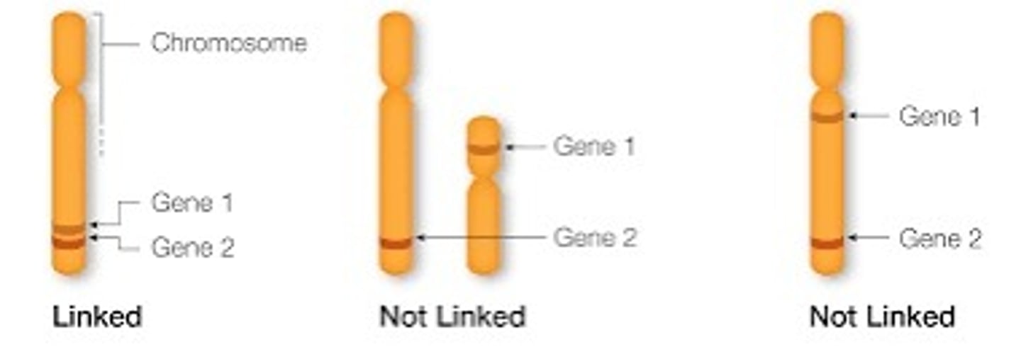
genetic linkage
genes on the same chromosome may be inherited together (linked) and belong to a linkage group
**genes on different chromosomes assort independently
⇒ frequency of allele recombination in offspring can be used to determine if genes are linked
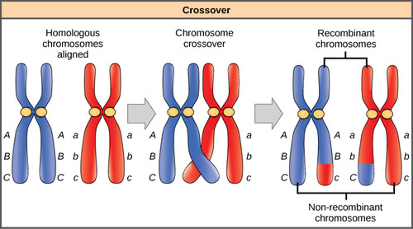
allele recombination frequency
how often two alleles on the same chromosome are separated during meiosis due to crossing over
⇒ proportion of offspring in which which new combinations of alleles appear
recombinant offspring
new associations of parental alleles are recombinants
⇒ offspring w/ new allele combinations different form parent
recombination
process by which homologous chromosomes exchange genetic information ⇒ mixing of parent alleles to generate new combinations on the gene
linked genes
genes on the same chromosome
• do NOT follow Mendel's laws b/c they are often inherited together, which violates the law of independent assortment
crossing over
homologous chromosomes exchange genetic material when each chromosome's chromatids perform breakage and crossing over
linkage in test crosses
linked genes are found by looking for deviation from the frequencies expected from independent assortment (chi square test)
• if alleles are not linked (chi square), all four possible combinations of traits will be present in equal numbers in the progeny
⇒ significant deviation in the ratio (more parental and fewer recombinant types) indicates linkage
test cross
one parent is homozygous recessive, one is heterozygous (aabb x AaBb ⇒ AaBb, Aabb, aaBb, or aabb)
• used determine which genes are linked allowing for ⇒ genetic map to be constructed for each chromosome
chi-square test cross (aabb X AaBb)
null: the genes are unlinked
• if the null hypothesis is true, we expect to see a phenotype ratio of 1:1:1:1 for the resulting offspring
• fig 13.7
• deviation from this ratio suggests linkage (reject the null)
chi-square dihybrid cross (AaBb X AaBb)
null: the genes are unlinked
• if the null hypothesis is true, we expect to see a phenotype ratio of 9:3:3:1 for the resulting offspring
• deviation from this ratio suggests linkage (reject the null)
analyzing chi-square and deviation
null: the genes independently assort
• use expected values determined by Punnett square in chi-square formula
• if p > 0.05, deviation between observed and expected is NOT significant
• if p ≤ 0.05, deviation is statistically significant ⇒ genes are linked
genetic map
• recombination frequency used directly as an estimate of how far apart genes are ⇒ find proportion of recombinants to total offspring
• measure is more accurate when alleles are close together and scoring large numbers of progeny (offspring) increases accuracy
• a 1% crossover rate is a genetic distance of 1 map unit (mu) which is also called a centimorgan (cM)
recombinants
offspring that are less frequent that you would expect with no linkage ⇒ when observed > expected
• recombination frequency of 50% means that genes are unliked
• two ways in which genes may be unliked are:
⇒ may be on separate chromosomes
⇒ may be far apart on the same chromosome
generating a linkage map
diagram showing relative position of genes on a chromosome based on recombination frequencies (estimating the crossover rate in a particular segment of a chromosome)
⇒ may not exactly match the physical
map because crossover is not equally probable at all
sites on the chromosome
genetic markers
"signposts" that are easily identifiable and comparable between different labs ⇒ scientists use other genetic markers to create a genetic map
• since number of physically observable genes in humans is small, genetic maps based on genes with a clear phenotype is not very useful
• Single Nucleotide Polymorphisms is an example (- loci on the genome where there is variance in the nucleotide at a specific spot, detectable by sequencing or another means)
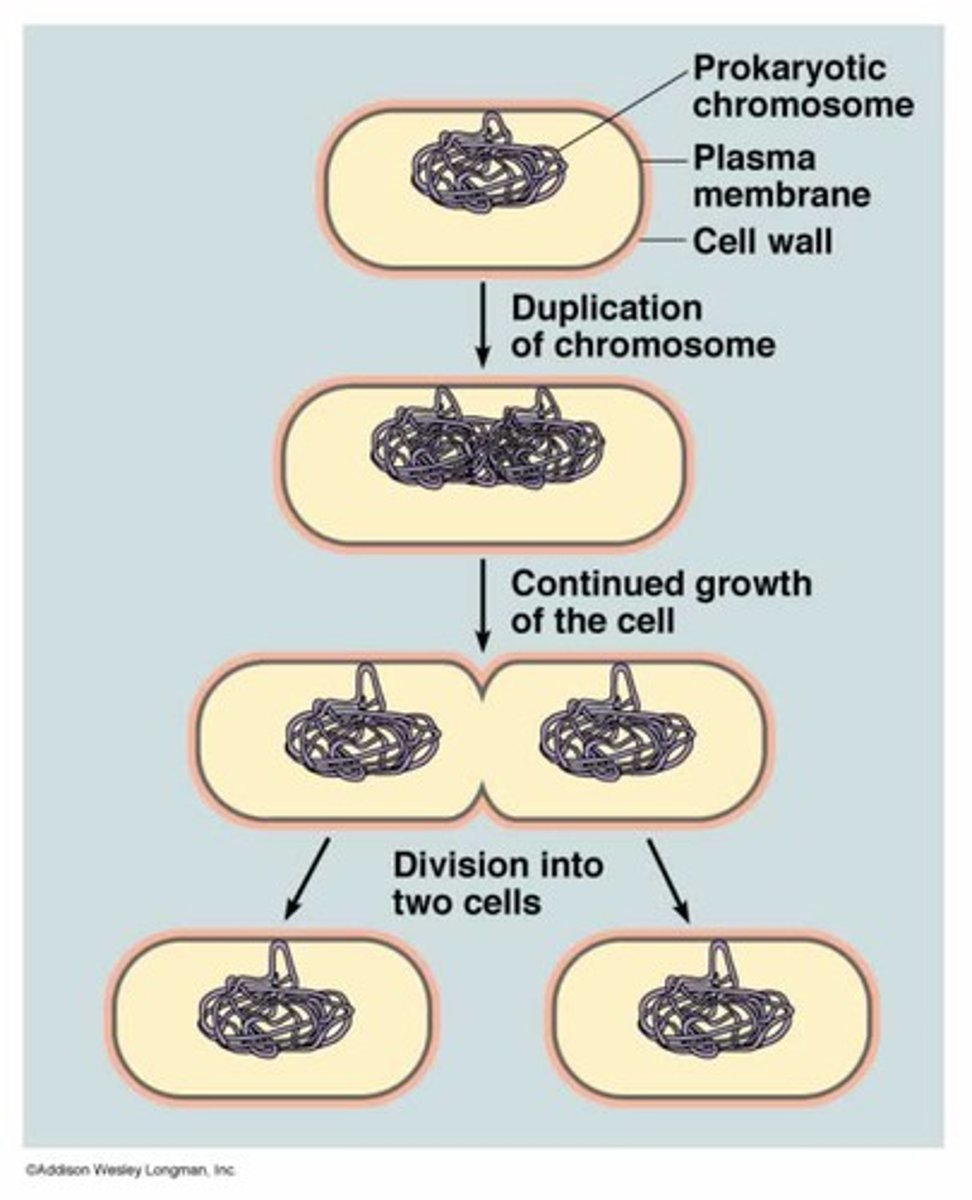
prokaryote cell cycle
binary fission
• replication of genetic material
• cell elongation
• simple pinching off of a new cell
⇒ mitochondria & chloroplasts also divide by binary fission inside eukaryotic cells
basic info about eukaryotic cell cycle
cell cycle requires significantly larger amounts of ATP
• interphase (growth 1, synthesis, growth 2) ⇒ more DNA & organelles to copy
• mitosis ⇒ involves nuclear division
• cytokinesis ⇒ cell division occurs
mitosis
duplication of nucleus:
• prophase
• prometaphase
• metaphase
• anaphase
• telophase
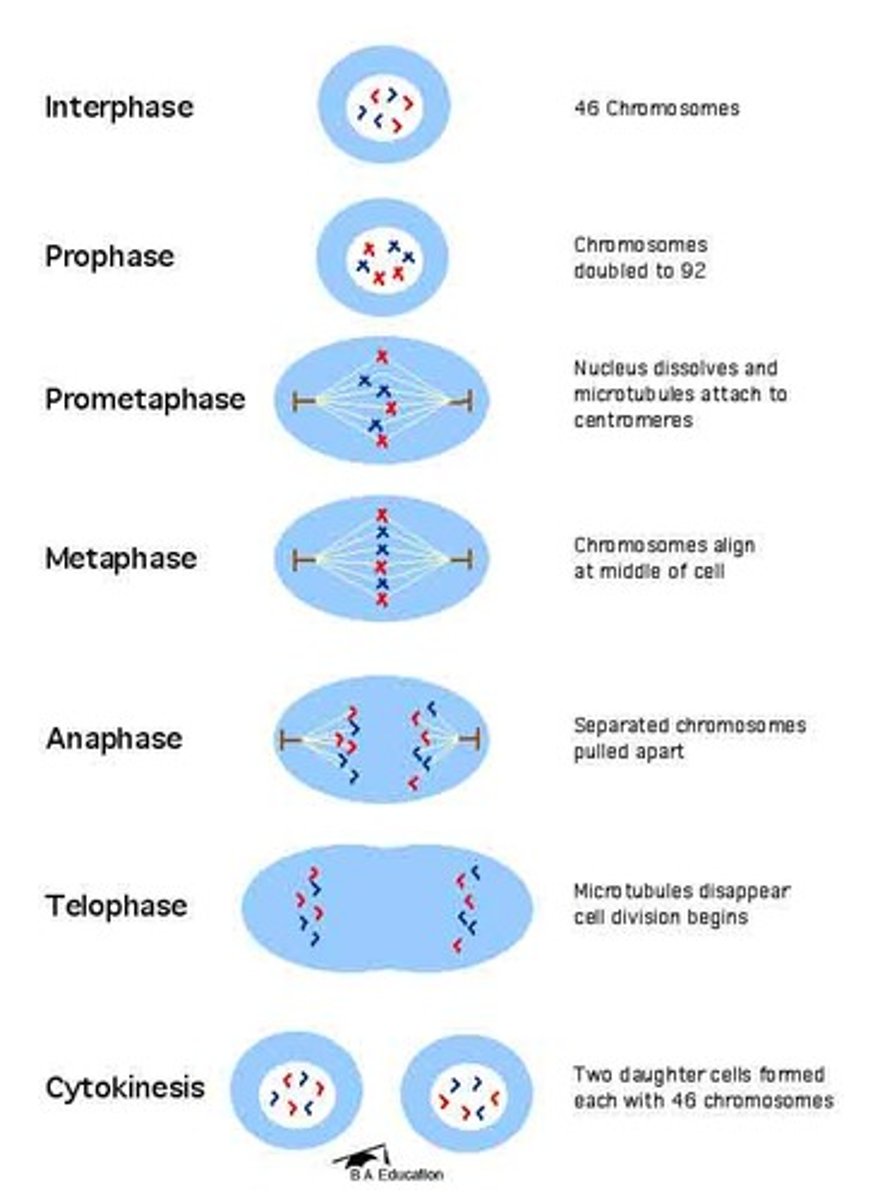
prophase
• chromosomes condense and become visible
• spindle fibers emerge from centrosomes
• nuclear envelope breaks down
• nucleolus disappears
prometaphase
• chromosomes continue to condense
• kinetochores appear at the centromeres
⇒ kinetochore is the protein helps becomes an attachment point for spindle fibers
• mitotic spindle microtubules attach to kinetochores
• centrosomes move toward opposite poles
metaphase
• mitotic spindle is fully developed, centrosomes are at opposite poles of the cell
• chromosomes are lined up at the metaphase plate
• each sister chromatid is attached to a spindle fiber originating from opposite poles
anaphase
• cohesin proteins (centromeres) binding the sister chromatids together break down
• sister chromatids (now called chromosomes) are pulled toward opposite poles
• non-kinetochore spindle fibers lengthen, elongating the cell
telophase
• chromosomes arrive at opposite poles and begin to decondense
• nuclear envelope material surrounds each set of chromosomes
• the mitotic spindle breaks down
cytokinesis
animal cells: a cleavage furrow separates the daughter cells
plant cells: a cell plate separates the daughter cells
mitotic spindle
• pulls apart sister chromatids
• created by centrosome and attaches to centromere
• kinetochore powers movement of chromosomes
problems with standard image of chromosomes
• both sides of the chromosome (sister chromatids) only exist after S phase
• chromosomes are only tightly wound and visible during mitosis
⇒ DNA is wrapped around molecules called histones
⇒ during interphase, DNA is wound loosely so that it can be transcribed
⇒ only during mitosis, the DNA condenses so that it is very tightly wound
cell differentiation
cells have different functions in the body but all cells have the same genetic material
• by turning on different genes, different cells can have very different shapes and functions
• a cell "decides" what kind of cell to be based on chemical signals from other cells
regulations of the cell cycle
four points during the cell cycle serve as checkpoints where the cell decides to proceed with the cell cycle
growth 1 checkpoint:
synthesis ⇒ growth 2 checkpoint:
mitosis checkpoint:
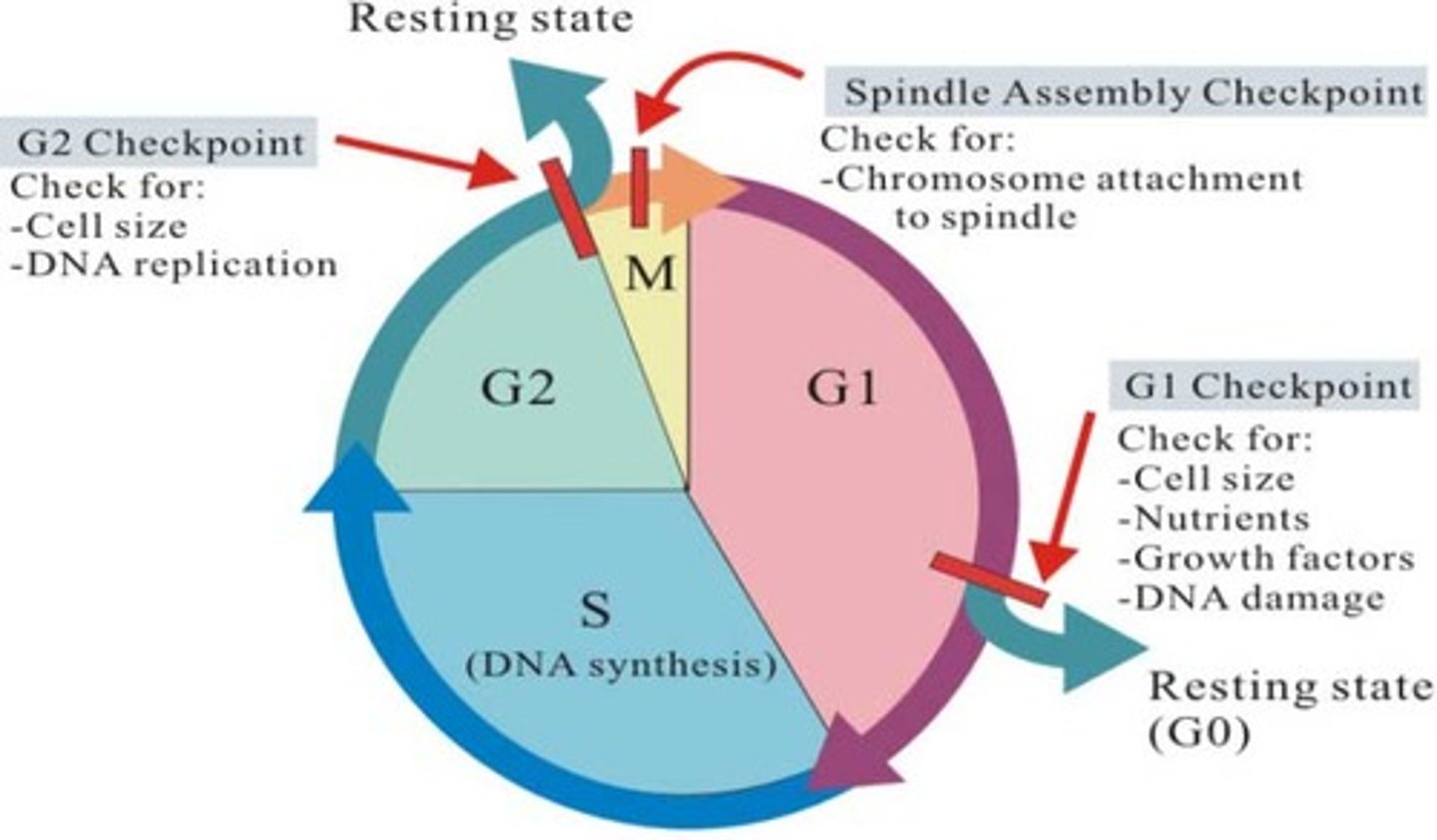
growth 1 checkpoint
• check size of cell
• make sure sufficient resources
• ensure no DNA damage
synthesis ⇒ growth 2 checkpoint
• check DNA "integrity"
mitosis checkpoint
• check spindle apparatus (metaphase -- ensures chromosomes are attached to the spindle fibers at opposite poles before anaphase begins)
proteins at checkpoints
to move from one stage to the next in the cell cycle, the cell must pass through a "checkpoint"
• checkpoint is regulated by proteins
• two classes of proteins are active at the checkpoints ⇒ cyclins, cyclin-dependent kinases
how cyclins and CDK's work
enzyme that turns on mechanisms needed for the cycle to move forward, dependent on the cyclin
• cyclin turns on CDK ⇒ CDK pushes through checkpoint
• activated via reverse phosphorylation by cyclins
* every checkpoint has its own cyclins and CDK's
kinase
an enzyme that activates other enzymes by adding a phosphate group
concentrations of cyclins and CDK's throughout cell cycle
red queen hypothesis
sex generates viability in offspring, which helps at least some offspring survive parasites (and competitors)
⇒ parasites evolve quickly, sex helps hosts "keep up"
meiosis
cell division that reduces the number of chromosomes in a cell by half to create four sex cells, or gametes (sperm and eggs)
genetic variation in meiosis
prophase I: independent assortment, crossing over
metaphase I: independent assortment
genetic variation in metaphase 1
independent assortment (of homologous chromosomes)
• the maternal and paternal chromosome pairs align randomly at the cell's equator meaning each resulting gamete can inherit a unique combination of maternal and paternal chromosomes
genetic variation in prophase 1
crossing over
• homologous chromosomes exchange genetic material
number of chromosomes & chromatids in each phase of mitosis
assume the cell has two pairs of homologous chromosomes
• interphase = 4 & 8 (after S phase)
• prophase = 4 & 8
• metaphase = 4 & 8
• anaphase = 8 & 8
• telophase = 8 & 8
• cytokineses = 4 per cell & 4
haploid (n) / diploid (2n) in each phase of mitosis
• interphase = diploid (2n)
• prophase = diploid (2n)
• metaphase = diploid (2n)
• anaphase = diploid (2n)
• telophase = diploid (2n)
• cytokineses = haploid (n) for each cell
number of chromosomes & chromatids in each phase of meiosis I
assume the cell has two pairs of homologous chromosomes
• prophase I = 4 & 8
• metaphase I = 4 & 8
• anaphase I = 4 & 8
• telophase I = 4 & 8
• cytokineses I = 2 per cell & 4 per cell
haploid (n) / diploid (2n) in each phase of meiosis I
• prophase I = diploid (2n)
• metaphase I = diploid (2n)
• anaphase I = diploid (2n)
• telophase I = diploid (2n)
• cytokineses I = haploid (n) for each cell
number of chromosomes & chromatids in each phase of meiosis II
assume the original cell had two pairs of homologous chromosomes
⇒ per cell
• prophase II = 4 & 4
• metaphase II = 4 & 4
• anaphase II = 4 & 4
• telophase II = 4 & 4
• cytokineses II = 2 per cell & 2 per cell
haploid (n) / diploid (2n) in each phase of meiosis II
• prophase II = haploid (n)
• metaphase II = haploid (n)
• anaphase II = haploid (n)
• telophase II = haploid (n)
• cytokineses II = haploid (n) for each cell
gametogenesis
the process of making haploid gametes (including division ie. meiosis and differentiation)
• common examples include
⇒ spermatogenesis (the formation of sperm in males)
⇒ oogenesis (the formation of eggs in females)
non-disjunction
error in meiosis chromosomes don't separate correctly
cancer and its characteristic
the processes regulating normal cell division are disrupted ⇒ cancer develops from changes that cause normal cells to acquire abnormal functions
• simply, it happens when changes leading to abnormal cell division occur to genes, making it a genetic disease
hallmarks of cancer
what cancer cells have in common
• evading apoptosis (not following programmed cell death)
• self-sufficiency in growth cells (doing growth without outside signals)
• insensitivity to anti-growth signals
• tissues invasion & metastasis (when it spreads throughout the body)
• limitless replicative potential (unlike other cells which have a limit)
• sustained angiogenesis (some tumors can cause blood vessels to grow into the tumor allowing the tumor to grow more)
balance of healthy tissue
healthy tissue depends on the careful balance of both cell birth and cell death
types of genes that can be altered leading to abnormal cell division
• tumor suppressor genes
• proto-oncogenes
• stability genes (dna repair genes)
tumor suppressor genes
genes responsible for slowing down cell growth and division
• when functioning normally, they serve to stop the progression of the cell cycle when cells are damaged
• when mutated and no longer functioning normally, damaged cells are allowed to continue to replicate
⇒ mutated genes have a recessive pattern of inheritance & are associated with family mutations
oncogenes
• called proto-oncogenes when functioning normally but called oncogenes in its mutated form
• has "dominant" mutations ⇒ meaning one copy is enough to encourage abnormal cell division
•. normally genes promote cell growth and division, when mutated unrestricted cell growth and division occurs
⇒ mutated genes have a dominant pattern of inheritance & are not associated with family mutations
proto-oncogenes
genes that code for proteins (proteins) that encourage cell division or prevent cell death
• they are typically involved in cell-signaling mechanisms
DNA repair genes
• in normal function, genes work to repair DNA damage and correct base-pairing errors, stabilizing the genome
• when mutated, no DNA repair occurs and mistakes are passed down to next generation
metastasis
mutations that allow cells to migrate to other parts of the body
• cancer progresses from benign (non-evasive) ⇒ malignant (locally evasive) ⇒ metastatic
angiogenesis
mutations that promote the formation of blood vessels within the tumor ⇒ means the cells will be able to grow easily
when a cancer cell is mutated
• cancer cell is able to evade the immune system
• cancerous cells compete for resources with other body cells
• mutations that allow them to gain more resources/survive better will become more common (natural selection)