Biochemistry Laboratory Practical: Enzyme Reactions and Assays
 Call Kai
 Learn
Learn Practice Test
 Spaced Repetition
 Match
Match Flashcards
 Knowt Play
1/86

There's no tags or description
Looks like no tags are added yet.
Name | Mastery | Learn | Test | Matching | Spaced | Call with Kai | Chat |
|---|
No analytics yet
Send a link to your students to track their progress
87 Terms
Microliter
1 L = 1x10^6 uL
1mL = 1000 uL
Suppose an entire class achieved high percent error values (>25%) for the molar absorptivity values of pNP, and also had high R2 values. What might this indicate about the experiment? (There is more than one correct answer; give TWO answers.)
the stock solution of pop is bad
or
the pH of the buffer is off
the absorbance of a compound is measured with
a spectrophotometer
- the wavelength at which a compound absorbs light is called the wavelength of maximal absorbance (lambda max)
-compare how much is in a sample compared to one that doesn't contain the sample (to blank the thing)
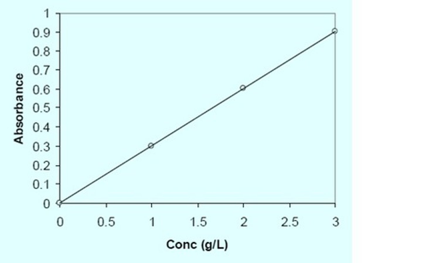
Beers law
A=ecl
y=mx+b
slope= extinction coefficient
- this standard curve can be used to find an unknown concentration
absorbance
e= extinction (absorption) coeffiecient
aka absorptivity (1/M*cm)
c= concentration
l= path length (1cm)
the amount of light absorbed is proportional to the concentration
pipette sizes and max-minimum volume
P20: 20-2uL
P200: 200-20 uL
P1000: (1mL) 1000-100uL
use a graduated glass pipette for 1-10mL
- use larger size if over 5mL
use a graduated cylinder if over 10 mL
Does the R2 value from your standard curve represent consistency, accuracy, or both, of your experiment?
consistency
Does the percent error of your molar absorptivity value represent consistency, accuracy, or both, of your experiment?
accuracy
Does the percent error of your assigned concentration value represent consistency, accuracy, or both, of your experiment?
accuracy
If a compound has a wavelength of maximum absorbance (λmax) of 500 nm, what will happen if the samples are measured at a higher wavelength (550 nm, for example)?
If samples are measured at a higher wavelength than the maximum, absorbances will be less than their actual value. Because absorbance of a light sample is proportional to the concentration of a the sample being measured, this value will also appear decrease. Therefore, your measured values of absorbance and concentration would be less than the actual values, increasing percent error.
Will the change in wavelength affect the R2 value of the graph? Why or why not?
No, even though a change in wavelength will decrease absorbance the data will stay consistent along the same linear fit line.
Will the change in wavelength affect the molar absorptivity calculated from the graph? Why or why not?
A change in wavelength deviating from the max wavelength decreases the absorbance while concentration remains constant. Given Beer's law (A= Ecl) if absorbance (A) decreases at a known concentration (c) and path length (l) is a constant, the molar absorptivity will also decrease. Therefore, molar absorptivity will change with a change in wavelength.
molarity units
M=mol/L
calculate percent error
= (your value - real value)/real value x 100
= (observed concentration- assigned concentration)/ assigned concentration
x 100
buffer
a solution of weak acid and its conjugate base that resists significant changes in pH upon additions of small quantities of acid or base
apparent dissociation constant of a weak acid
Ka= [H][A-}/[HA]
Henderson Hasselbach equation
relates the pH of a buffer to the different buffer species in solution
pH = pKa + log [A-]/[HA]
pKa=pH when [A-]=[HA]
pKa=
pKa=-logKa
titration curve of carbonate
- the flat part of the curse is the buffer range (ability to effectively absorb H or OH- ions from added bases/ acids)
-usually given by the pKa of the acid +/- 1
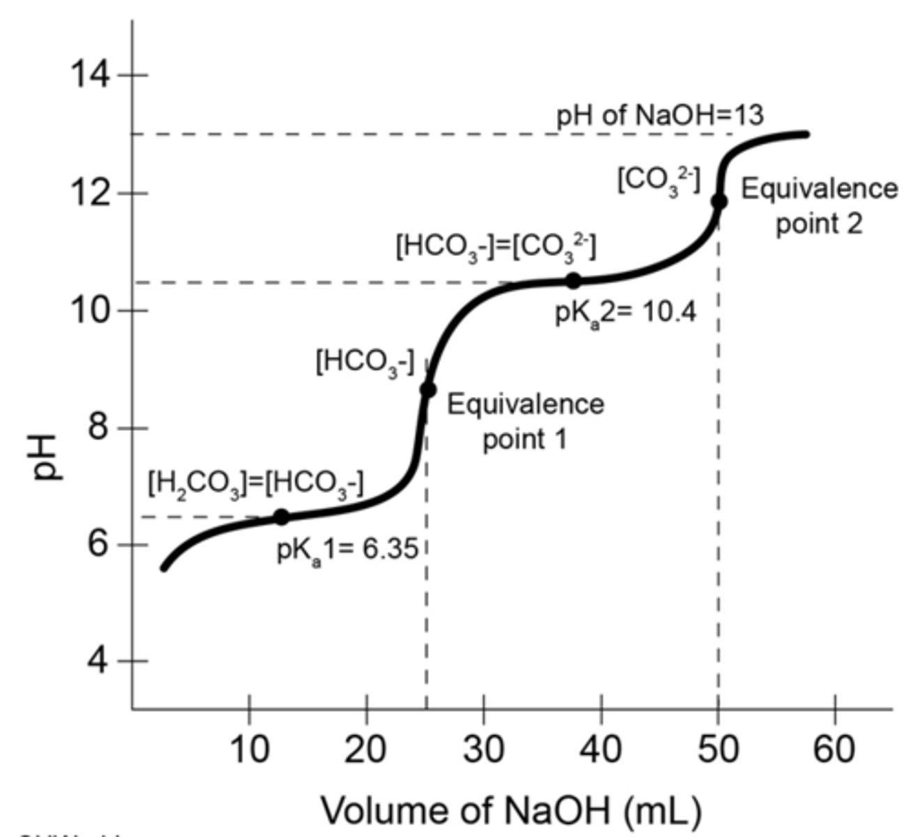
buffering capacity
the total molar amount of the weak acids and its conjugate base making up the buffer. This determines the number of H that can be absorbed or donated
- the concentration of the buffer is the total concentration of both acid and its conjugate base
amino acids are
polyprotic
-two titratable groups at least (NH3+ and COOH; R group)
pH=
pH= -log[H+]
If you have 1 mole of a diprotic weak acid in solution, how much strong base much be added to reach the first equivalence point in a titration curve (when all of the first group has been deprotonated)?
1 mol
If you have 1 mole of a diprotic weak acid in solution, how much strong base much be added to reach the pH that equals the pKa of the second titratable hydrogen?
1.5mol
For a diprotic buffer with pKas of 4.7 and 7.8, what would the buffering range(s) be?
pH 3.7-5.7 and pH 6.8-8.8
Is water a good buffer? Why does water change pH so suddenly when NaOH is added?
Water is not a good buffer because it doesn't have any weak acid and conjugate base pair present in its solution to buffer the added NaOH. Therefore, water would simply give up its proton so it can't compensate for a small pH change with added base.
Why does an amino acid have multiple pKa values? How many molar equivalents of base are required to titrate the amino acid?
An amino acid will have multiple pKa values when it has more than one proton that can be protonated/ deprotonated with the addition of a base or acid. To completely remove the first proton from all of the acid a ratio of 1:1 molar equivalents of base are needed. For the second pKa value, another molar equivalent of base is needed to completely remove the second proton. To have an equal ratio of acid to conjugate base only .5 molar equivalents of the base are needed (pKa will equal pH at this titration).
deprotonation order
low-> high pH
first the carboxyl group, then the amino group
-R group can depend after carboxyl group
What is the biochemical term for a protein with two different polypeptide chains? (Synonyms for polypeptide chains are protomers and subunits.)
heterodimer
a protein complex made up of four identical subunits which are associated but not covalently bound.
A homotetramer is a protein complex made up of four identical subunits which are associated but not covalently bound. Conversely, a heterotetramer is a 4-subunit complex where one or more subunits differ.
The amino acid sequence of a protein constitutes what level of structure?
primary
Why is the SARS COV 2 spike protein considered a homotrimer? Answer in your own words.
A homotrimer is composed of three identical units of polypeptides. The SARS COV 2 spike protein contains three identical promoters that each contain a receptor binding domain for the ACE-2 receptor
The polar noncovalent interactions that hold the subunits of ACE-2 together is an example of:
quaternary protein structure
Protein concentration and Absorptivity lab
What is the purpose of the BSA protein used in the Biuret assay of this week's lab? Select one.
Standard for absorbance of Cu complex in assay
- under alkaline conditions Cu2+ will be reduced to Cu+ and form a coordination complex with the nitrogen in the peptide bonds
-the solution changes from a light blue to a purple
-aa are clear
Why does the Biuret assay give similar results for all proteins, even if the amino acid sequences are different? Select one.
The assay is based on binding of Cu ions to peptide bonds, which all proteins have
- usually UV absorbance of proteins depends on the presence of aromatic amino acid side chains
Which of these best summarizes today's experimental design? Select one.
Measure concentration of unknown by Biuret assay, then measure absorbance of different dilutions at 280 nm
how can you find an undiluted sample's concentration
using the absorbance of the unknown and the standard curve you can calculate it
Absorbance spectra of the complex at different protein concentrations
Abs (y axis) and wavelength(nm) on x axis
- curve for each sample looks like a hill, increasing absorbance as the sample concentration increases
-peak is always near 540 nm wavelength (x axis)
- max absorbance of .5 does not change with increasing concentration (below until maxes out)
beers law is inaccurate....
for absorbance values over 1
y=mx+b will not be true because slope will not be a straight line
you must dilute the solution to improve accuracy
absorptivity is different
when measured at different wavelengths, so all measurements/ absorption values of concentrations need to be recorded at the max wavelength (same value) for max absorption
how did you determine the max wavelength of the solution?
THE HIGHEST ABSORBANCE FROM THE ABSORPTION SPECTRA OF THE COMPLEx determined the max wavelength (540nm)
if a solution with a certain amount of glucose in it has water added to it, what stays/changes?
-concentration
- grams of glucose in the solution
- mols of glucose in the solution
decrease
stay the same
stay the same
view #5 in packet
In another colorimetric protein assay is the Bradford assay, in which a non polar, hydrophobic dye binds directly to proteins in solution, causing color change from red to blue. Since there is no chemical reaction occurring, the color changes is immediate. Give one advantage and one disadvantage of this method compared to the buret assay
Advantage: there is no chemical reaction occurring so the speed of the color change is immediate making the procedure more efficient
Disadvantage: The dye is hydrophobic and will only bind to the hydrophobic side chains, which not every amino acid has. This will decrease absorptivity compared to the Biuret assay
velocity or rate of reaction
the rate or velocity of an enzyme reaction measured as the amount of substrate that is converted to product in a given amount of time
- depends on enzyme and substrate (organic starting material). concentration, and conditions like pH and temp
Michaelis menton equation
V=Vmax[S]/Km+[S]
rate of formation of a product with a constant enzyme concentration and with increasing substrate concentration
enzyme kinetics saturation curve
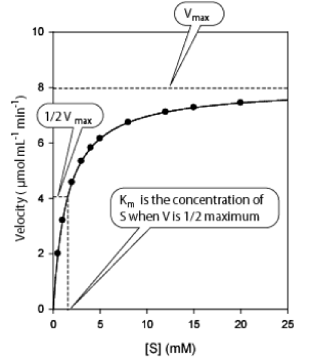
vmax equation
Vmax = kcat [E]
kcat is
turnover number: number of molecules of substrate consumed per enzyme per second and the total enzyme concentration
Michaelis menton constant
a measure of substrate concentration at which the enzyme works at half it's maximum speed
Km
small= high affinity
large= low affinity and more substrate is needed
why is an enzyme pH dependent
ionization of
-amino acid R groups in the active site
- binding groups on the substrate
- groups on the enzyme involved in substrate binding
can also alter conformation
We will measure the absorbance of which molecule in the kinetics experiment?
oxidized dye
To measure velocity of glucose oxidase, we are going to set up a coupled enzyme assay. What reagents/chemicals need to be added to water and oxygen in the cuvette for the assay to work? Consider what is present in the assay mix, as well as other solutions added to the cuvette?
glucose oxidase
reduced dye
glucose
peroxidase
glucose+ O2+ H2O---glucose oxidase----> H2O2 + gluconic acid
reduced dye (colorless)+ H2O2---peroxidase--> oxidized dye
the color change allows the first reaction to be monitored
Which enzyme has a faster Vmax?
peroxidase over glucose oxidase.
(less bonds to break/ smaller molecule?)
Why do we say the two enzyme-catalyzed reactions taking place in your test tube are "coupled"?
peroxidase utilizes the hydrogen peroxide produced by glucose oxidase as a substrate
Based on your graph, what is the optimal pH for glucose oxidase? Note that the optimal pH may be between pH values that you tested. Explain how you determined the optimal pH.
The optimal pH for glucose oxidase is about 7. Looking at our graph, absorbance was the highest for pH 7. This means the reaction produced the most product at a pH of 7. Because one of the products of the glucose oxidase reaction is H2O2, more of the reduced dye will be oxidized by peroxidase producing a stronger color with more absorption.
Our assay had two reactions, one catalyzed by glucose oxidase, and the second by peroxidase. What must be true about the maximum possible rates (Vmax) of the two enzymes to insure that the velocity we measure corresponds to glucose oxidase activity, and not peroxidase activity?
Glucose oxidase has a slower vmax the peroxidase, so it is the rate limiting step of the reaction. The amount of product peroxidase can use to create color directly depends on what is produced in the first reaction by glucose oxidase.
double reciprocal plot
1/V = Km/Vmax(I/[S])+ 1/Vmax
![<p>1/V = Km/Vmax(I/[S])+ 1/Vmax</p>](https://knowt-user-attachments.s3.amazonaws.com/b9056260-03f9-438c-9456-ec31474ce606.png)
Suppose you don't know the concentration of glucose in a can of soda, but want to find it by using the glucose oxidase reaction from today's lab. Describe how you would go about finding the unknown glucose concentration, using the same reagents and equipment from today's lab, and using your data from today. What would you measure and how would your data from today's lab help you?
With the soda, we could create various dilutions of the soda by diluting it with water as we did in the original experiment part C. Adding the dilutions into the glucose 2 mL oxidase assay we would measure absorbance. Using these absorbances, we can compare the values to our original saturation plot to find each specific glucose concentration for the dilutions and the original soda.
Carbonated drinks usually have a pH that is rather low, around 2-3. How might this complicate your experiment from the last question, and why?
This would complicate our previous experiment because glucose oxidase has optimal function at a pH 7, meaning any pH less than this will slow the rate of reaction. If the rate of reaction is slowed, there will be less H2O2 produced for preoxidase to react with and create a colorful solution, so absorbance will appear to be very low. According to the first graph in pH vs velocity, anything below about a pH of 4 will not produce any enzyme activity, so there could be an issue as the concentrations of soda increase in the previous experiment.
the gel used in PAGE
polyacrylamide gel prevents large molecules from moving quickly through the matrix allowing for a separation by size
- the resolving portion is about 7-18% to create separation of proteins 10-250 kDa
-stacking gel with wells is only about 4%
PAGE
polyacrylamide gel electrophoresis
SDS PAGE
allows the proteins to be separated purely by size
- uniform shape and charge to mass ratio
-achieved by unfolding the proteins and coating them with a negative charge
BUT first need to
1) break weak-non covalent interactions between proteins and their subunits
- SDS binds to protein backbones unfolding and coating the protein with a negative charge º The polypeptide charge of the backbone will be negligible because of the negatively charged coat, so all of the proteins will be standardized
-unfolded with heat (boiled)
-also added to gels to prevent refolding
2)covalent disulfide bonds within or between protein subunits
- ß-mercaptoethanol (BME) reduces disulfide bonds
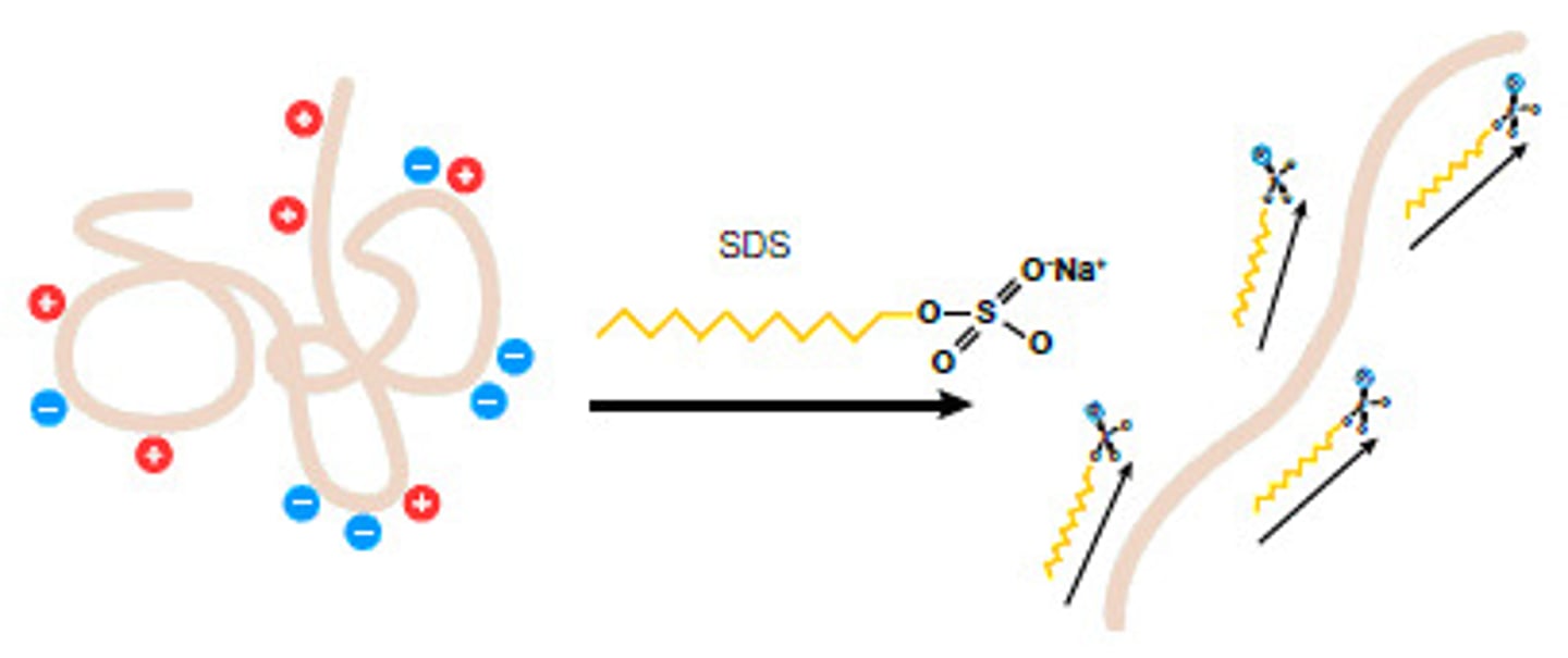
reducing conditions vs non-reducing SDS PAGE
SDS PAGE ran with BME and SDS
only SDS used: disulfide bonds remain present but non covalent interactions are broken
plotting sds page
a linear relationship between log molecular weight (y) vs distance migrated for the protein standards
- deviates from linear if too large a range of molecular weights is used, so standards need to be close to the size of the protein of interest
purpose of bromophenol blue
a low weighted tracking dye to help indicate the progression of the electrophoresis
- helps determine when to turn off the power or the smallest proteins will run off the bottom of the gel
-does not stain the protein
-afterwards, coomassie brilliant blue (CBB) dyes the proteins on the gel
ºadded with acid methanol to fix the protein
-to remove excess stain microwave it in the solution; proteins are insoluble in this solution
Why must a gel be stained?
most proteins are not colorful or visible
the intensity of the band is related to
the amount of protein in the band
you can't say that two different proteins with the same intensity are present in the same amounts because proteins vary in positively charged aa and hydrophobic areas
In SDS-PAGE, why do all of the proteins move towards the anode?
Each protein is covered with a negative charge from from SDS. The gel electrophoresis contains a positive charge at the bottom and a negative charge at the top, so following the electrical current the negatively charged proteins will move to the bottom of the positive end of the gel.
reducing vs non reducing conditions
If the band in Gel 2, Lane D is at 30 kDa, estimate the number and size of the subunits making up the native protein D.
Using both gel images, if the band in Gel 1, Lane B is at ~120 kDa, estimate the number and size of the subunits making up the native protein B.
If the bands in Gel 2, Lane C are at ~20 and ~45 kDa, estimate the number and size of the subunits making up the native protein, assuming that previous gel filtration studies have shown the size of the native protein is 110 kDa.
For lane D in both gels they each have one band at 30 kDa. We can assume that the size of the subunits are the same, but there is no way to tell the number of subunits because there is no variation in either gel as they line up in one band.
The band in gel 1 has multiple subunits at 120 kDa without being run with a reducing agent. Once they are reduced, two bands form at smaller sizes of about 50 kDa and 70 kDa, which add up to the original 120 kDa before separation. Therefore, we can assume there are two subunits that make up native protein B.
We can assume there are 3 subunits for lane C. Gel 1 has two sizes of about 20 and 90kDa. When a reducing agent is added in Gel two, it is likely that the 90 kDa broke into two smaller subunits at about 45 kDa, because there are no other sizes present in the gel besides 20 kDa. Therefore, a subunit of 20 kDa and two subunits of 45 show that there are subunits present.
ELISA stands for
enzyme-linked immunosorbent assay
What reagent will be chemically modified during the assay to produce visible color change?
OPD
You are measuring the concentration of which molecule using the ELISA?.
immunoglobin
Which of the following are reasons that ELISA is a sensitive method that can detect very low concentrations of a target antigen? Select all that apply.
The antibody and antigen exhibit very tight and specific binding.
The linked enzyme produces many colored molecules per antigen molecule.
Your standard curve will contain the log of concentration in ng/mL on the x axis, and absorbance on the y axis. Suppose you get an absorbance value for an unknown sample and find an x value of 2 using your standard curve. What is the concentration in ng/mL that the absorbance value corresponds to?
2= log[IgA]
[IgA]=100
steps of the ELISA assay
1) a plastic well is coated with a capture antibody specific for the desired antigen (IgA)
2) the wells must be blocked to prevent non-specific binding (used BSA) to any exposed plastic
3) antigen is added (IgA) and binds to the capture antibody while other molecules remain in solution
4)washed then a second, enzyme (HRP) labeled antibody is introduced and binds the captured antibody to form a sandwich (detection antibody)
5) excess washed away and the colorless substrate is added (OPD). to be converted into a colored product that is quantified with the spectrophotometer
why is a standard curve used
allows the concentration of the analyze to be determined accurately
why is the ELISA sensitive and specific
-sensitive assay can be measured in small quantities
-specificity: antibodies bind antigens in a specific manner and very strongly
The enzyme coupled reaction allows a single enzyme to generate hundreds of thousands of colorful products, increasing sensitivity
Briefly explain your choice (1:100 or 1:500 values or both) that you used to calculate the final average. You should refer back to the dynamic range of your experiment in the previous question.
My 1:100 absorbance values were 1.077, 1.036, and .979 while my 1:500 absorbance values were .742, .756, and .807. According to the standard curve, my dynamic range is between .5-.9 which the absorbances of 1:500 fit into.
In preparation of the ELISA plates, an albumin protein was used to "block" the plate after the first antibody was immobilized on the plate. Explain why this is important.
Because the primary antibody isn't the only molecule that can bind to the wells through hydrophobic interactions, the wells need to be blocked to prevent nonspecific binding after the addition of the primary antibody. Our antigen along with other proteins and the secondary antibody can all bind, which is why this step is important. This will prevent false readings from other proteins that we don't want to measure and only measure the protein bound to our antibody.
What would happen if you omitted a step in the procedure, and forgot to wash the plate after the enzyme-coupled antibody was applied? What would the ELISA plate look like, and why?
If we forgot to wash after the enzyme-coupled antibody was applied, there would be much more enzyme present in the well than there would be if the plate was washed. Washing the plate with TBT removes the enzyme-coupled antibodies that are not attached to the protein-primary antibody complex attached to the plate. If excess enzymes are present, the addition of OPD substrate would result in many more oxidized OPD molecules creating a very colorful product. This will greatly increase the absorption spectrum and concentrations.
How do antioxidants work to prevent cell damage? Select all that apply.
The react with free radicals to make a relatively stable and nonharmful product.
They reduce the probability that free radicals will react with vital biomolecules like DNA and cell membrane lipids.
What antioxidant will you be using to make a standard curve in this experiment?
ascorbic acid
What reagent will be chemically modified during the assay to produce visible color change?
ABTS radical
steps of antioxidant assay
1. Add 1000 uL of ABTS radical to the cuvette
2. Place cuvette containing ABTS in spectrometer
3. press 'Play' in Logger Pro
4. remove spec from cuvette, leaving spectrometer running
5. add antioxidant solution, invert to mix
6. Place cuvette containing ABTS and antioxidant in spectrometer
to detect antioxidants in solution we will measure the absorbance before and after the addition of antioxidant solution. The magnitude of the change in absorbance should be proportional to the concentration of antioxidant
Was green tea or your own antioxidant a better antioxidant solution? Briefly explain how you determined your answer. Your answer should include reference to your standard curve, and any dilutions you performed.
Green tea was a better anti oxidant than our purple gatorade. We measured the changes in absorbance for 1:10 dilutions which was 1.03 and in purple gatorade it was .27. When adding these changes in absorbance for y to our standard curve formula (y = .068x +.23) we can find the amount of equivalents each beverage had. Because green tea has a higher number of equivalents, it had a higher concentration of antioxidants in solution. In addition, because green tea had a larger change in absorbance, we know that more free radicals were reduced.
Why did the absorbance of the ABTS solution decrease after an antioxidant was added?
Absorbance for the free radical solution is at 414 nm because it is a colored solution. When the radical is reduced by the presence of anti oxidants, the product ABTS+ does not contain a color, so absorbance will decrease as more antioxidants are added.
Would ascorbate (vitamin C) or tocopherol (vitamin E) be more effective at reacting with lipid radicals such as the one shown above? The structures for these vitamins are shown in Figure 1 in the lab manual. Explain your answer, including specific references to differences between the structures of ascorbic acid and tocopherol.
Vitamin E would be more effective at reacting with lipid radicals because it has a long hydrocarbon tail. Vitamin E and the lipid above are both hydrophobic in polar solutions like our cells and will interact via van Der Waals and hydrophobic interactions. This will make it more effective and likely to run into a lipid free radical whereas vitamin C is a polar molecule and will have few interactions with lipids based on its polar structure.
Why do you think we used ascorbate as the standard in this experiment, instead of tocopherol (vitamin E)? Consider how their structures would affect their solubility in the assay.
We used ascorbate because it is more likely to interact with ABTS free radicals. Tocopherol is a hydrophobic molecule so it would likely clump in the polar solution that was used and have little interaction with the free radical. Ascorbate is a polar molecule that is water soluble in solution so it can assimilate and effectively neutralize free radicals in water-based environments.