27 Heart
 Call Kai
 Learn
Learn Practice Test
 Spaced Repetition
 Match
Match Flashcards
 Knowt Play
1/41

There's no tags or description
Looks like no tags are added yet.
Name | Mastery | Learn | Test | Matching | Spaced | Call with Kai | Chat |
|---|
No analytics yet
Send a link to your students to track their progress
42 Terms
Atria
receiving chambers w weak muscular walls
Ventricles
pumping chambers w thick muscular walls
General circulation
right atrium → right ventricle → pulmonary trunk → pulmonary arteries → lungs → pulmonary veins → left atrium → left ventricle → aorta → body → IVC/SVC → right atrium
Heart surfaces
Anterior: sternocostal surface
covers most of R ventricle
Inferior: diaphragmatic surface
covers most of L ventricle + some R ventricle
Left: pulmonary surface
covers most of L ventricle
occupies cardiac area of L lung
Heart borders
Right: R atrium + SVC + IVC
Left: L ventricle + L auricle
Inferior: R ventricle
Superior: L/R atria + ascending aorta + pulmonary trunk + SVC

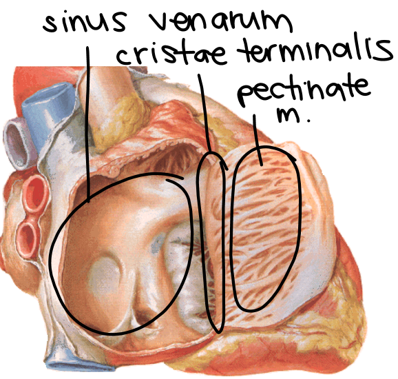
Right atrium posterior part
derived from right horn of sinus venosus
contains sinus venarum (smooth walls)

Right atrium anterior part
derived from atrium proper
contains pectinate muscles

Cristae terminalis
heart tissue btwn smooth + rough atrial walls
Coronary sinus
drains coronary veins
Fossa ovalis
embryonic remnant of foramen ovale
allowed blood to go directly from right atrium to left atrium bc lungs not operational during fetal development

Triangle of Koch
location of AV node + nodal bundles
Atrial septum defect
hole btwn L + R atria
trabecula carnea
rough irregular walls in right ventricle
makes up inflow tract of right ventricle
Conus arteriosus (infundibulum)
outflow tract of right ventricle that leads to pulmonary trunk
papillary muscles
type of trabeculae carnea that pulls chordae tendineae
anterior papillary muscle
posterior papillary muscle
septal papillary muscle
septomarginal trabecula (moderator band)
trabeculum bridging lower portion of interventricular septum + base of anterior papillary muscle for cardiac conduction
tricuspid valve (right AV valve)
valve w 3 leaflets to prevent backflow of blood
Left atrium characteristics
smaller + thicker walls than right atrium
forms anatomical base of heart
receives blood from foramina venarum minimarum + 4 pulmonary veins
Left ventricle characteristics
thickest walls for powerful expulsion pump for entire body
contains trabeculae carnae + papillary muscles + chordae tendinae
bicuspid valve (mitral valve; left AV valve)
valve w 2 leaflets that prevent blood backflow btwn L atrium + ventricle
interventricular septum
membranous + muscular wall btwn R + L ventricles
Aortic valve characteristics
3 semilunar cups btwn aorta + L ventricle
Auscultatory areas for valves
Aortic: 2nd intercostal space just right-lateral to sternum
Pulmonary: 2nd intercostal space just lateral left-lateral to sternum
Tricuspid: 5th intercostal space just left-lateral to sternum
Mitral: 5th intercostal space at L midclavicular line
Sinoatrial node (SA node)
site of initiation of each cardiac cycle; pacemaker
in sinus venarum side of cristae terminalis
Atrioventricular node (AV node)
gives rise to AV bundle (Bundle of His)
near coronary sinus
Right AV bundle pathway
passes right fibrous trigone towards apex of right ventricle + papillary muscles
Left AV bundle pathway
passes right fibrous trigon along side of interventricular septum + papillary muscles
Layers of heart
epicardium
subserous fat
myocardium
endocardium
Functions of cardiac skeleton (4)
insulation
creates electrical discontinuity btwn atria + ventricles
attachment for myocardium
prevent bulging as blood is pumped
provides stable + deformable base for valve leaflet attachment
Pulmonary trunk characteristics
divides at TV5
covered in pericardium
Aorta sections
ascending
arch
descending
Branches from ascending aorta
right coronary a.
left coronary a.
branches from aortic arch
brachiocephalic a.
right common carotid a.
right subclavian a.
left common carotid a.
left subclavian a.
Right coronary a. supply
most of RV + RA
small part of LV
inferior half of IV septum
part of LA
most of SA/AV nodes
Branches off right coronary a.
atrial + ventricular a.
right marginal a.
posterior interventricular a. (posterior descending a.; PDA)
SA nodal a.

Left coronary a. supply
most of LV + LA
strip of anterior RV
2/3 of IV septum
Branches off left coronary a. (left main trunk; LMT)
anterior interventricular a. (left anterior descending a. LAD)
goes towards apex of heart
left marginal a.
circumflex a.

Venous drainage of the heart
great cardiac v.
runs w left anterior descending a.
middle cardiac v.
runs w posterior descending a.
small cardiac v.
runs w R marginal a.
anterior cardiac veins
drains into R atrium
vena cordis minimae
small veins that drain directly into heart

Most common sites of occlusion in coronary arteries
50% anterior interventricular LAD
30% right coronary artery
20% left circumflex artery
GVA innervation spinal levels
T1- T4
Regions most susceptible to deceleration trauma
aortic valve
ligamentum arteriosum
aortic hiatus
Coronary artery anastomoses
Right coronary a. ←→ circumflex a.
left anterior descending a. ←→ posterior descending artery.
and/or within the interventricular septum