Ch. 6 - Nervous System
 Call Kai
 Learn
Learn Practice Test
 Spaced Repetition
 Match
Match Flashcards
 Knowt Play
1/44

There's no tags or description
Looks like no tags are added yet.
Name | Mastery | Learn | Test | Matching | Spaced | Call with Kai | Chat |
|---|
No analytics yet
Send a link to your students to track their progress
45 Terms
brain
-cerebrum, cerebellum, brain stem
-consumes 20% of the total body oxygen
cerebrum
highest part
contains the 4 lobes
brain’s largest component.
• A longitudinal fissure divides the cerebrum into two hemispheres.
• Verbal functions (reading, writing, speaking, and calculating) are the domain of the left hemisphere, while artistic or creative activities (drawing and creating rhythm) is the domain of the right hemisphere.
• The element that joins the two hemispheres together is the corpus callosum. It is composed of white matter (the largest in the brain) and has high myelin content for faster neural transmission.
cerebellum
balance control, equilibrium, posture, and muscle coordination
brain stem
-regulates vital functions such as respiration, heart rate, blood pressure, and consciousness
-serves as a relay center for sensory and motor pathways
spinal cord
• The spinal cord extends from the inferior part of the brain stem to approximately the level of the first or second lumbar vertebra (L1–L2) in adults. Its main function is to carry messages between the spinal nerves and the brain.
• Spinal nerves exit the spinal cord through openings (intervertebral foramina) between the vertebrae.
• Spinal nerves supply nerves to different organs or areas in the body.
• The spinal cord carries messages between the spinal nerves and the brain.
meninges
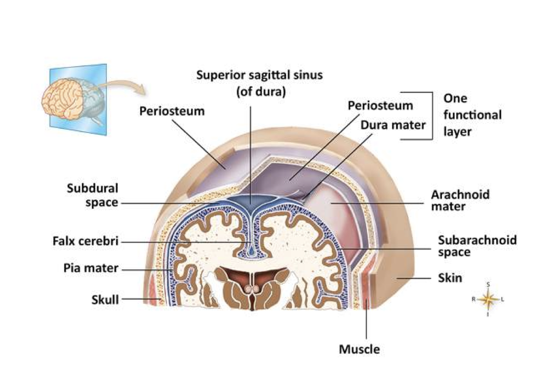
• Dura mater is a tough membrane and the outer layer of the meninges.
• Below the dura mater is the subdural space, and above it is the epidural space.
• Arachnoid is the middle membrane that looks like a spider web.
• Subarachnoid space is present below the arachnoid membrane that allows cerebrospinal fluid to
circulate. CSF contains glucose, proteins, and chloride.
• Pia mater is the innermost, thinnest layer of the meninges. It is highly vascular and supports the brain’s blood vessels.
Cranial Nerves
• Begin from the brain’s underside portion
• Connect to the sensory organs and muscles of the face and neck to transmit information back and forth
spinal nerves
• Use spaces between the vertebrae as pathways to enter and leave the spinal canal
• Transmit information to and from the brain using the pathway through the spinal cord
• Consist of sensory (stimuli transmission from skin and organs to CNS) and motor fibers (stimuli transmission from CNS to skeletal muscles)
• Autonomic nerves regulate important body functions such as breathing and digestion
Autonomic Nervous System (ANS)
sympathetic & parasympathetic
Carries information to and from the brain through the spinal cord
Manages homeostatic processes (body temperature control, etc.)
Cerebrovascular disease (CVD)
-type of CNS disorder
-the blood vessels that supply blood to the brain are affected. It involves disorders of the blood vessels supplying the brain, such as blockage or rupture, which can impair blood flow.
-ex: TIA, CVD
transient ischemic attack (TIA)
-type of CVD in which there is a temporary reduction in blood supply to a part of the brain without permanent damage
-also known as a mini stroke
-symptoms include numbness in the face, arm, or leg or on one side of the body. Patients may also experience difficulty while communicating or understanding others, diplopia, difficulty in walking, and vertigo.
Cerebrovascular accident (CVA)
-type of CVD
-caused by rupture or occlusion of a blood vessel in the brain
-Symptoms include slurred speech, unexplained confusion, sudden and severe headaches, and difficulty swallowing. CVA can also cause paralysis on one side of the body.
dementia
o Progressive memory loss
o Disorientation about person, time, and place
o Neglect of personal hygiene, nutrition, and safety
o Personality changes
o Inability to follow simple directions
Alzheimer’s
-common disorder categorized under dementia and mainly affects the older population
-Symptoms include slow and progressive loss of recent memory; inability to recognize people, places, and events; and disorientation
Epilepsy
-recurrent abnormal electrical impulses in the brain called seizures
-A seizure is a temporary disturbance in the brain’s electrical activity.
Partial seizures
-divided into simple and complex seizures
simple partial: do not cause loss of consciousness, but may cause the patient to repeat certain actions, such as shaking a leg or arm, resembling a twitch or tremor
complex partial: cause staring and repeated movements, like swallowing or lip smacking
-result from abnormal electrical activity in one part of the brain
Generalized seizures
-involve more parts of the brain
absence epilepsy
patient stares with the eyes open, while being unconscious
Encephalitis
-inflammation of the brain most commonly caused by viral infections
-The patient may experience stiff neck, headache, muscle aches, malaise, and flu in the mild stage. Severe conditions bring about symptoms including fever, delirium, seizures, and coma
meningitis
-inflammation of the meninges that may be due to bacterial, viral, or fungal infections.
-Symptoms include headache, flu-like symptoms, nuchal rigidity, and seizures.
concussion
-mild brain injury
-associated with loss of consciousness that can last from seconds up to several minutes
-may be followed by disorientation
contusion
-caused by a more severe brain injury
-Symptoms include headache, nausea, vomiting, vision disturbances, and sensitivity to light.
open brain injury
-occurs when the skull is fractured or displaced; an object may penetrate into the skull, resulting in the fractured skull.
Multiple Sclerosis
• Autoimmune disease affecting the myelin sheath, resulting in inflammation and deterioration leaving nerve fibers exposed and scattering the nervous system message as it travels down the axon
• Causes damage to nerve cell bodies and axons in the brain, spinal cord, and optic nerve.
• Symptoms include numbness, paresthesia, diplopia, ataxia, and bladder control problems
Parkinson’s Disease
• A chronic, progressive, and debilitating condition, Parkinson’s disease affects men more than women and accounts for tens of thousands of new cases annually.
• A deficiency of the neurotransmitter dopamine in the brain, caused by a combination of genetic and environmental factors, is believed to cause this disorder.
• Symptoms include tremors of the extremities, jaw, and face; slow movements; impaired coordination; and a mask-like expression.
• Comprehensive medical history and neurologic examination help to diagnose this condition.
Brain Tumors
Signs and symptoms depend on location and type of the tumor
Initial symptoms include headaches, dizziness, and diplopia.
• Brain tumors may cause increased intracranial pressure.
• History, neurologic examination, and imaging studies help to identify tumors.
Amyotrophic Lateral Sclerosis (ALS)
• Also known as Lou Gehrig’s disease
• Cause not known; results in destruction of motor neurons that control voluntary muscles
• Symptoms include small, local, involuntary muscle contraction in the forearm and hands.
Bell’s Palsy
• Temporary facial paralysis
• Occurs due to inflammation and edema of cranial nerve VII (caused by a viral infection, such as herpes simplex)
• Symptoms include facial twitching, eyelid drooping, and excessive tearing of the affected eye
Peripheral Neuropathy
• Idiopathic (unknown cause) and a condition of peripheral nerve dysfunction
• May be caused by conditions such as diabetes mellitus, human immunodeficiency virus (HIV) infection, and nutritional deficiencies
• Symptoms include muscular weakness, pain, and sensory disturbances such as burning, numbness, and tingling
primary goals of the neurological examination
determining:
-the presence of neurologic dysfunction
-the part of the neurological system that is affected
-the type and extent of the issue.
This provides the physician a variety of possible etiologies.
components of neurological examination
• Assessing the patient’s emotional status, intellectual performance, cognitive ability, and behavior
• Observing the patient’s grooming and mannerisms
• Evaluating the patient’s ability to communicate (use of speech, language, and writing skills)
cranial nerve and peripheral nerve evaluation
MA responsibility in a neurological examination
observe the patient for any abnormalities such as slurred speech and
unintelligible conversation
cranial nerve and peripheral nerve evaluation
-There are 12 cranial nerves, and the proper functioning of each cranial nerve is determined by performing simple tests
-For example, testing cranial nerve number I involves determining the patient’s ability to identify recognizable odors (olfaction). Testing cranial nerve number V involves determining the patient’s ability to differentiate between hot and cold substances.
Peripheral Nerve Examinations
-includes assessment of motor function such as muscle strength, gait, and movements
-health care professional measures and compares the diameters of the upper arms and the calves of the legs to diagnose muscle atrophy.
-Motor function can be examined by performing Romberg’s test, particularly in terms of steadiness. This test is done by asking the patient to stand straight with his or her feet together and the eyes closed.
Sensory Examinations
-determining the patient’s ability to identify or feel superficial sensations.
-Reflex testing (such as deep tendon reflexes and the Babinski reflex) is part of the neurologic examination, separate from sensory testing.
-The Babinski reflex is a reflex extension evoked by stroking the sole of the foot with a reflex hammer or another dull instrument.
Deep tendon reflexes
• Biceps reflex–to assess the flexion of the elbow
• Triceps reflex–to assess the extension of the arm
• Patellar reflex–to assess the extension of the leg
• Achilles reflex–to assess the flexion of the foot
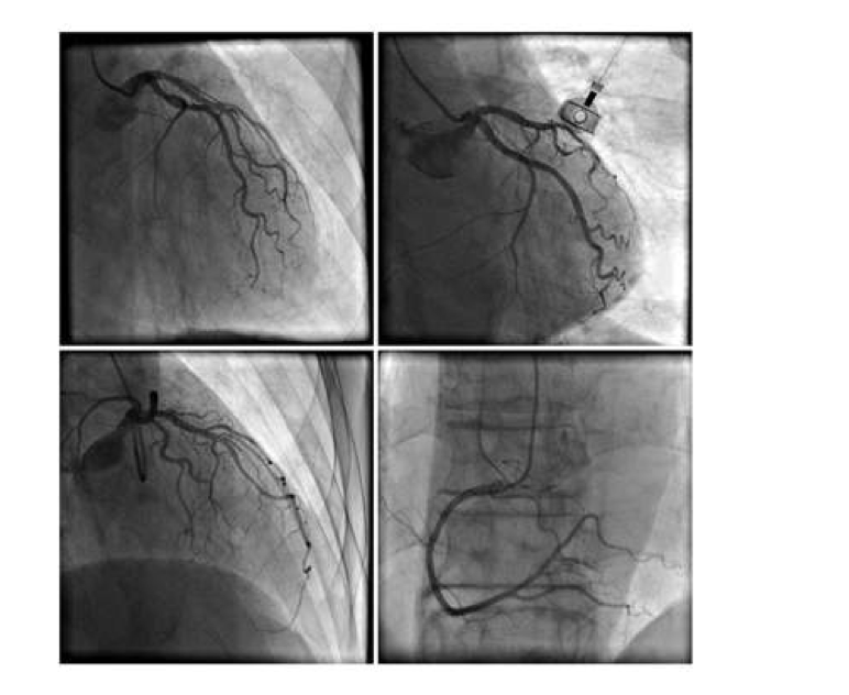
arteriography
• Helps in the detailed viewing of vertebral and carotid arteries, cerebral arterial circulation
• Highlights presence of leaking vessels, aneurysms, and occluded blood vessels
• Performed through injection of a contrast medium into the artery through a catheter
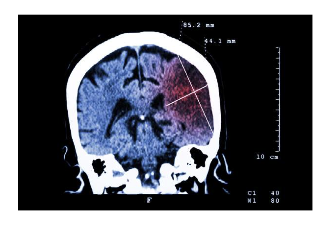
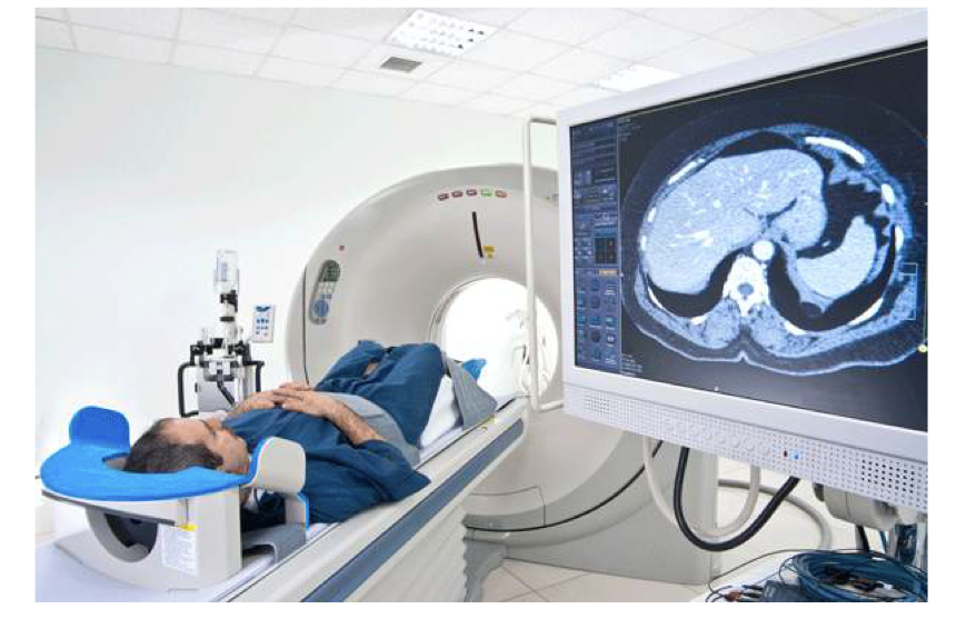
Computed Tomography Scan
• Creates visuals of multiple, serial, radiographic sections; differentiates between bones and soft tissue
• Uses X–rays to visualize the multiple transverse sections of the test structure
Electroencephalography
• Records the brain wave activity to diagnose seizure types and determine effectiveness of seizure-related pharmaceutical treatment
• Performed by attaching electrodes to the scalp to record the brain’s electrical activity
Lumbar Puncture
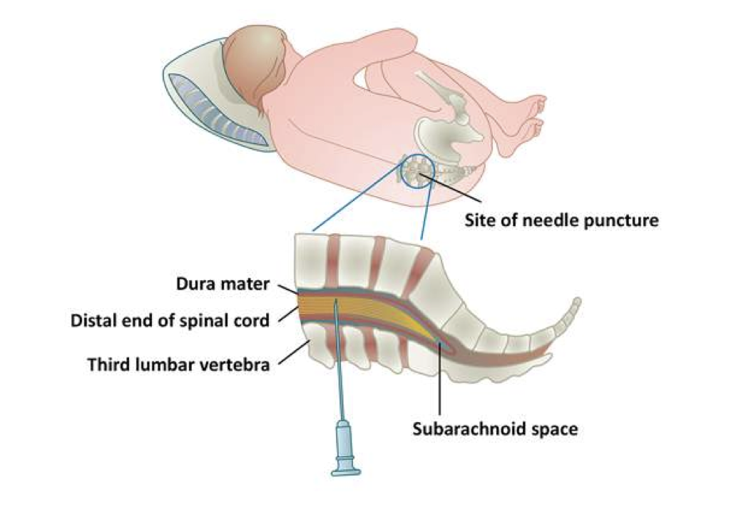
• A lumbar puncture measures cerebrospinal fluid pressure and helps detect increased intracranial pressure, infection, or bleeding.
• Checks for infection of inflammation of CNS or to detect increased intracranial pressure or bleeding
• Invasive process; requires insertion of a special needle into the subarachnoid space to collect the cerebrospinal fluid (CSF) for analysis
Magnetic Resonance Imaging (MRI)
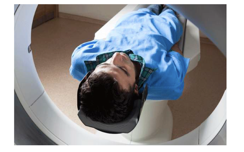
• Aids in viewing of multiple, serial, and radiographic sections; produces detailed images of brain, spinal cord, and surrounding tissue
• Uses magnetic fields and radio waves to produce detailed images of the brain and spinal cord
Positron Emission Tomography (PET)
• Identifies the areas of increased metabolic activity, vascular abnormalities, and space-occupying lesions
• Involves injecting a radioactive isotope and scanning the brain to locate the areas of isotope concentration
Assisting During a Neurological Examination & Diagnostic Procedures
-Main functions include preparing the instruments required for examination and helping the patient assume the required position for the diagnostic tests.
1. Gather and prepare the equipment.
2. Sanitize the hands.
3. Greet and identify the patient. Explain the procedure.
4. Ask the patient to disrobe and wear an exam gown with the opening in the back.
5. Help the patient to change positions during the examination.
6. Record the results as indicated by the physician.
Preparing a Patient for Electroencephalogram
1. Greet the patient and explain the procedure.
Tell the patient the significance of the procedure, how it is performed, and what the patient needs to do during the procedure.
Inform the patient that the electrodes are safe and will not cause pain. Reassure the patient that there is no danger of electrical shock.
Go over the dietary requirements (avoiding stimulants such as coffee, chocolate, or sodas and avoid fasting and maintain normal meals unless instructed otherwise) before the procedure.
Explain that the test requires a baseline EEG during which the patient should avoid any kind of movement.
If the provider orders for stimulation examination, ask the patient to view flickering lights that stimulate the brain.
Offer to answer questions or doubts.
Verify the patient’s understanding and document the education in the patient’s record.
Assisting During a Lumbar Puncture
1. Assemble the equipment and sanitize the hands.
Identify the patient and describe the steps in the procedure.
Ensure the informed consent form is present in the patient’s record.
For a lumbar puncture, assist the patient to get into the left side-lying fetal position. Use a pillow for the patient’s head and between the knees (if required).
Conduct sterile skin preparation of the patient’s lumbar region.
Provide the lumbar puncture kit on an instrument stand for the physician’s use.
Perform cleansing procedure of the rubber top of the anesthetic vial and hold it in position as soon as the provider is ready to begin.
Help the patient to hold still while inserting a spinal needle.
Collect the specimen tubes and label them as required.
10. Fill the laboratory requisition form with the required information. Prepare the specimens so they are
ready to go to the laboratory.
11. Monitor the patient. Provide liquids as instructed.
12. Record the procedure in the patient’s file.