MSK: Exam 1
 Call Kai
 Learn
Learn Practice Test
 Spaced Repetition
 Match
Match Flashcards
 Knowt Play
1/173

There's no tags or description
Looks like no tags are added yet.
Name | Mastery | Learn | Test | Matching | Spaced | Call with Kai | Chat |
|---|
No analytics yet
Send a link to your students to track their progress
174 Terms
What the definition of anatomy
To cut up (via Greek & Latin)
The science of the shape and structure of organisms and their parts
Anatomy is the setting (structure) in which the events (functions) of life occur
What are the 3 types of anatomy
Systemic
Regional
Clinical
Functions of the skeletal system
Stability of limbs and thorax
Protection for vital organs
Attachment site for muscles and ligaments
Blood cell production — continuous supply of new blood cells
Mineral reservoir
What is the articular system
Joints: sites where movement occurs
Function of the muscular system
Produce movement
Maintain posture
Production of heat
Function of the nervous system
Regulate body activities
Oversee coordination of all systems
Sensory and motor functions
Glandular secretions
Function of the circulatory system
Distribute O2 and nutrients via cardiovascular (CV) system
Carry CO2 and wastes from cells
Regulate temperature
Function of lymphatic system
Return protein and plasma from interstitial fluid to CV system
Protects against disease
Definition of the sagittal plane; What does “mid-sagittal mean”
Divides body into left and right sections
Body divided into equal right and left halves - aka “median plane”
Definition of the frontal plane
Divides body into front (anterior) and back (posterior) sections - aka “coronal plane”
Definition of the transverse plane
Divides body into upper and lower sections - aka “horizontal plane”
What does motion occur in (relate to plane & axis)
Motion occurs in a plane and about an axis
How does an axis pass through a plane
Perpendicularly
What movement is the mediolateral axis associated with
Flexion/Extension
What movement is the longitudinal axis associated with
Rotation
What movement is the anteroposterior axis associated with
Abduction/Adduction
Which plane is associated with the mediolateral axis
Sagittal
What movement is the longitudinal axis associated with
Transverse
What movement is the anteroposterior axis associated with
Frontal
What does parietal and visceral mean in directional terms
Parietal: pertaining to forming outer wall of body cavity
Visceral: pertaining to covering of organ in ventral cavity
Difference between flexion and extension; Which plane does this joint action occur in
Flexion: decrease in angle
Extension: increase in angle
Sagittal
Difference between abduction and adduction; Which plane does this joint action occur in
Abduction (ABD): moving away from midline of body - take away
Adduction (ADD): moving towards midline of body - add
Frontal
Difference between internal and external rotation; Which plane does this joint action occur in
Internal Rotation: Anterior surface moves toward midline
External surface: Anterior surface moves away from midline
What does circumduction mean
The combination of flexion/extension and abduction/adduction
What are the two main parts of the skeletal system and what makes up those parts
Axial:
Head, neck, trunk (spine & sacrum)
Appendicular:
Limbs
Girdles (shoulder & pelvis)
What are the different classification of bone shapes
Long
Short
Flat
Irregular
Sesamoid
Examples of long bone
Femur
Tibia
Fibula
Humerus
Radius
Ulna
What makes up the macrostructure of long bones (the regions)
Epiphysis - proximal & distal parts
Metaphysis
Diaphysis - long shaft of bone
What makes up the macrostructure of long bones and where is it
Periosteum: outer covering & highly innervated
Endosteum: inside (surrounds medullary canal)
Medullary Canal: most inner part
Articular (hyaline) cartilage: at the ends of the bone
Example of short bones
Carpal bones
Example of flat bone
Parietal bone ( flat bones are typically in the skull)
Example of irregular bone
Vertebra
Example of sesamoid bone and what differentiates it
Patella - held in place by tendinous structures
What are the two types of bone on the structural level
Cortical (compact)
Trabecular (spongy)
Characteristics of cortical bone
80% of the skeleton
Haversian Systems
Slow turnover rates
Characteristics of trabecular bone
Higher turnover rate
Elastic
Structural Differences in Cortical vs. Trabecular Bone
Cortical
Solid structure with few spaces
Medullary Cavity
Trabecular
Porous space
Trabeculae
Honeycomb
Red Marrow
Functional Differences in Cortical vs. Trabecular Bone
Cortical
Strength for weight-bearing/attachments
Max strength is diaphysis
Trabecular
Grows along lines of stress
Lightweight scaffolding
Types of Bone - Tissue Level, and which is more mature
Woven bone
Lamellar — more mature
Types of Bone Cells and their Function
Osteocytes
Ion exchange
Maintain bone tissue
Mechanosensory (respond to stresses)
OsteoBLASTS
Bone FORMATION
Are stimulated by stress
OsteoCLASTS
BREAKDOWN of bone
Osteogenic cells
Undifferentiated cells in which osteoblasts are derived
What stimulates more production of osteoblasts
Stress
What is the best treatment for osteopenia and osteoporosis
Training/Lifting weights (more stress on bones)
Microstructures of compact bone
Osteon: functional unit of compact bone
Haversion Canal: contains blood vessels and nerves that serve cortical bone (aka central canal) - vertical
Volkmann’s Canals: similar to haversion canals, BUT horizontal
Lamella: concentric rings of tissue
Lacuna: small cavity containing an osteocyte
Canaliculi: microscopic canals between adjacent lacunae

Microstructures of Trabecular Bone
Spicules (mineralized tissue)
Basic unit of structure
Trabecular - reference name in bone
Lattice work for marrow spaces
Scaffolding for maximum support in places of high stress
Contain osteocytes
Red Marrow: RBC/WBC/platelet production
Often found in flat bones and end of long bones
Yellow Bone Marrow Characteristics
Typically inside the medullary canal of long bones
High fat content
WBC production
Characteristics of the Nutrient Artery System
Nutrient Artery: Branches from major arteries and enters through periosteum
Poor arterial supply → poor bone growth/healing
Nutrient Foramina: path through the diaphysis to medullary cavity
Supplies bone marrow, cancellous bone, and deep compact tissue
Function of the metaphyseal-epiphyseal system
Arteries supplying the joints
Supplies the end of long bones
Where is the nerve supply in bones
Periosteum — very rich with sensory nerve endings
Characteristics of Intramembranous Ossification
Occurs as fetus
Undifferentiated mesenchymal cells differentiate into osteoblasts
Deposit organic matrix
Occurs in flat bones
Characteristics of Endochondral Ossification
As fetus, cartilage make mold of bone
Osteoblasts build around center of diaphysis in utero
Bone replaces cartilage from birth to adult
Primary & secondary ossification centers
Bone healing after fracture
Two types of bone growth and development
Intramembranous
Endochondral
How does length and diameter increase during Endochondral Ossification and what are bone growth factors controlled by
Length: grow from center toward the end
Diameter: bone is added to outside, osteoclasts remodel from inside
Bone growth controlled by hormones
Where do the centers form in primary vs secondary ossification
Primary: diaphysis
Secondary: epiphysis
When the epiphyseal plates are closed, what does that mean
The bones are fully grown
How does the bone maintain and repair itself
Constant remodeling of bone through osteoclast and osteoblast activity (10%/yr)
Maintains normal bone mass
Repair of daily wear and tear
Wolff’s Law
SAID principle
Specific Adaptation to Imposed Demands
Tissue remodels along the lines of greatest stress
Mechanisms of remodeling bone
Activation
Osteocytes
Resorption
Osteoclasts
Formation
Osteoblasts
Characteristics of the skeletal system during aging
90% peak bone density: F=18yrs, M=20yrs
Peak bone density ~ 30yrs
>30yrs: Resorption > Formation
Can offset with exercise
Menopause: significant increase in resorption due to hormonal changes
Low estrogen levels
Also happens in younger women with amenorrhea or oligomenorrhea
Osteopenia vs. Osteoporosis
Osteopenia: bone mineral density 1.0-2.5 SD below normal healthy 30yr
Osteoporosis: bone mineral density >2.5 SD below normal healthy 30yr
Mechanism of Stress Fracture
Osteoclast activity > Osteoblast activity
rate of increase in the volume of stresses
rate of increase in the intensity of stresses
What are Salter-harris Fractures and describe the different levels
Fractures through open epiphyseal plates (often in younger athletes)
Type 1: Through growth plate
Type 2: Through growth plate and metaphysis (most common)
Type 3: Through growth plate and epiphysis
Type 4: Through all 3 elements
Type 5: Crush injury of growth plate (compression fracture)
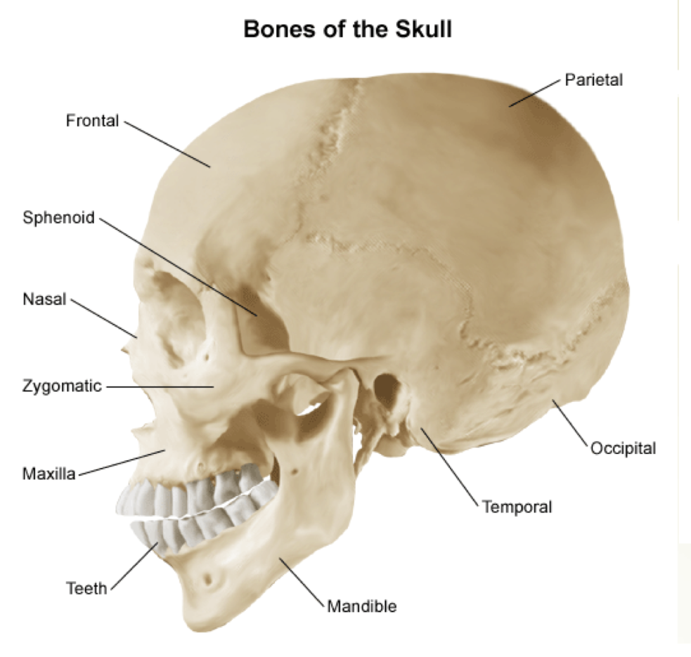
Major Regions of the Skull
Frontal
Parietal
Occipital
Temporal
Mandible
Maxilla
Nasal
Zygomatic
Sphenoid
The Spine (Vertebrae): # and type of vertebrae
7 - Cervical
12 - Thoracic
5 - Lumbar
5 - Sacral
4 - Coccygeal
Which vertebrae is the biggest and has the largest body
Lumbar vertebrae
How many ribs are there
24
Which ribs are true, false, and floating
True: 1-7
False: 8-10
Floating: 11-12
Which vertebrae are the ribs attached to
Thoracic
What attaches the ribs to the sternum
Cartilage
What make up the shoulder complex
Clavicle
Scapula
Glenoid
Bones that make up the arm
Humerus
Ulna
Radius
Radius vs. Ulna
Radius:
thumb side
Moves during pronation
Ulna:
pinky side
Doesn’t move during pronation
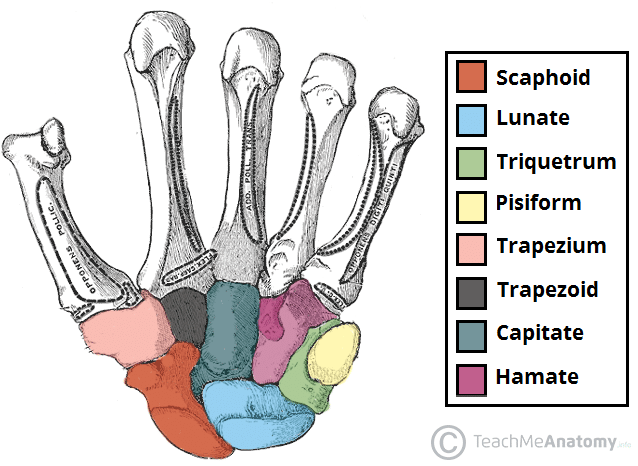
Names and order of carpal bones
Some Lovers Try Positions That They Can’t Handle
Scaphid
Lunate
Triquetrum
Pisiform
Trapesium
Trapezoid
Capitate
Hamate

How many metacarpals, phalanges, and phalanx
Metacarpals: 5 (numbered I to V)
Phalanges: 5
Phalanx: 14
2 in digit I (thumb) — proximal and distal
3 in digits 11 to V — proximal, middle, and distal
Parts of the pelvis what do they form when put together
Illium
Ischium
Pubis
Acetabulum

Bones in the leg
Femur
Tibia: big toe side
Fibula: pinky side
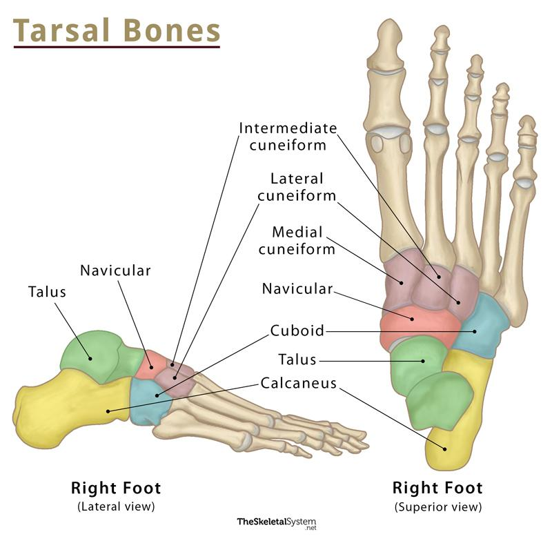
Name and order of the tarsals
Talus
Calcaneus
Navicular
Cuboid
Cuneiforms
Lateral
Intermediate
Medial
How many metatarsals, phalanges, and phalanx
Metatarsals: 5
Phalanges: 5
Phalanx: 14
2 on 1st phalange (big toe) — proximal and distal
3 on 2-5 Phalanges — proximal, middle, and distal
What does arthrology mean and what is a joint
The study of joints
Where two or more bones articulate with one another
What is the function of joints
Allow efficient movement patterns
Provide stability to the bony skeleton
What is the Structural Classification of Joints
Synovial
Fibrous
Cartilaginous
What is the Functional Classification of Joints
Synarthrosis
Amphiarthrosis
Syndesmosis
Synchondrosis
Symphysis
Diarthrosis
Hinge
Pivot
Condyloid
Ellipsoid
Saddle
Ball and Socket
Plane (gliding)
What are the two types of synarthroidal joints
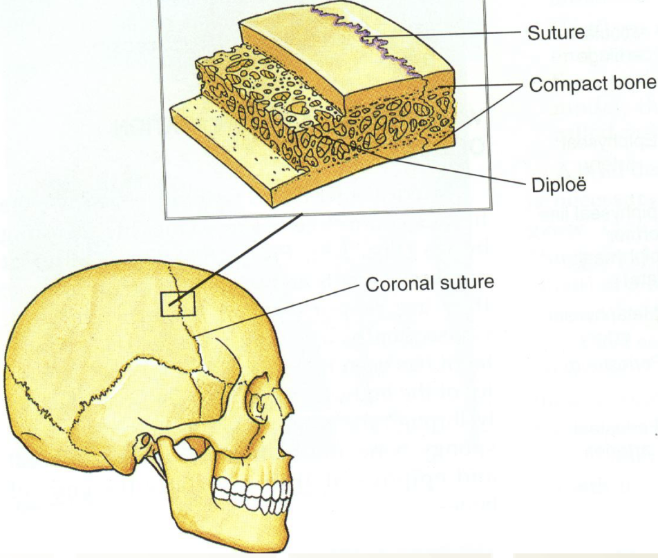
Sutures
Gomphoses
Characteristics sutures (joint def.)
Immovable fibrous joints
Fibrous periosteum forms between bones
Joints of the skull
Characteristics of gomphoses
Attachment of a tooth in its socket
Tooth held in place by fibrous periodontal ligament
Allows tooth to move slightly while chewing

What are two types of amphiarthroidal joints
Ligamentous
Cartilaginous
Characteristics of ligamentous joints
Slightly moveable
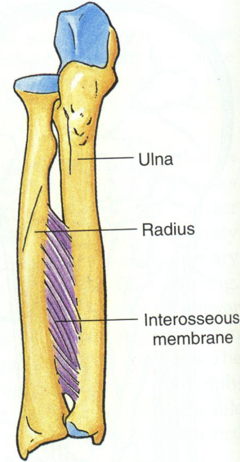
Syndesmosis (fibrous joint where two adjacent bones are strongly united by ligaments or a broad sheet of connective tissue)
Ligamentous interosseous membrane provides stablility
Characteristics of cartilaginous joints
Slightly moveable
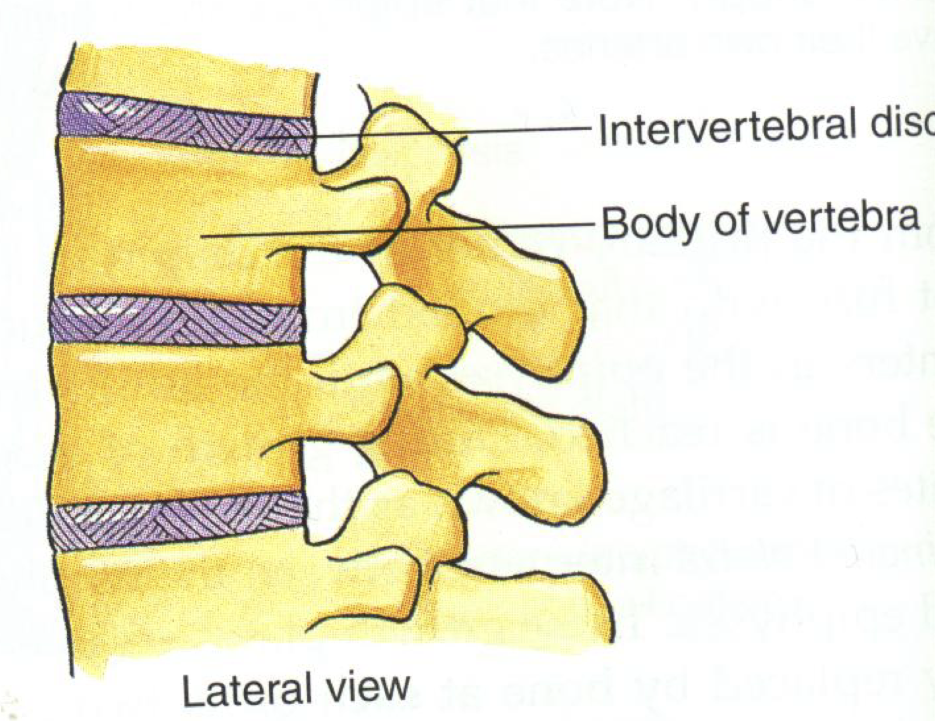
Bones separated by fibrocartilaginous disk
Joints reinforced by ligaments
Intervertebral discs
Pubic symphysis
Characteristics of Diarthroidal joints
Synovial joints (has synovial fluid to lubricate joint)
Very mobile
Ligamentous joint capsule
Synovial membrane
Synovial fluid
Articular cartilage
Need all points in blue to be considered a true diarthroidal joint
Characteristics of gliding joints and examples
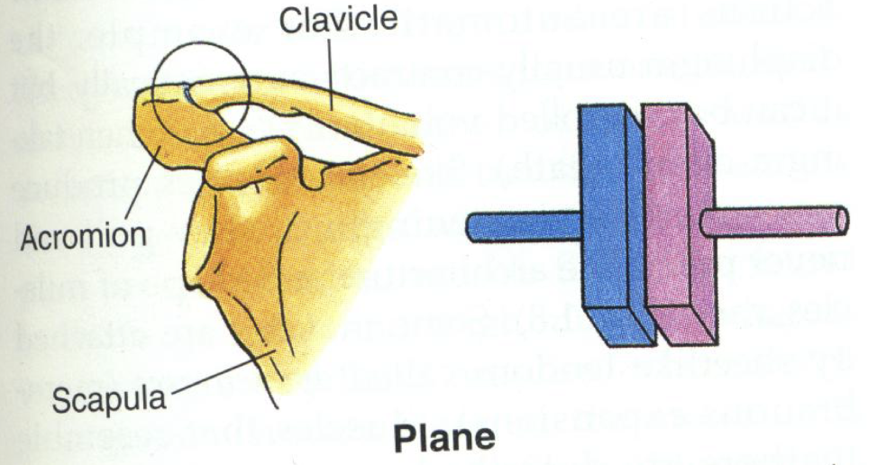
aka “plane” joint
Nonaxial
can move superior/inferior, anterior/posterior, or rotate
Examples:
Acromioclavicular joint
Intercarpal and intertarsal joints
Characteristics of hinge joints and examples
aka “ginglymus”
Uniaxial
Motions of flexion and extension are allowed (just sagittal)
Example:
Ulnohumeral joint

Characteristics of pivot joints and examples
aka “trochoid”
Uniaxial
only rotation
Example:
Atlandtoaxial joint (between C1 & C2 vertebrae)
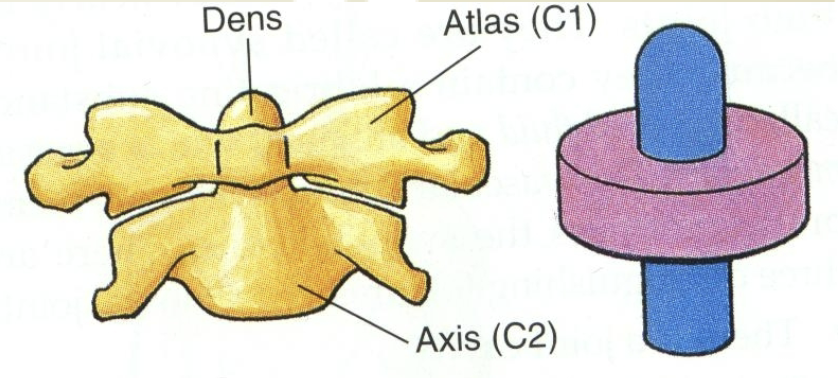
Characteristics of ellipsoid joints and examples
biaxial
flexion/extension
abduction/adduction
circumduction
rotation not possible
convex:concave surfaces
ex: glenoid (convex) & humerus (concave)
Example:
Radiocarpal joint

Characteristics of condyloid joints and examples
Biaxial
Flexion/extension
Rotation
Convex:concave surfaces
Example:
Metacarpophalangeal joint
Tibiofemoral joint

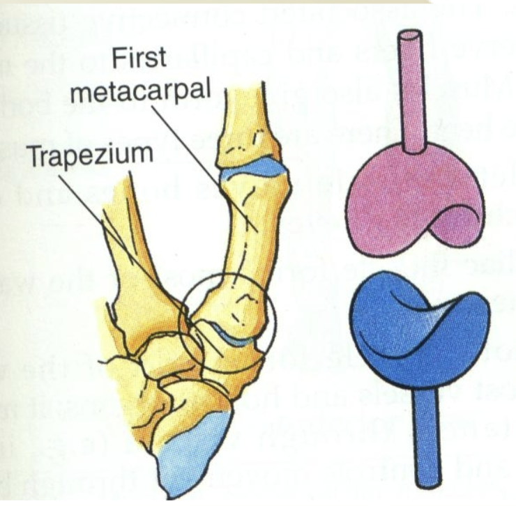
Characteristics of saddle joints and examples
Biaxial
flexion/extension
abduction/adduction
rotation
circumduction
Both bones have saddle shapes
saddles fit together
Example:
Carpometacarpal joint of thumb
Characteristics of ball and socket joints and examples
Triaxial
flextion/extension
abduction/adduction
rotation
circumduction
Example:
Hip
Glenohumeral

What do ligaments do
Connect bone to bone
Two functions of ligaments
Mechanical function
hold adjacent bones together
Sensory function
provide feedback to nervous system regarding joint motion and joint position (proprioception)
What is the difference between an extrinsic and intrinsic ligament
Extrinsic:
outside joint capsule
Intrinsic:
inside joint capsule

What is the ligament structure
Primary cells are fibroblasts
Most prevalent fibrous component is collagen
Type I is most prevalent
Proteoglycans also present
Similar to structure to tendon, but more elastic
2/3 of extracellular matrix is water
Bundles of collagen fibers comprise ligaments
Bundles can be arranged:
In parallel
Obliquely
Spiral arrangement
Arrangement of fibers is dictated by ligament function
What is collagen and the characteristics of the 3 types
The primary structural protein of the body — different types depend on structure and function
Type I
Dense fibrils
High tensile strength
ex: tendon & ligaments
Type II
>50% dry wt of hyaline cartilage
Less dense fibrils
ex: articular cartilage and nucleus of disc
Type III
Immature collagen
least tensile strength
associated with Type I
ex: vessels, skin

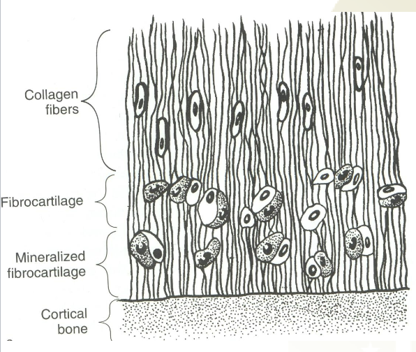
What is the structure of ligament attachment to bone
4 Zones: (gets more stiff when closer to bone)
Ligament (collagen)
Fibrocartilage
Calcified fibrocartilage
Cortical bone
Sharpey’s Fibers
Ligamentous fibers which penetrate fibrocartilage
Some ligamentous fibers also blend with periosteum