MHHS2401- Musculoskeletal System
 Call Kai
 Learn
Learn Practice Test
 Spaced Repetition
 Match
Match Flashcards
 Knowt Play
1/136

Earn XP
Description and Tags
Clinsci year 1- session 2
Name | Mastery | Learn | Test | Matching | Spaced | Call with Kai | Chat |
|---|
No analytics yet
Send a link to your students to track their progress
137 Terms
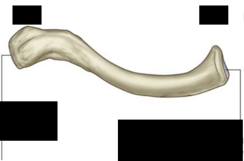
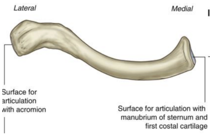
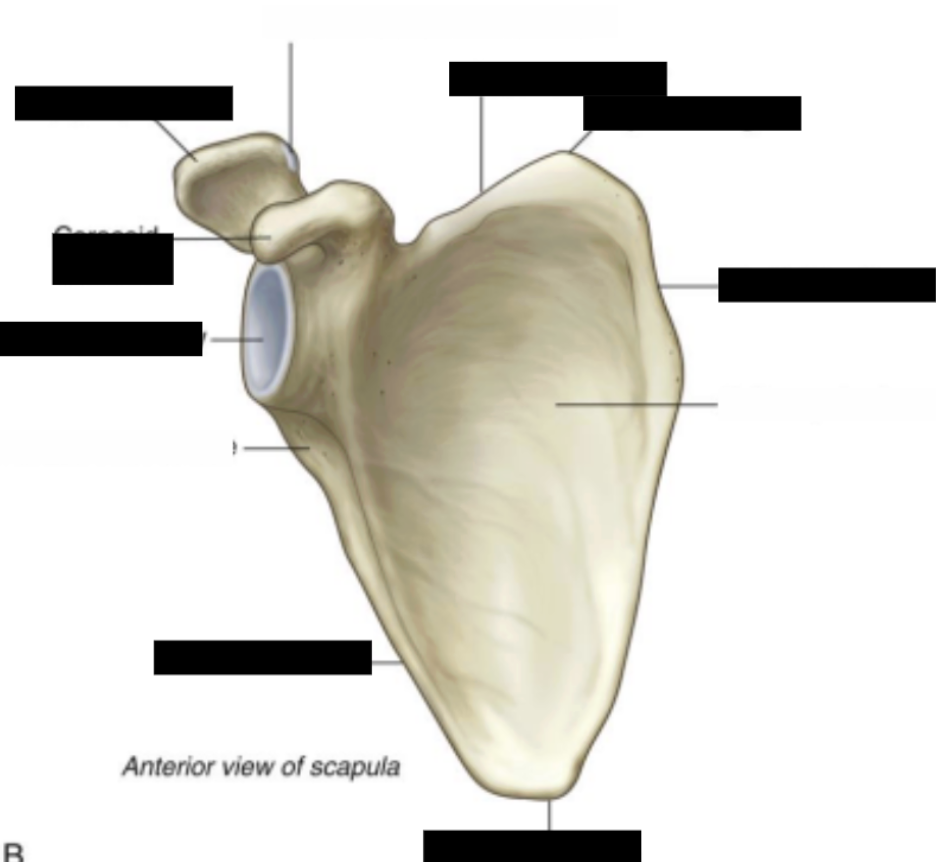
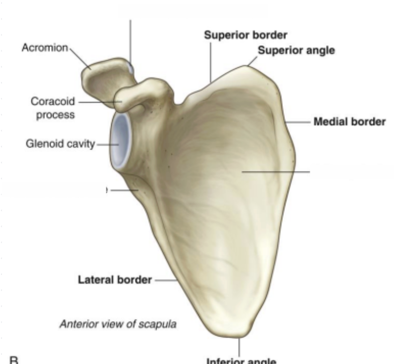
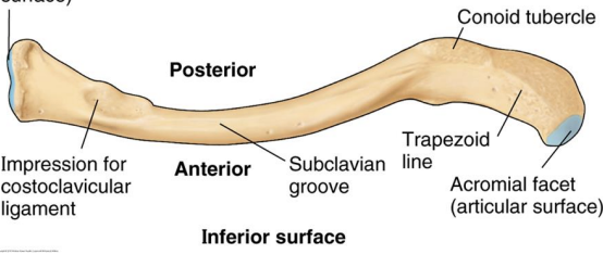
costoclavicular ligament
The impression on the medial inferior surface of the clavicle is the site for the attachment of the:
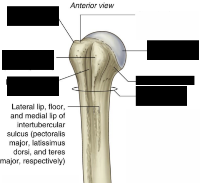
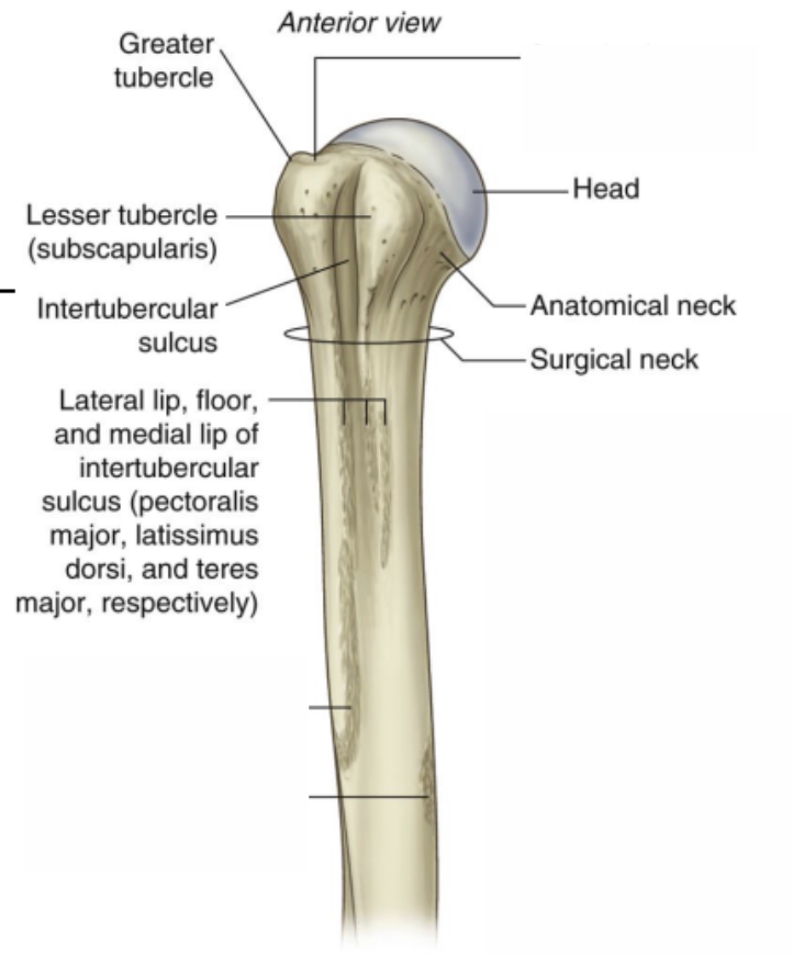
The other name for the intertubercular sulcus is ___
Bicipital groove
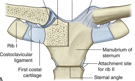
what joint is Sternoclavicular classified as
saddle
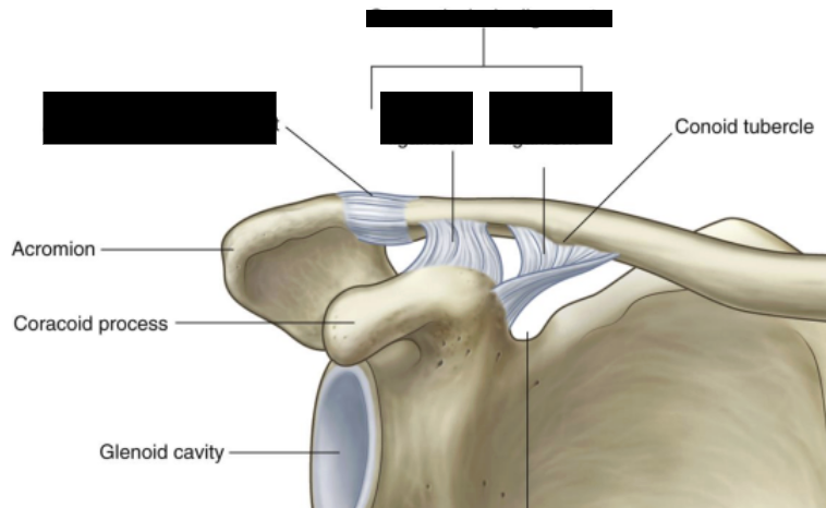
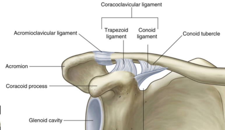
1- Coracoclavicular Ligament
2- Trapezoid Ligament
3-conoid ligament
4-acromioclavicular ligament
Shoulder Joint- Glenoid labrum
deepens Glenoid cavity
ring of fibrocartilage
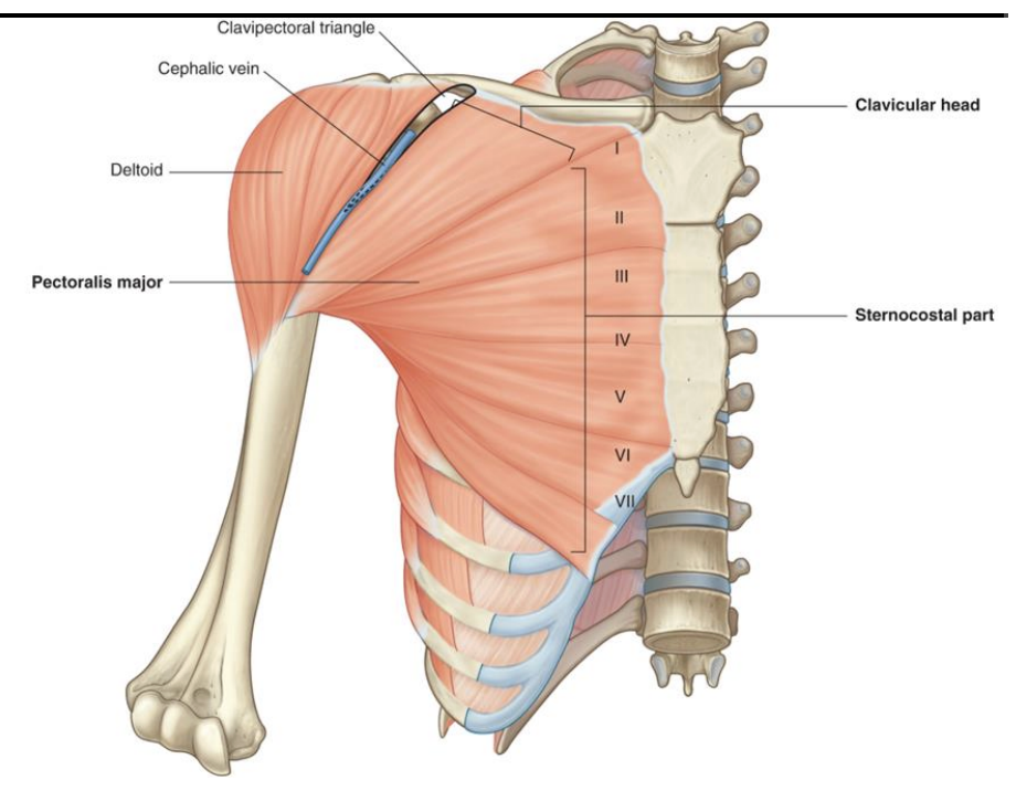
Anterior muscle group: Pectoralis Major
two origins: Clavicle and sternocostal
Distal attachment: Lateral lip of bicipital groove (Intertubercular sulcus)
Action: Helps move humerus through adduction and flexion
Nerve Supply: Lateral and medial pectoral nerves
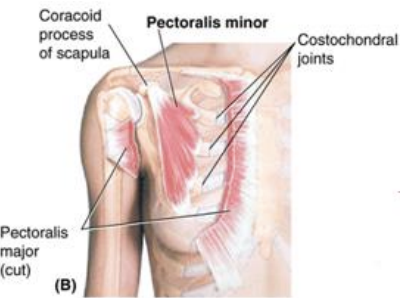
Pectoralis Minor
Origin: Ribs 3 → 5
Distal Attachment: Coracoid process of scapula
Action: Stabilizes scapula and helps in respiration
Nerve Supply: Medial pectoral nerve
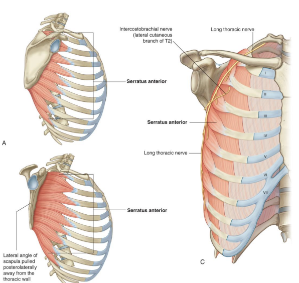
Serratus Anterior Muscle (Boxer Muscle)
Origin: Ribs 1 → 8
Distal Attachment: Anterior (costal) surface of medial scapular border
Action: protracts scapula and holds it to thoracic wall when punching, rotates scapula
Nerve Supply: Long Thoracic nerve
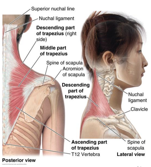
Trapezius
Origin: C7 → T12
Distal Attachment: Lateral part of clavicle and acromion and spine of scapula
Action: Elevates scapula (Superior fibers), retracts scapula (Middle fibers), depresses scapula (Inferior Fibers)
Nerve Supply: Spinal root of CNXI- motor fibers and cervical nerves C3 and C4 (sensory)
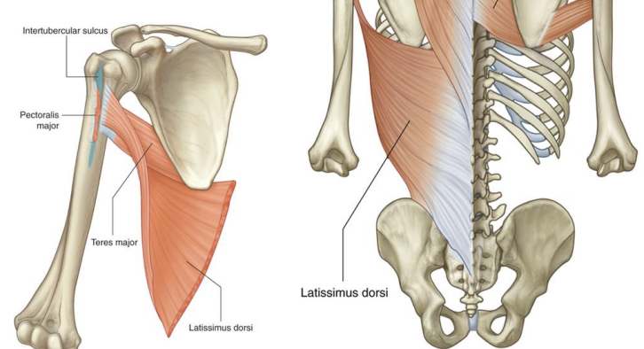
Latissimus dorsi
Origin: T7 → T12
Distal Attachment: Floor of intertubercular groove (Bicipital groove)
Action: movement of humerus and helps body with climbing
Nerve Supply: Thoracodorsal nerve
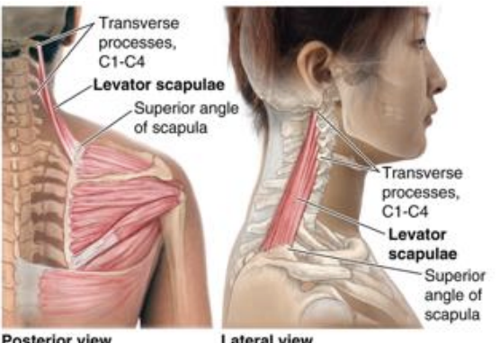
Levator scapulae
Origin: C1 → C4
Distal Attachment: superior part of medial border of scapula
Action: elevates scapula and tilts glenoid cavity inferiorly
Nerve Supply: Dorsal scapular nerve
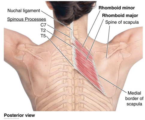
Rhomboid minor and major
Origin: Major = T2 → T5, minor = C7 and T1
Distal Attachment: medial border of scapula
Action: retract and rotate scapula
Nerve Supply: Dorsal scapular nerve
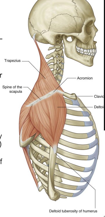
Deltoid Muscle
Origin: lateral part of clavicle,, acromion and spine of scapula
Distal Attachment: deltoid tuberosity
Action: Anterior fibers = flexes arm, middle fibers = abduction of arm, posterior fibers = extend arm
Nerve Supply: axillary nerve
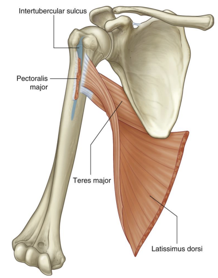
Teres major
Origin: Inferior lateral border of scapula
Distal Attachment: medial lip of intertubercular groove
Action: abduction and medial rotation of at at shoulder joint
Nerve Supply: lower subscapular nerve
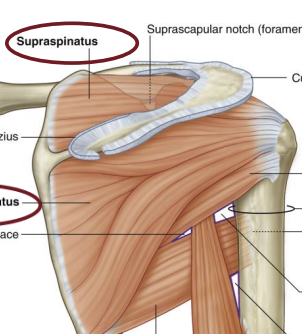
Rotator Cuff Muscle: Supraspinatus
Origin: Supraspinous fossa
Distal Attachment: Greater tubercle
Action: Abduction of arm
Nerve Supply: Supscapular nerve
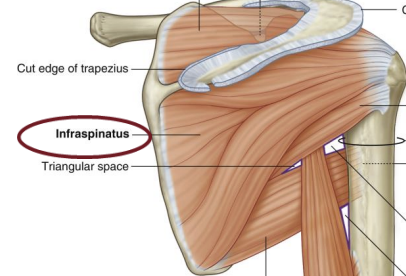
Rotator Cuff Muscle: Infraspinatus
Origin: Infraspinous fossa
Distal Attachment: Greater tubercle
Action: Lateral rotator of shoulder
Nerve Supply: subscapular nerve
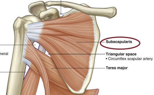
Rotator Cuff Muscle: subscapularis
Origin: Superior part of lateral border of scapula
Distal Attachment: Lesser tubercle
Action: Medial rotator of humerus
Nerve Supply: upper and lower subscapular nerve
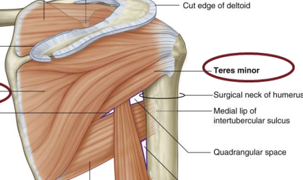
Rotator Cuff Muscle: Tres minor
Origin: Lateral border of scapula
Distal Attachment: Greater tubercle
Action: Lateral rotator of shoulder
Nerve Supply: Axillary nerve
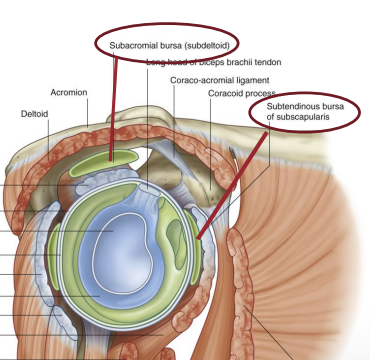
Subtendinous Bursa and subacromial bursa (subdeltoid bursa)
Where tendon rubs against bone
acts as cushion
Bone structure- Osteoprogenitors
Stem cells that differentiate into osteoblasts
Bone structure- Osteoblast
For bone formation
Release bone matrix
Bone structure- Bone matrix (Osteoid / hydroxyapatite)
Osteoid: Organic, mainly collagen
Hydroxyapatite: inorganic, calcium + phosphate
Bone structure- Osteocytes
get stuck in bone matrix
former osteoblasts
Bone structure- Osteoclast
Bone resorption
Proteases break down osteoid
HCL break down hydroxyapatite
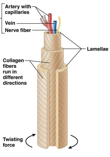
Bone structure- Lamellae
layers of bone
Bone structure- Osteons (Haversian system) / central canal (Haversian canal)
Osteons: lamellae form osteons
Central canal: empty space where blood supply and nerves come
Bone structure- Spongey bone (Trabecular bone)
Irregular lamellae forming trabecular
NOT RANDOM- align along stress lines and develop fro micro fractures caused by weight bearing
Blood supply comes from Endosteal capillaries
Bone structure- Compact bone
Fibers not parallel to resist torsional (twisting) forces and gives strength
collagen and hydroxyapatite
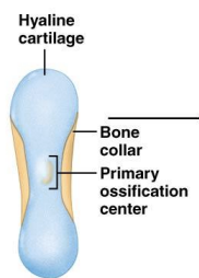
Formation- What happens in week 9 utero when starting ossification in long bone?
Hyaline cartilage model developed bone collar and primary ossification centre
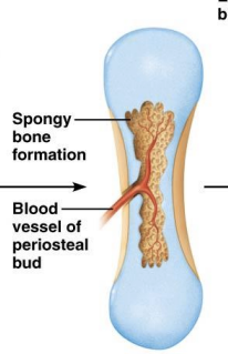
Formation- Structure during 3 months of utero?
cartilage matric deterioates
spongey bone formation begins
Formation- Structure during birth?
medullary cavity
secondary ossification centres apear in epiphyses
Formation- Growth plate
between epiphysis and diaphysis
articular cartilage
Formation- Longitudinal bone growth
ends at the end of puberty
happens at growth plate between head and shaft of long bone
Formation- Endochondral ossification
where chondrocytes lay down cartilage foundation and osteoblast replace it with trabecular bone matrix
Remodelling and Hormones- RANK-ligand (RANKL)
secreted by osteoblast
Activated osteoclast
Remodelling and Hormones- OPG (osteoprotegerin)
Secreted by osteoblast
Blocks osteoclast activation
Allows for less resorption
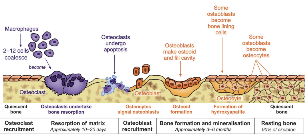
Remodelling and Hormones- squence
osteoclast recruitment
Rsorption of matrix
osteoblast recruitment
bone formation and mineralisation
resting bone (90% of bone most the time)
Remodelling and Hormones- apoptosis
Osteoclasts undergo when they die
Remodelling and Hormones- Appositional growth
Growth in bone diameter
occurring at periosteum of diaphysis
Remodelling and Hormones- PTH
increased blood Ca
Secreted from parathyroid gland
Remodelling and Hormones- Calcitriol
made in kidney
Stimulated by PTH
increases blood Ca
Remodelling and Hormones- Calcitonin
Secreted by Thyroid gland
decreases blood Ca
promotes bone formation
Remodelling and Hormones- PTH continuous and Intermittent exposure
Continuous exposure: catabolic (↑RANKL ↓OPG → resorption)
Intermittent exposure: Daily low-dose → Anabolic → ↑osteoblast differentiation, ↓osteoblast apoptosis → formation
Remodelling and Hormones- Vitamin D3 biosynthesis
Diet
Liver
Kindey (PTH induced)
Remodelling and Hormones- HGH
Allows for formation of bone
↑osteoblast proliferation, ↑apposition, ↑longitudinal growth.
Remodelling and Hormones- TH
Allows for bone resorption
↑osteoblast differentiation, ↓osteoblast proliferation, ↑osteoclast activity.
Remodelling and Hormones- Glucocorticoids (eg. cortasol)
Allows for resorption
↑RANKL, ↓OPG.
Remodelling and Hormones- Estrogen
Allows for bone formation
↓RANKL, ↑OPG, ↑osteoclast apoptosis (effect observed in females).
Remodelling and Hormones- Andogens
Allows for bone formation
↓RANKL, ↑OPG, ↑apposition, ↑longitudinal growth.
Disorders- Osteoporosis
Metabolic
Osteoclast hyperactivity
Due to endocrine supression
Disorders- Osteomalacia
metabolic
Abnormal bone mineralisation due to vitamin D deficiency
Disorders- Rickets
same as osteomalacia
happens in kids
Disorders- Brown tumour
metabolic
Caused by hyperparathyroidism
Vascular growths in bone
Disorders- Osteogenesis Imperfecta
Genetic
Defective collagen type 1
Disorders- osteopetrosis
Genetic
Defective osteoclast
Thick but weak bone
no medullary cavity
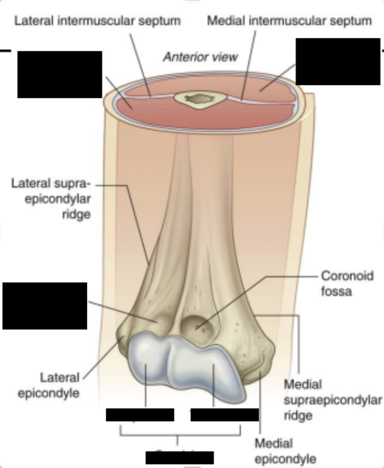
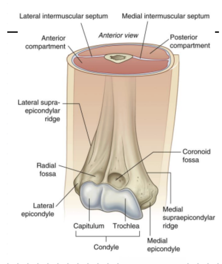
Bony features
Elbow- Joint type and movments?
synovial hinge Joint
flexion and extension only
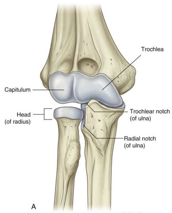
Elbow- Articular surfaces (3)
Capitulum + trochlea
Head of radius
Trochlear notch of ulna
Elbow- Fibrous capsule
weak anteriorly and posteriorly
Thickened medially and laterally to form collateral ligamnets
Elbow- Collateral ligaments
Lateral (radial) collateral ligament
Medial (ulnar) collateral ligament (3 bands; anterior, posterior and oblique)
Elbow- Carrying angle
angle between acis of arm and forearm in anatomical position
Greater in females (> 15% degrees)
Males (10-15 degrees)
disappears when supinated
Elbow- Students elbow
Olecranon bursitis get inflamed
Elbow- Proximal radioulnar joint
synovial pivot joint
articular surfaces: Head of radius and radial notch of ulna
anular ligament of radius holds it together and allows pronation and supination
Arm muscle- intramuscular septum
divided arm muscles into anterior and posterior
Anterior Arm muscle- 3 muscles that make up anterior compartment
biceps brachii
brachialis
coracobrachialis
nerve supply: musculocutaneous nerve
Anterior Arm muscle- Biceps brachii
Long head Origin: Supraglenoid tubercle of scapula
Short head Origin: coracoid process of scapula
Insertion: Radial tuberosity
Action: Flexor at elbow and powerful supinator of forearm
Nerve supply: musculocutaneous
Anterior Arm muscle- Coracobrachialis
Origin: coracoid process of scapula
Insertion: Mid shaft of humerus
Action: Weak flexor and adductor of arm
Nerve supply: musculocutaneous
Anterior Arm muscle- Brachialis
Origin: Mid-shaft of humerus (deep to biceps)
Insertion: Ulnar tuberosity
Action: Chief flexor of forearm
Nerve Supply: musculocutaneous
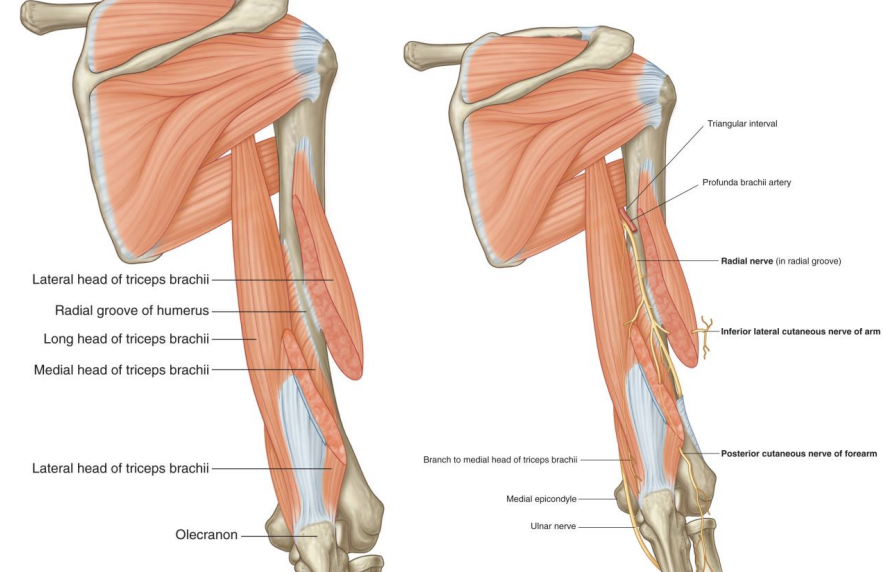
Posterior arm muscle- Triceps brachii
Nerve supply: Radial Nerve
Origin: Long head- infraglenoid tubercle of scapula, medial head- posterior humerus (inferior to radial groove), lateral head- posterior humerus superior to radial groove
Insertion: Olectanon process
Action: stabilizes humeral head when abducts arm and chief extensor of forearm
Distal radioulnar joint- Interosseous membrane
structure that connects radius and ulna
stabilises bone
provides attachment for deep forearm muscle
allows for passage of blood vessels
Distal radioulnar joint- Intermuscular septa
divides forearm into compartments:
-→posterior: extensors / supernators
→ anterior: flexors / pronators
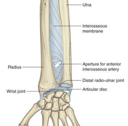
Distal radioulnar joint- what kind of joint / articular surfaces
synovial pivot joint
articular surfaces include:
-→Head of ulna
-→ulnar notch of distal radius
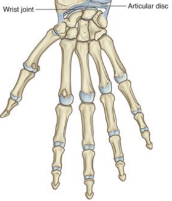
Distal radioulnar joint- Fibrocartilaginous articular disc
unites distal radioulnar joint
separates wrist joint cavity
Injuries- Posterior dislocation of elbow joint
caused by fall on outstretched hands with elbow flexed
distal humerus driven through weak anterior part of fibrous capsule
Injuries- Pulled elbow
dislocation of radial head from sudden pulling of upper limb
distal attachment of anular ligament at neck of radius broken
Injuries- golfer’s elbow (medial epicondylitis)
Overuse of flexor-pronator muscles
causes microtrauma
Anterior forearm- 3 layers
Superficial (4 muscles)
→ common attachment is medial epicondyle of humerus
Intermediate (1 muscle)
deep (3 muscles)
innervated by median nerve
Anterior forearm- Nerve supply
innervated by median nerve
Except flexor carpi ulnaris and medial half of flexor digitorum profundus
-→ both supplied by ulnar nerve
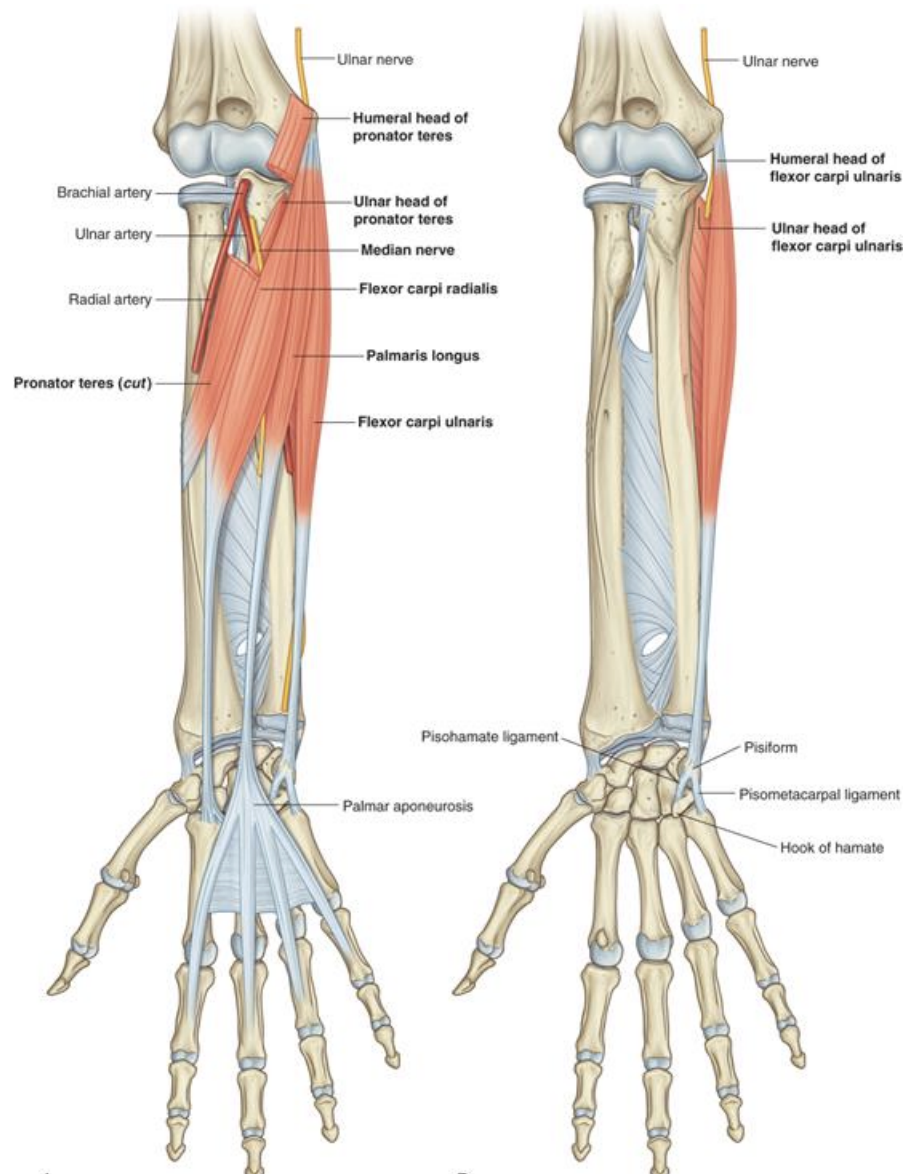
Anterior forearm (superior)- Flexor carpi radialis
insertion: base of 2nd metacarpal
action: abducts and flexes hand at wrist
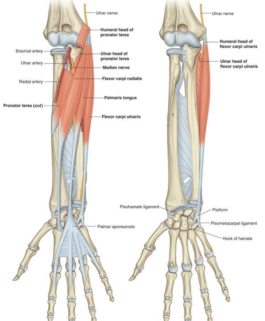
Anterior forearm (superior)- Pronator teres
insertion: Mid shaft of radius
action: pronates forearm
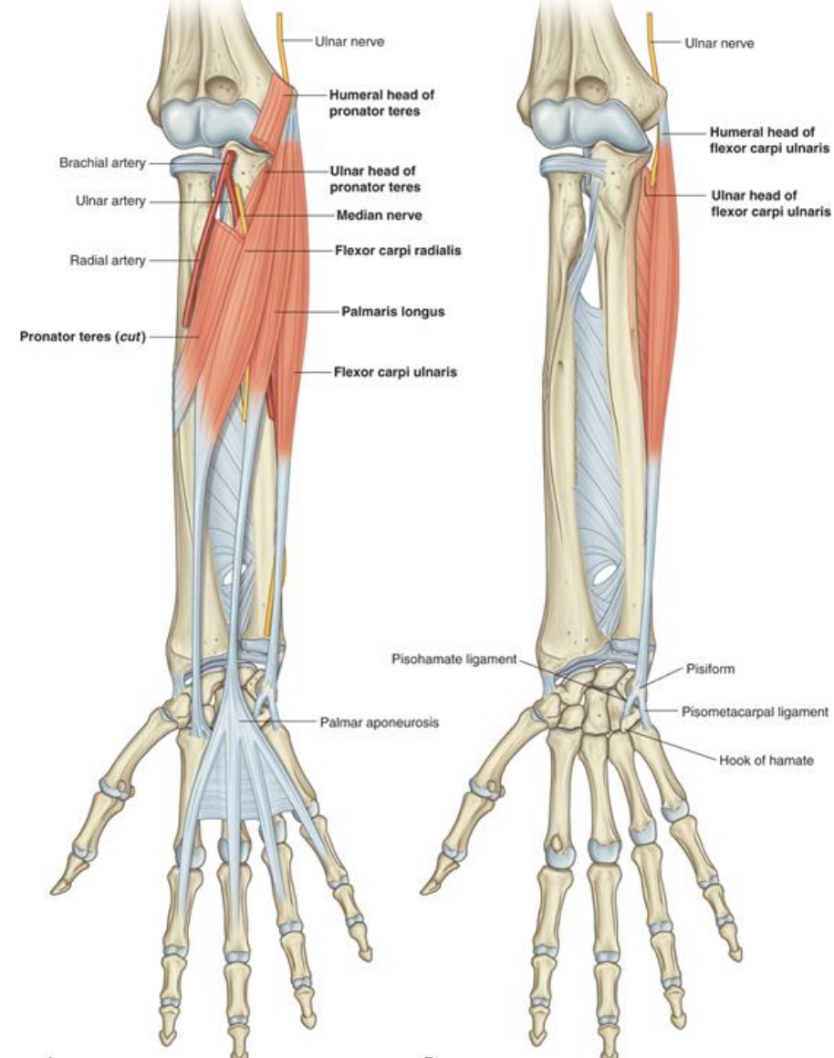
Anterior forearm (superior)- Palmaris longus
absent in 14% of people
blends in with palmar aponeurosis
flexes hand at wrist
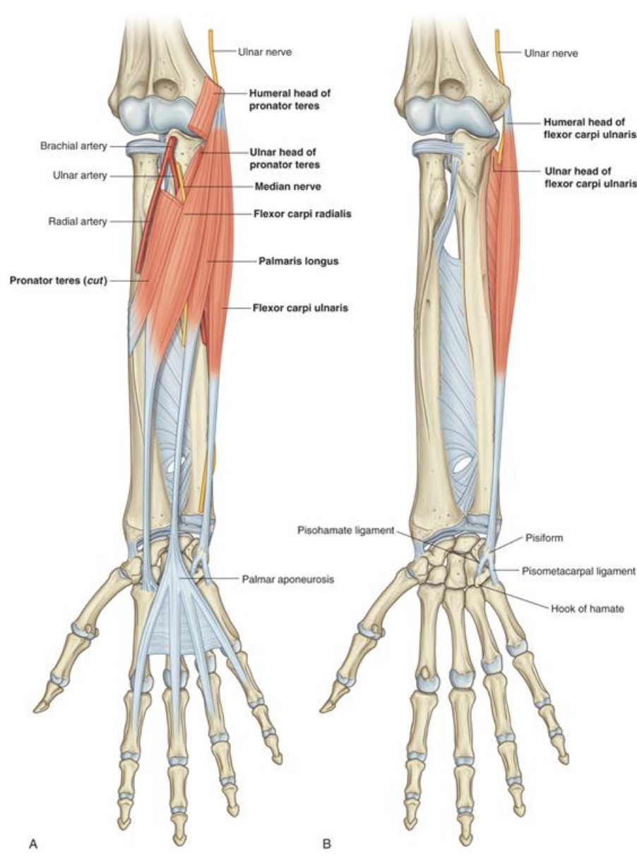
Anterior forearm (superior)- Flexor carpi ulnaris
insertion: pisiform, hook of hamate and 5th metacarpal
action: flexes and adducts hand at wrist
Nerve supply: ulnar nerve
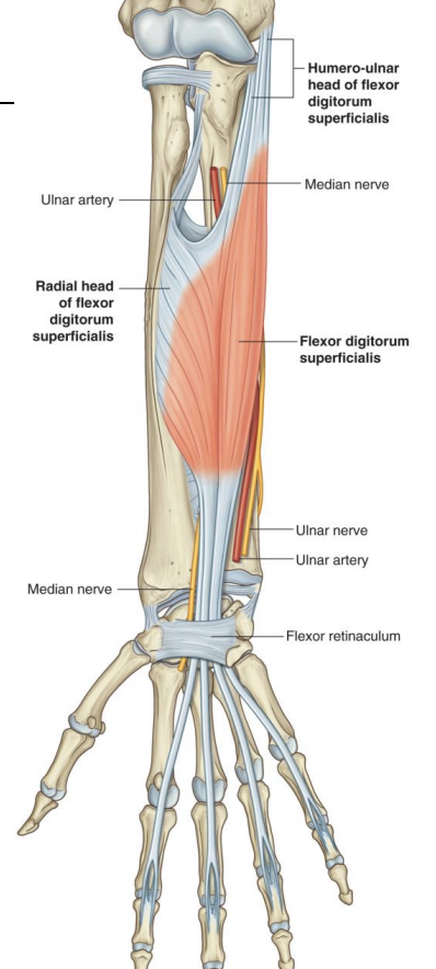
Anterior forearm (Intermediate)- Flexor digitorum superficialis
origin: medial epicondyle of humerus
insertion: middle phalanges of the medial 4 digits
action: flexes middle phalanges at PIP joints and flexes proximal phalanges at MCP joint
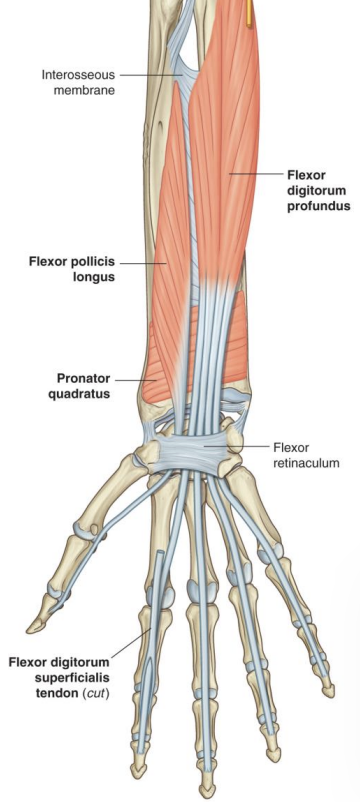
Anterior forearm (Deep)- Flexor pollicis longus
Action: Flexes phalanges of thumb
insertion: base of distal phalanx of thumb
origin: anterior surface of radius + interosseous membrane
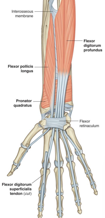
Anterior forearm (Deep)- flexor digitorum profundus
Action: Flexes distal phalanges at DIP joints, MCP joints and assists hand flexion
insertion: distal phalanges of medial 4 digits
origin: proximal anterior surface of ulna
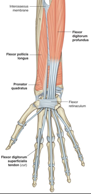
Anterior forearm (Deep)- pronator quadratus
Attachment: distal anterior surfaces of radius and ulna
origin: Pronates forearm
Posterior Forearm Muscle- Superficial Group
Origin: Lateral Epicondyle of Humerus
Nerve Supply: Radial Nerve
6 muscles:
→ Brachioradialis
→ Extensor carpi radialis Longus (Longer) (ECRL)
→ Extensor carpi radialis brevis (shorter) (ECRB)
→ Extensor digitorum
→ Extensor digiti minimi
→ Extensor carpi ulnaris
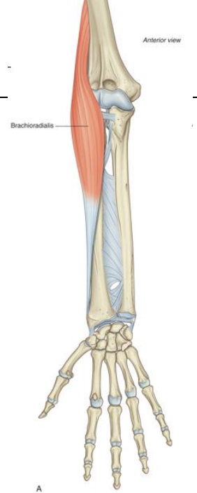
Posterior Forearm Muscle- Brachioradialis
Insertion: Lateral surface of distal radius
Action: Flexor of forearm at elbow
→ only posterior muscle that doesn’t cross wrist joint
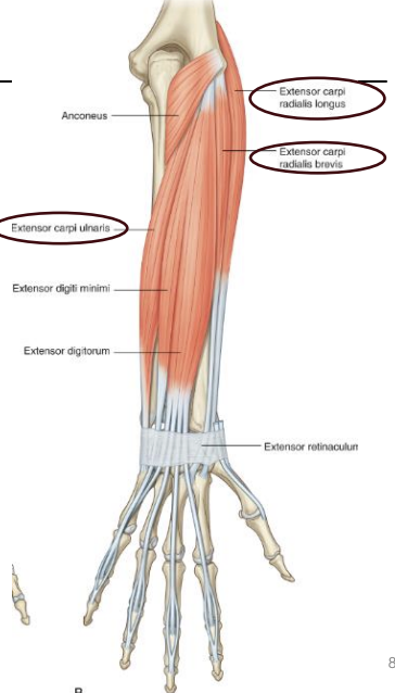
Posterior Forearm Muscle- Extensor carpi radialis Longus (ECRL) / Extensor carpi radialis brevis (ECRB)
Action: Wrist extensors, produce abduction at wrist, limited about 15 degrees
Nerve: Deep branch of radial nerve
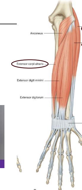
Posterior Forearm Muscle- Extensor carpi ulnaris (ECU)
Action: Wrist Extensor, adduction at wrist joint
Nerve: Deep branch of radial nerve
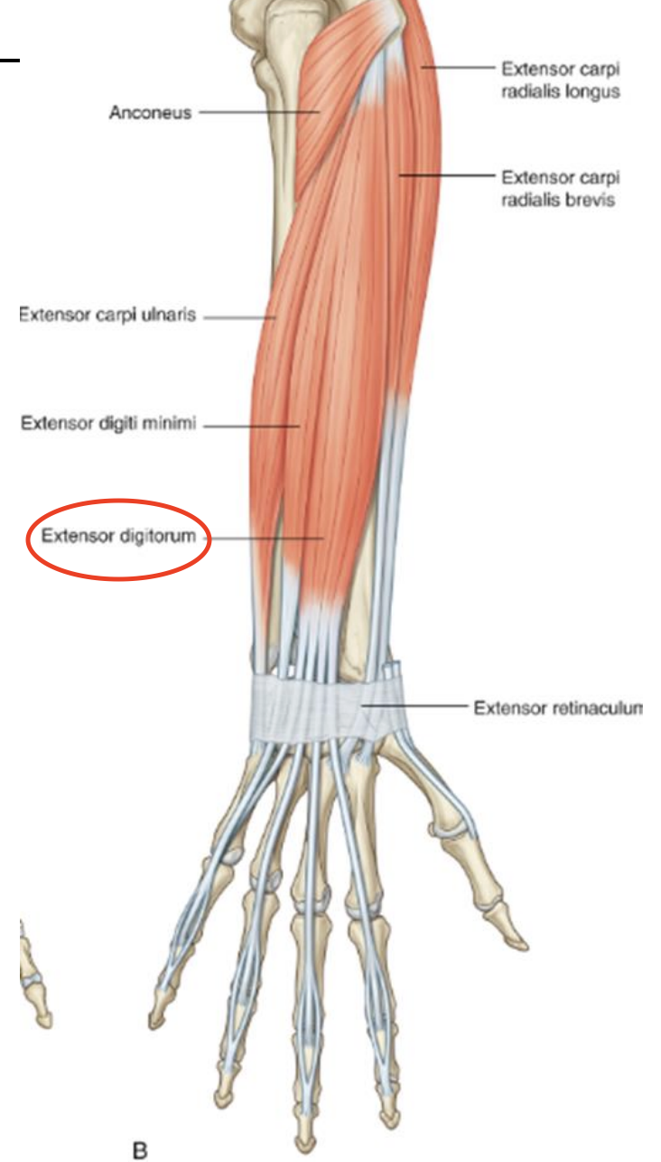
Posterior Forearm Muscle- Extensor digitorum
Insertion: Extensor expansion of medial 4 digits
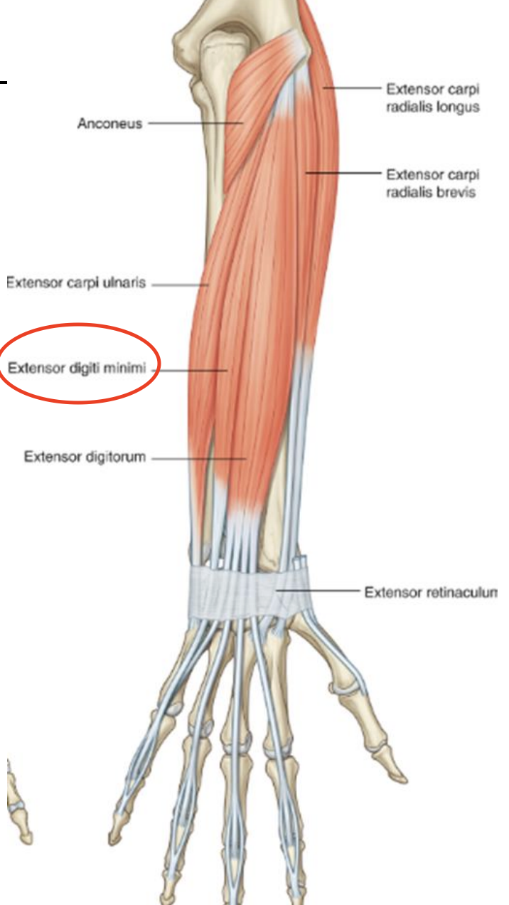
Posterior Forearm Muscle- Extensor digitorum
Action: primary extending at Metacarpal joint
Insertion: extensor expansion of 5th digit
Posterior Forearm Muscle- Extensor Expansion
Divides into medial band and two lateral bands
Lumbrical and dorsal interossei muscle attatch to medial band
Posterior Forearm Muscle- Extensor retinaculum
holds extensor tendons in place at wrist
Posterior Forearm Muscle- Synocial Tendon sheaths
Helps reduce friction against bone and tendons
Posterior Forearm Muscle- Deep Group
5 muscles
→ Supinator
→Abductor pollicis longus
→ Extensor pollicis bevis
→ Extensor pollicis longus
→ Extensor indicis
Posterior Forearm Muscle- Supinator
Location: Cubital fossa
insertion: Proximal radius
Origin: Lateral epicondyle of humerus
Action:
Nerve: Radial nerve