Intro to Pathology and Morphological Diagnoses
 Call Kai
 Learn
Learn Practice Test
 Spaced Repetition
 Match
Match Flashcards
 Knowt Play
1/91

There's no tags or description
Looks like no tags are added yet.
Name | Mastery | Learn | Test | Matching | Spaced | Call with Kai | Chat |
|---|
No analytics yet
Send a link to your students to track their progress
92 Terms
Health
the normal condition of the body, in which all cells, tissues, organs, and systems are structurally intact and functioning properly
Disease
any detectable deviation from the normal range
Three forms of evidence for disease:
1.) symptoms
2.) signs
3.) lesions
Four main aspects of disease:
1.) etiology
2.) pathogenesis
3.) morphologic changes
4.) functional consequences
etiology
anything that can disrupt the normal functioning of a cell, tissue, organ, or system; what is "causing" the disease
etiology is aka...
the cause of the disease
Two types of etiology:
1.) intrinsic
2.) extrinsic
intrinsic etiology
cause of disease from inside the body; uncontrollable individual-specific factors predisposing the host to disease
Three types of intrinsic etiology
1.) primary
2.) secondary
3.) idiosyncratic
primary intrinsic etiology
genetic mutations in germ or somatic cells that arise spontaneously either during embryogenesis (congenital) or after birth (acquired)
examples of a primary intrinsic etiology
-hip dysplasia in german shepherds
-brain tumors in boxers
secondary intrinsic etiology
Physiologic abnormalities related to age, sex, species; modify the expression of a disease
examples of secondary intrinsic etiology
-age --> younger animals more affected by infectious disease, older animals more affected by neoplasia
-color --> deafness in white cats
-sex --> mammary cancer in female dogs, urinary obstruction in male cats
-species specific --> swine get hog cholera, horses get laminitis
idiosyncratic intrinsic etiology
a patient-specific disease; often related to enzymopathy (inherited or acquired)
idiosyncratic intrinsic etiologies may be triggered by ________ factors
extrinsic
extrinsic etiology
cause of disease from outside the body
Two types of extrinsic etiology
1.) animate agents
2.) inanimate causes
extrinsic etiology: animate agents
aka pathogens and their products (fungi, bacteria, parasites, viruses, etc)
extrinsic etiology: inanimate causes
energy (radiation, thermal), physical forces (pressure, trauma), surgical intervention, xenobiotics (chemicals, drugs, toxins)
Pathogenesis
the sequence of events from the start to the end of a disease
Pathogenesis encompases both ______ and _______ to a disease
mechanisms and responses
Lesions
a visible manifestation of disease; the structural or morphologic effects of a disease
Three levels that lesions are visualized:
1.) gross pathology
2.) histopathology
3.) ultrastructural
gross pathology
macroscopic; seen with the naked eye
histopathology
microscopic; seen using a light microscope
ultrastructural
cell organelles; seen with an electron microscope
Lesions are identifiable as a departure from ________
normal
Lesions usually fall into three general categories:
1.) something has been added
2.) something has been taken away
3.) something has been altered
Lesions where something has been added
The tissue contains something not normally present (cells, fluid, pigment, microbial agents, foreign bodies, etc)
Lesions where something has been taken away
The tissue looks different with normal elements gone (atrophy, hypoplasia, degeneration, necrosis)
Lesions where something has been altered
The normal tissue components are present but not normal (malformations, dysplasia, metaplasia, neoplasia)
metaplasia
when one tissue type turns into another tissue type
Examples of attributes of lesions:
-color
-consistency
-contours
-distribution
-duration
-extent
-location
-margins
-shape
-size
-texture
Pathognomonic lesions
A lesion so distinctive and unique that it can only be caused by a single etiologic agent or disease process
Pathognomonic lesions have very high _______
specificity (if present, it confirms a disease)
*ex: diamond shaped rash confirms swine erysipelas
Necropsy
post mortem dissection of an
animal to determine the cause of death
Autopsy
the post mortem dissection of a human being to define the cause of death
Biopsy
the surgical removal of a representative sample of tissue from a suspicious lesion for a diagnosis
Prognosis
prediction of a future outcome for the patient (excellent, fair, guarded, poor)
Diagnosis
determination of the nature or cause of a disease
Three types of diagnosis:
1.) morphologic diagnosis
2.) etiologic diagnosis
3.) disease diagnosis
morphologic diagnosis
An orderly, detailed description of a lesion; a summary of the lesion, but generally does not describe what is causing the lesion
Five components for a morphologic diagnosis:
1.) affected structure (cell, tissue, organ)
2.) distribution
3.) nature of lesion (process)
4.) severity
5.) duration
Four ways to describe distribution:
1.) focal
2.) multifocal
3.) locally extensive
4.) diffuse

focal
Limited to one site
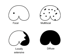
focally to locally extensive
All of the tissue in a zone is affected

multifocal
multiple sites

multifocal to coalescing
Many lesions present that appear to be growing together or fusing
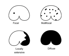
diffuse
involves the entire tissue/organ; no normal tissue is left
Two types of the nature of a lesion (process)
1.) non-inflammatory
2.) inflammatory
non-inflammatory process
does not involve the body's immune-mediated inflammatory response
Six examples of a non-inflammatory process:
1.) degeneration
2.) pigmentation
3.) mineralization
4.) disturbances of growth
5.) neoplasms
6.) displacement of viscera
degeneration
the deterioration of tissues
pigmentation
deposited pigment or color in a tissue
mineralization
calcium depositions in the tissue
disturbances of growth
atrophy, hypertrophy, hyperplasia, etc
neoplasms
abnormal growths of new tissue that are classified as benign or malignant
How are neoplasms named?
Organ + name of neoplasm
Ex: Kidney: Lymphosarcoma -or- Renal lymphosarcoma
displacement of viscera
volvulus, torsion
inflammatory process
does involve the body's immune-mediated inflammatory response
What suffix is added to the end of an organ when inflammation is involved?
"itis"
Five descriptors for severity of a lesion:
1.) minimal
2.) slight/mild
3.) moderate
4.) marked/moderately severe
5.) severe
Four descriptors for duration of a lesion:
1.) acute
2.) subacute
3.) chronic
4.) chronic-active
acute
hours to a day or so
subacute
one or a few days
chronic
several days to weeks, months, or years
chronic-active
chronic duration but ongoing inciting stimulus
Example of a morphologic diagnosis
Acute, severe, diffuse, suppurative encephalitis
What descriptor must be used for a lesions on paired organs?
unilateral or bilateral
Three paired organs:
1.) Adrenal glands
2.) Kidneys
3.) Repro organs (testis, ovaries, etc)
etiologic diagnosis
A diagnosis that names the cause or agent of the disease; comprised of two words- the causative agent (etiology) and the site (organ or tissue) (i.e., mycobacterial enteritis)
Three descriptors of an etiologic diagnosis
1.) agent or cause
2.) location
3.) process
Examples of an etiologic diagnosis
-Microsporidial encephalitis
-Parvoviral enteritis
disease diagnosis
A diagnosis that states the "name of the disease" (ex Johne's disease)

The term for the distinctive target lesions pictured that are characteristic of Histomonas meleagridis in a bird is:
A Pathogenesis
B Prognostic
C Predictived
D Pathognomonic
Pathognomonic
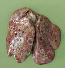
What is the distribution of this lesion?
A Focal
B Locally extensive
C Multifocal to coalescing
D Diffuse
Multifocal to coalescing
Which of the Following is an Etiologic
Diagnosis?
A Intestine: Severe, diffuse, granulomatous enteritis
B Intestinal histoplasmosis
B Intestinal histoplasmosis
*A is a morphological diagnosisf
Which of the Following is the Morphologic Diagnosis?
A Clostridial enteritis
B Small intestine: Enteritis, necrohemorrhagic, acute, diffuse,
marked, with vasculitis, edema, and numerous bacilli
C Clostridium perfringens type C
B Small intestine: Enteritis, necrohemorrhagic, acute, diffuse,
marked, with vasculitis, edema, and numerous bacilli
*A is etiologic, C is the etiology

Case 1: What organs are present?
What two organs are present here?
a Liver and Gallbladder
b Stomach and Intestine
c Heart and Lung
d Spleen and Lymph node
c Heart and Lung
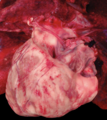
Case 1: Has anything been added or removed?
a added
b removed
a added
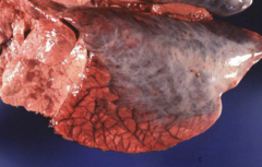
Case 2: What is the Lesion Distribution?
a Focal
b Locally Extensive
c Multifocal/Coalescing
d Diffuse
b Locally Extensive

Case 2: Has something been Added or Removed?
a Added
b Removed
a Added

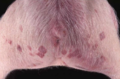
Case 3: What is the lesion distribution?
a Focal
b Multifocal/Coalescing
c Locally Extensive
d Diffuse
b Multifocal/Coalescing

Case 3: What do you think the Duration is for this Lesion?
a Acute
b Subacute
c Chronic
a Acute
*Lesions are bright to dark red (hemorrhage), a chronic hemorrhage would be green/blue/brown as blood is broken down (almost like a bruise), no fibrosis present
Case 4:

Case 5:

Case 5:

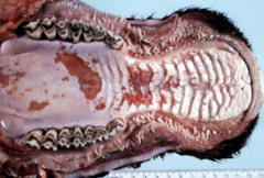
Case 6:
Case 6:

Case 6:
What type of Diagnosis is the following? Patient presents with chronic, severe, multifocal granulomatous pneumonia.
A Etiologic diagnosis
B Disease diagnosis
C Condition
D Morphologic diagnosis
D Morphologic diagnosis
Which of the following is an etiologic diagnosis?
A Pulmonary lymphoma
B Johne's disease
C Diffuse granulomatous pneumonia
D Mycotic rumenitis
D Mycotic rumenitis