Nervous System Info Pt. 2
 Call Kai
 Learn
Learn Practice Test
 Spaced Repetition
 Match
Match Flashcards
 Knowt Play
1/152

There's no tags or description
Looks like no tags are added yet.
Name | Mastery | Learn | Test | Matching | Spaced | Call with Kai | Chat |
|---|
No analytics yet
Send a link to your students to track their progress
153 Terms
Meninges
Membranes that protect the brain and spinal cord; lie between bone and soft tissue
Dura Mater
Tough, dense connective tissue that is the outermost meninge
Arachnoid Mater
Middle meninge, web-like; space contains CSF
Pia Mater
Inner most meninge; attached to surface of brain and spinal cord; contains blood vessels and nerves; nourishes CNS
Interventricular Foramina
Connects the third to the lateral ventricles
Cerebral Aqueduct
Connects the third and fourth ventricles
Choroid Plexuses
Secrete CSF, special capillaries of pia mater covered by ependymal cells
Arachnoid Granulations
Absorbs CSF after it exchanges substances
140 ml
Average volume of CSF
Forebrain
Prosencephalon
Midbrain
Mesencephalon
Hindbrain
Rhombencephalon
Forebrain
Divides into the telencephalon and diencephalon
Hindbrain
Divides into metencephalon and myelencephalon
Mesencephalon
Persists throughout development (use the fancy name)
Forebrain
Becomes the cerebrum, basal nuclei, and diencephalon
Midbrain
Mesencephalon remains as …
Hindbrain
Becomes cerebellum, pons, and medulla oblongata
Telencephalon
Anterior portion of forebrain; gives rise to the lateral ventricles, cerebrum, and basal nuclei
Diencephalon
Posterior portion of the forebrain; gives rise to the third ventricle, thalamus, hypothalamus, posterior pituitary gland, and pineal gland
Mesencephalon
Gives rise to the cerebral aqueduct and midbrain (fancy name)
Metencephalon
Anterior portion of hindbrain; gives rise to the fourth ventricle, cerebellum, and pons
Myelencephalon
Posterior portion of the hindbrain; gives rise to the fourth ventricle and medulla oblongata
Falx Cerebri
Separates cerebral hemispheres
Corpus Callosum
Connects cerebral hemispheres
Gyri
Ridges in the brain
Sulci
Shallow grooves on the surface of the brain
Fissures
Deep grooves in the surface of the brain
Longitudinal Fissure
Separates the cerebral hemispheres (fissure)
Transverse Fissure
Separates the cerebrum from the cerebellum
Cerebral Cortex
Contains abt 75% of all neuron cell bodies in NS; thin layer of gray matter on outside of cerebrum
White Matter
Contains bundles of myelinated axons that connect cell bodies to other portions of the NS
Cutaneous Sensory Area
In parietal lobe; interprets sensations on skin
Wernicke’s Area or sensory speech area
Found in temporal/parietal lobe; usually left hemisphere; understanding and formulating language
Visual Area
Found in occipital lobe, interprets vision
Auditory area
Found in temporal lobe, interprets hearing
Sensory area for taste
Found near the base of the central sulcus, includes part of insula
Sensory area for smell
Arises from centers deep within the temporal lobes
Association Areas
Regions that are not primarily motor or sensory, connect the cerebral cortex; analyze and interpret sensory information; provide memory, reasoning, verbalization, judgement, and emotions
Frontal Lobe Association Areas
Concentration, planning, complex problem solving; emotional behavior, judging consequences
Parietal Lobe Association Areas
Understanding speech, choosing words to express thoughts and feelings
Temporal lobe association areas
Interpret complex sensory experiences, stores memories of visual scenes, music, and complex patterns
Occipital lobe association areas
Analyze and combine visual images with other sensory experiences
Insula
Translates sensory info into proper emotional responses
Primary motor areas or motor cortex
Found in frontal lobes, controls voluntary muscles, most nerve fibers cross over in brainstem
Broca’s area
Anterior to primary motor cortex, usually in left hemisphere, controls muscles needed for speech
Frontal Eye field
Above Broca’s area, controls voluntary movements of eyes and eyelids
Short-term memory
Neurons connected in a circuit that is constantly stimulated; when impulse flow ceases, so does memory
Long-term memory
Changes structure or function of neurons, makes new synaptic connections by increasing branching of processes
Long-term potentiation
Increase in neurotransmitter release and effectiveness of synaptic transmission upon repeated stimulation
Basal nuclei
Masses of gray matter deep within cerebral hemispheres; produce dopamine and control voluntary movement
Nucleus, putamen, globus pallidus
3 parts of basal nuclei
Thalamus
Gateway for sensory impulses ascending to cortex; receives all sensory impulses (except smell); channels impulses to appropriate part of cortex
Hypothalamus
Maintains homeostasis by regulating visceral activities
Limbic system
Consists of several structures in various brain parts; controls emotional responses, feelings, survival oriented behavior; reacts to potentially life-threatening upsets
Midbrain
Contains bundles of fibers that join lower parts of brainstem and spinal cord with higher part of brain
Cerebral peduncles
main motor pathways that connect cerebrum to lower nervous system
Corpora quadrigemina
Centers for visual and auditory reflexes
Red nucleus
Role in postural reflexes
Pons
Relays impulses between medulla oblongata and cerebrum and from cerebrum to cerebellum; regulates rhythm of breathing
Medulla Oblongata
Conducts ascending and descending impulses between brain and spinal cord; contains cardiac, vasomotor, respiratory, and nonvital control centers
Reticular Formation
filters incoming sensory info, passing some to cortex and discarding the rest; arouses cortex into wakefulness; decreased activity cause sleep
Slow wave
Person is tired, decreasing activity of reticular formation, restfulness and dreamlessness, reduced BP and RR, 3 stages
REM
HR and RR irregular, dreams
Cerebellum
Contains dentate nucleus; integrate sensory info abt the body position, coordinates muscle activity and posture
Falx cerebelli
Separates cerebellum
Vermis
Connects cerebellum halves
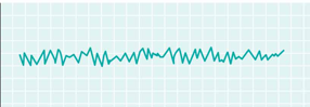
Alpha
Type of wave; awake, resting eyes closed
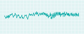
Beta
Type of wave; active mental activity, under tension
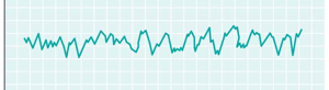
Theta
Type of wave; mostly in children, in some adults in sleep or stress
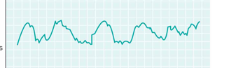
Delta
Type of wave mostly in sleep
Alpha
Beta
Theta
Delta
Cervical Enlargement
Supplies nerves to upper limb; part of spinal cord
Lumbar Enlargement
Supplies nerves to lower limbs; part of spinal cord
Conus Medullaris
Tapering region of spinal cord below lumbar enlargement
Filum Terminale
Cord of connective tissue that anchors spinal cord to coccyx
Cauda Equina
Group of lumbar and sacral nerves extending downward from conus medullaris in vertebral canal
Anterior Median fissure and posterior median sulcus
2 grooves that extend whole length of spinal cord
Funiculi
White matter in spinal cord is arranged into …
Horns
Gray matter in the spinal cord is arranged into …
Monosynaptic reflex
Contains 2 neurons: sensory and motor
One synapse in spinal cord
Polysynaptic
Contains sensory neuron, interneuron, and motor neuron
Reciprocal innervation
Withdrawal reflex; flexors contract, extensors are inhibited
Ascending tract
Conducts sensory impulses to brain
Descending tracts
Conduct motor impulses from the brain to motor neurons and effectors
Fasciculus gracilis and cuneatus
2 tracts; ascending; conduct sensory impulses associated with the sense of touch, pressure, and body movement from skin, muscles, tendons, and joints to the brain; located in the posterior funiculi
Spinothalamic tract
Located in lateral and anterior funiculi; ascending; conducts sensory impulses associated with the senses of pain, temperature, touch, and pressure from various body regions to the brain
Spinocerebellar tracts
Located in lateral funiculi; ascending; conducts sensory impulses required for coordination of muscle movements form muscles of lower limbs and trunk to cerebellum
Corticospinal tracts
Descending tract; located in lateral and anterior funiculi; conducts motor impulses associated with voluntary movements from brain to skeletal muscles
Reticulospinal tracts
Descending tract; located in lateral and anterior funiculi; conducts motor impulses associated with the maintenance of muscle tone and activity of sweat glands from brain
Rubrospinal tracts
Descending tract; located in lateral and anterior funiculi; conducts motor impulses associated with muscular coordination from the brain
Somatic NS
Cranial and spinal nerves that connect CNS to skin and skeletal muscles
Autonomic NS
Cranial and spinal nerves that connect CNS to viscera
Endoneurium
Loose connective tissue surrounding individual axons
Perineurium
Loose connective tissue surrounding fascicles
Epineurium
Dense connective tissue that surrounds a group of fascicles
Nerve
Bundle of axons in the PNS