ANTH 327 Prac 1
 Call Kai
 Learn
Learn Practice Test
 Spaced Repetition
 Match
Match Flashcards
 Knowt Play
1/47

There's no tags or description
Looks like no tags are added yet.
Name | Mastery | Learn | Test | Matching | Spaced | Call with Kai | Chat |
|---|
No analytics yet
Send a link to your students to track their progress
48 Terms
Axial skeleton vs Appendicular skeleton
Axial skeleton: skull, vertebral column, rib cage
Appendicular skeleton: limbs, shoulder girdle, pelvic girdle
Median/saggital plane
A vertical plane that divides the body into equal right and left halves.
Transverse/horizontal plane
A horizontal plane that divides the body into upper (superior) and lower (inferior) parts.
Frontal/coronal plane
A vertical plane that divides the body into front (anterior) and back (posterior) sections.
Anterior (ventral) vs Posterior (dorsal)
Anterior: Front facing
Posterior: Back facing
Superior vs Inferior
Superior: Toward the head or upper part of a structure
Inferior: Away from the head or toward the lower part of a structure
Medial vs Lateral
Medial: Toward the midline of the body
Lateral: Away from the midline of the body
Proximal vs Distal
Proximal: Closer to the point of attachment or origin of a limb or structure
Distal: Farther from the point of attachment or origin of a limb or structure
dorsal-palmar vs dorsal-plantar
Dorsal-palmar: Referring to the back (dorsal) and palm (palmar) surfaces of the hand
Dorsal-plantar: Referring to the top (dorsal) and sole (plantar) surfaces of the foot
What is circumduction movement?
A limb movement combining flexion, extension, abduction, and adduction in a circular path.
What is abduction movement? What is adduction movement?
Abduction is the movement of a limb away from the midline of the body
Adduction is the movement of a limb toward the midline of the body.
What is flexion movement? What is extension movement?
Flexion is the bending of a joint that decreases the angle between two bones
Extension is the straightening of a joint that increases the angle between two bones
What is the gliding movement?
Gliding movement is the sliding of one bone over another, as seen in the wrist bones
What is the rotation movement?
Rotation movement is the turning of a bone around its own long axis, such as when the head shakes "no."
What is supination? What is pronation?
Supination is the rotation of the forearm or foot so that the palm or sole faces upward or forward
Pronation is the rotation of the forearm or foot so that the palm or sole faces downward or backward
What is origin?
The point where a muscle or tendon is anchored to a bone, typically remaining stationary when the muscle contracts.Attachment site on bone that is more stable during movement
What is insertion?
The point where a muscle or tendon attaches to the bone that moves when the muscle contracts; it is typically less stable than the origin.
What are tendons?
Fibrous tissue that connects muscle to bone
What are ligaments?
Fibrous tissue that connects bone to bone
What is articulations?
The point where two or more bones meet, forming a connection that allows for movement, stability, or both
What are the various types of synovial joints?
Synovial joints
Spheroidal
Hinge
Saddle-shaped
Planar
Pivot
Condyloid
Waht is the synovial joint?
A type of joint that is surrounded by a fluid-filled capsule, allowing for smooth movement between the bones it connects.
What is spheroidal joint (aka ball and socket joint)?
A type of synovial joint where a spherical or rounded head fits into a cup-like cavity, allowing movement in multiple directions, including rotation
Hip
Shoulder
Sternoclavicular
What is a hinge joint?
A type of synovial joint that allows movement primarily in one plane, similar to the motion of a door hinge
Elbow
Knee
Finger/Toes
What is a saddle-shaped joint?
A type of synovial joint where the articulating surfaces are shaped like a saddle, allowing movement back and forth and side to side but limited rotation
Thumb
What is a planar joint?
A type of synovial joint where flat or slightly curved surfaces slide past one another, allowing limited gliding movements
Wrist
Ankle
What is a pivot joint?
A type of synovial joint that allows rotational movement around a single axis
Atlas
Axis
What is the condyloid joint?
A type of synovial joint where an oval-shaped condyle fits into an elliptical cavity, allowing movement in two planes, such as flexion-extension and abduction-adduction.
Wrist joint
What are cartilaginous (synchondroses) joints?
Joints where the bones are connected entirely by cartilage, allowing limited movement
Pubic symphysis
Growth plates
Rib cage
What are fibrous (syndesmoses) joints?
Joints where bones are connected by dense connective tissue, allowing little to no movement
Cranial sutures
Distal tibiofibular articulation
What is the make up of bone?
Organic matrix: 30% of wt but 90% of organic is collagen
Inorganic matrix: 70% of wt but made up of hydroxyapatite crystals
What marrow produces red and white blood cells?
Red bone marrow
What marrow has deposits of fat? Where is the medullary cavity found in the bone?
Yellow bone marrow; the medullary cavity is found in the shafts of long bones.
What are the types of bones?
Compact (cortical)
Subchondral
Spongy (trabecular)
Diploe
Marrow
Trabeculae
What is the difference between periosteum and endosteum?
The periosteum is a dense layer of vascular connective tissue that covers the outer surface of bones
Endosteum is a thin membrane lining the inner surfaces of the bone, including the medullary cavity
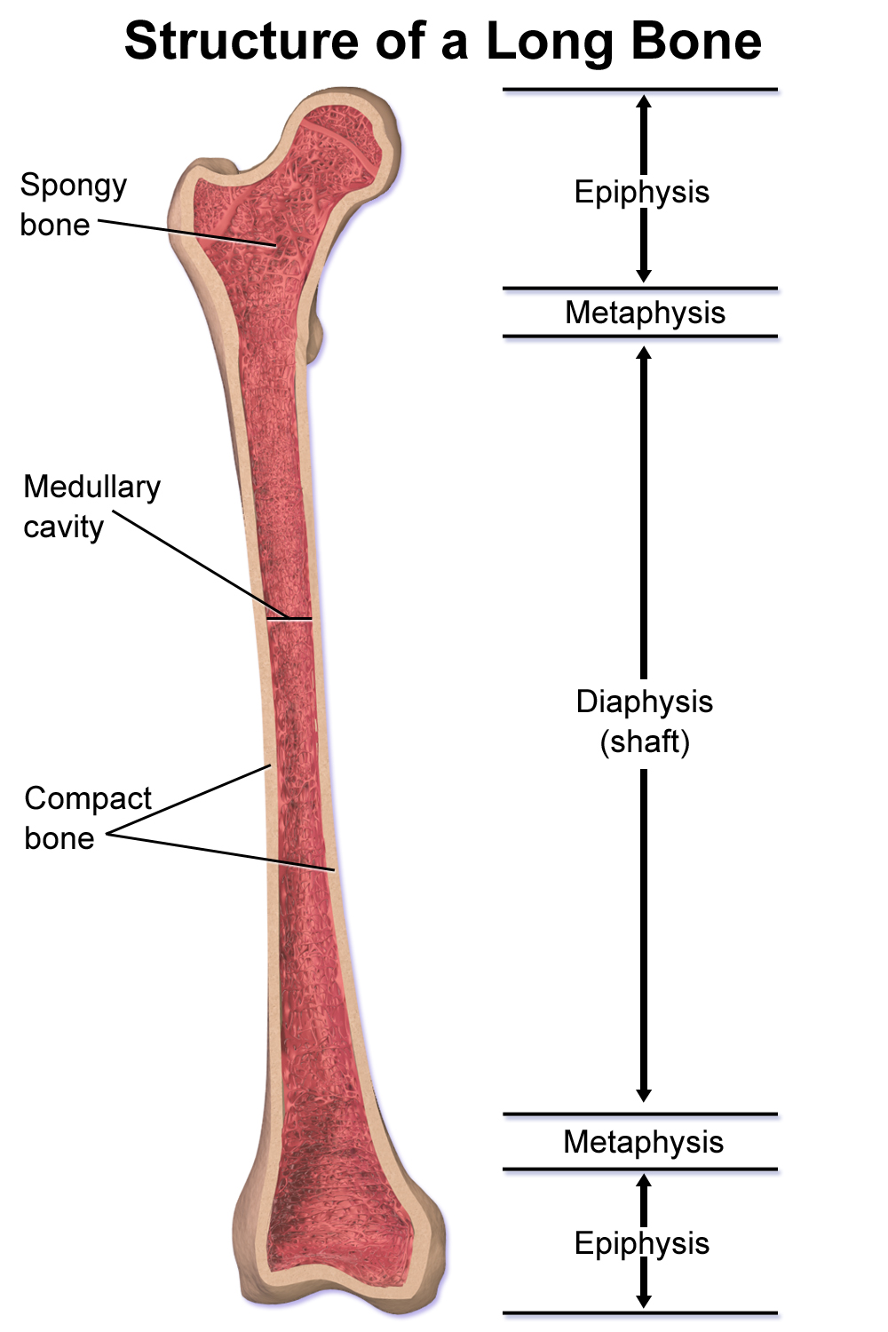
What are the long bone components?
Diaphysis – the shaft or central part of a long bone
Epiphysis – the rounded ends of a long bone, which articulate with other bones
Metaphysis – the region between the diaphysis and epiphysis, containing the growth plate in children
Articular cartilage – smooth tissue covering the epiphysis to reduce friction in joints
Medullary cavity – central cavity within the diaphysis containing bone marrow
What is osteon?
The basic unit of compact bone with concentric rings around a central canal containing blood vessels and nerves.
What is haversian canal?
What is lamella(e)?
What is lacuna(e)?
What is canaliculi?
• Haversian canal – a central channel in an osteon containing blood vessels and nerves
• Lamella(e) – concentric rings of bone matrix around the Haversian canal
• Lacuna(e) – small spaces housing osteocytes within the bone
• Canaliculi – tiny canals connecting lacunae, allowing nutrient and waste exchange between osteocytes
Primary vs secondary osteons?
Primary osteons form during initial bone growth with fewer lamellae
Secondary osteons arise later through remodeling with more lamellae around a larger Haversian canal
What are the different types of bone cells?
Osteocytes: Mature bone cell that maintains the bone matrix within lacunae
Osteoblast: Matrix-synthesizing cell responsible for bone growth, found at edge of bone
Osteoclast: Bone-reabsorbing cell
Osteoblasts vs Osteoclasts
Osteoblasts: Responsible for forming/building bone
Osteoclasts: Responsible for dissolving bone
What does Wolff’s law state?
Every change in the form or function of a bone is followed by adaptive changes in its internal architecture and its external shape
Intramembranous vs Endochondral bone growth?
Intramembranous bone growth: Bone develops directly from mesenchymal tissue without a cartilage stage.
Endochondral bone growth: Bone forms by replacing a hyaline cartilage model, typical in long bones.
What are the stages of bone growth from fetus to adolescence?
1. Formation of cartilage model during fetal development
2. Ossification begins and primary ossification centers form in the diaphysis
3. Secondary ossification centers develop in the epiphyses after birth
4. Rapid bone growth during childhood and adolescence
5. Epiphyseal plates close and bones reach adult size
Appositional growth vs Longitudinal growth?
Appositional growth: Increase in bone thickness or diameter through the addition of new bone tissue on existing surfaces, mainly at the periosteum.
Longitudinal growth: Increase in bone length through the activity of the epiphyseal plates, where cartilage is replaced by bone tissue.
Where does bone remodeling occur?
In local groups of osteoblasts and osteoclasts, aka bone multicellular units (BMU)
What are the stages of bone remodeling & healing?
- Inflammation (Hematoma formation): Blood clot forms, and inflammatory cells remove debris while releasing signaling molecules
- Soft Callus (Fibrocartilaginous) Formation: Fibrocartilage develops around the fracture site to stabilize the bone
- Hard Callus Formation: The soft callus is replaced by woven bone, strengthening the structure
- Bone Remodeling: Woven bone is gradually replaced by lamellar bone, restoring original shape and strength
Modeling vs Remodeling
Modeling: bone shaped by osteoblasts and osteoclasts independently, mainly during growth or stress
Remodeling: old bone replaced by new via coordinated osteoblast and osteoclast activity, maintaining strength and mineral balance