Lymphatic System Overview for NPTE Exam Preparation
 Call Kai
 Learn
Learn Practice Test
 Spaced Repetition
 Match
Match Flashcards
 Knowt Play
1/87

There's no tags or description
Looks like no tags are added yet.
Name | Mastery | Learn | Test | Matching | Spaced | Call with Kai | Chat |
|---|
No analytics yet
Send a link to your students to track their progress
88 Terms
Lymph
Interstitial fluid that enters the lymphatic system.
Lymphatic Organs
Includes nodes, tonsils, thymus, spleen, and marrow.

Major Lymph Nodes
Includes submaxillary, cervical, axillary, and more.
Flow of Lymph
Transported from capillaries through collecting vessels.

Lymphatic Load
Amount of lymphatic fluid transported by the system.
Transport Capacity
Maximum fluid amount lymphatic system can handle.
Lymphedema
Chronic disorder with abnormal lymph fluid accumulation.
Primary Lymphedema
Congenital or hereditary causes of lymphedema.
Secondary Lymphedema
Result of injury to lymphatic system components.
Pitting Edema
Indentation persists after pressure on swollen tissue.
Brawny Edema
Hard palpation indicates severe interstitial swelling.
Weeping Edema
Severe lymphedema with fluid leakage from sores.
Severity of Lymphedema
Described by changes in skin and subcutaneous tissue.
Venous Insufficiency
Condition affecting lymphatic load and transport capacity.
Lymph Node Removal
Can reduce transport capacity of the lymphatic system.
Adverse Effects
Negative outcomes from lymphatic system dysfunction.
Rehabilitation
Therapeutic interventions for lymphedema management.
Differential Diagnosis
Process of distinguishing lymphedema from other conditions.
Non-Pharma Management
Treatment strategies excluding pharmaceuticals for lymphedema.
Lymphatic Trunks
Major vessels that collect lymph from nodes.
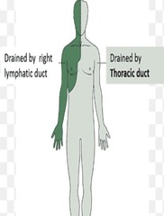
Lymphatic Collecting Ducts
Final pathways for lymph before entering veins.
Subclavian Veins
Where lymphatic fluid is returned to the bloodstream.
Stemmer Sign
Indicates stage II or III lymphedema.
Positive Stemmer Sign
Skin on fingers/toes cannot be pinched.
Stage 0 Lymphedema
No edema, occasional heaviness reported.
Stage I Lymphedema
Soft, pitting edema; reversible with elevation.
Stage II Lymphedema
Fibrosclerotic tissue; non-pitting 'brawny' edema.
Stage III Lymphedema
Severe changes; includes papillomas and infections.
Lymphedema Stages
Progressive stages from latency to elephantiasis.
Edema Grading Scale
Measures severity of edema from 1+ to 4+.
1+ Edema
Mild indentation;
2+ Edema
Moderate indentation; 1/4-to-1/2-inch pitting.
3+ Edema
Severe indentation; 1/2-to-1-inch pitting.
4+ Edema
Very severe indentation; >1 inch pitting.
Lipedema
Bilateral condition affecting lower extremities.
Lipedema Characteristics
Affects proximal areas; spares distal feet.
Lipedema Symptoms
Sensitive skin; bruises easily and is painful.
Brawny Edema
Hard swelling; non-pitting type of edema.
Fibrosclerotic Tissue
Thickened tissue due to chronic lymphedema.
Hyperkeratosis
Thickened skin; common in advanced lymphedema.
Papillomas
Skin growths associated with severe lymphedema.
Clinical Presentation
Signs and symptoms observed during physical examination.
Lymphedema Diagnosis
Identified by positive Stemmer sign and swelling.
Pressure Sensitivity
Lipedema skin is sensitive to touch.
Lymphedema
Unilateral or bilateral swelling, often asymmetric.
Lipedema
Bilateral symmetric swelling, primarily in lower extremities.
Stemmer sign
Positive in lymphedema, negative in lipedema.
Cellulitis
Common complication in lymphedema, rare in lipedema.
Pain on pressure
Absent in lymphedema, present in lipedema.
Distal edema in foot
Present in lymphedema, absent in lipedema.
Girth Measurements
Circumferential limb measurements for comparison.
Volumetric Measurements
Water displacement method for limb volume assessment.

Bioimpedance Measurements
Resistance measurement indicates extracellular fluid levels.
Indirect lymphography
Imaging technique to visualize lymphatic system.
Lymphoscintigraphy
Identifies lymphatic insufficiency during rest and exercise.

MRI/CT
Shows honeycomb patterns in soft tissue.
Ultrasonography
Assesses soft tissue thickening and fibrosis.
Fluorescent microlymphography
Detailed imaging of lymphatic vessels.
Doppler US
Differentiates lymphatic issues from venous insufficiency.
Hard lymph nodes
Indicates potential malignancy, requires physician referral.
Soft lymph nodes
Less concerning, may not require referral.
Radical mastectomy
Can cause secondary lymphedema.

Pre/Post Surgery Assessment Tool
Bioelectric impedance for lymphedema evaluation.
Stage of lymphedema
Positive Stemmer sign indicates early fibrosis.
Bilateral lower extremity edema
Characteristic of lipedema, no pedal edema.
Tender lymph nodes
Palpable and tender nodes indicate possible issues.
Lymphedema
Swelling due to lymphatic system blockage.
Goal of Treatment
Minimize lymphedema or return to latency stage.
Interstitial Pressure
Increased by external forces like compression therapy.
Dynamic Pressure Changes
Altered by breathing or muscle contractions.
Manual Lymphatic Drainage (MLD)
Technique to clear lymphatic congestion.

Proximal to Distal
Order of treatment in MLD technique.
Compression Therapy
Uses bandages to manage lymphedema phases.
Phase I Compression
Active reduction phase using low-stretch bandages.
Low-Stretch Bandages
Provide low resting pressure, high work pressure.
Distal-to-Proximal Direction
Compression applied from extremities towards trunk.
Active ROM Exercises
Range of motion exercises with compression garments.
Skin Care
Prevent skin breakdown and infection in lymphedema.
Stemmer Sign
Positive test indicates lymphedema presence.
Circumferential Measurement
Best method to measure upper arm edema.
Proximal Areas First
Clear trunk and axilla before limbs.
Distal Strokes
MLD strokes directed towards lymph nodes.
Exercise Program
Developed to improve strength and mobility.
Furosemide
Medication for managing fluid retention.
Edema Management
Lymphedema does not decrease with elevation.
Poor Grip Strength
Indicates weakness in affected limb.
Exercise Direction
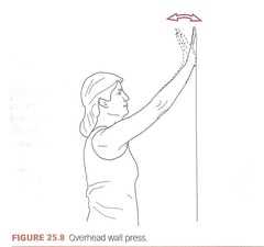
Upper extremity exercises performed proximal to distal.
High-Stretch Bandages
Not recommended for lymphedema management.